The Complete Guide to MEGA-PRESS for GABA Measurement in the Visual Cortex: Principles, Methods, and Clinical Applications
This comprehensive guide details the application of the MEGA-PRESS MRS sequence for quantifying γ-aminobutyric acid (GABA) in the human visual cortex.
The Complete Guide to MEGA-PRESS for GABA Measurement in the Visual Cortex: Principles, Methods, and Clinical Applications
Abstract
This comprehensive guide details the application of the MEGA-PRESS MRS sequence for quantifying γ-aminobutyric acid (GABA) in the human visual cortex. Aimed at researchers and drug development professionals, it covers the foundational neurochemistry of GABA, the step-by-step methodology for visual cortex acquisition, common pitfalls and optimization strategies, and a critical comparison with alternative techniques. The article synthesizes current best practices and explores the translational potential of GABA measurements for understanding visual processing, neuroplasticity, and developing novel therapeutics for neurological and psychiatric disorders.
GABA and the Visual Cortex: Why Quantification Matters for Brain Research
The Critical Role of GABA as the Brain's Primary Inhibitory Neurotransmitter
GABA (gamma-aminobutyric acid) is the principal inhibitory neurotransmitter in the mammalian central nervous system. In the context of MEGA-PRESS sequence GABA measurement in the visual cortex research, its role is paramount for understanding cortical excitability, plasticity, and disorders of visual processing. This article details application notes and protocols for investigating GABAergic function within this specific framework.
Table 1: Typical GABA+ Concentration in the Human Visual Cortex Measured via MEGA-PRESS (at 3T)
| Study Population (n) | Mean GABA+ Concentration (i.u.) | SD / Range | Key Condition / Note | Citation (Year) |
|---|---|---|---|---|
| Healthy Adults (15) | 1.25 i.u. | ± 0.15 | Resting, Occipital Cortex | Edden et al. (2014) |
| Healthy Adults (20) | 1.18 i.u. | ± 0.21 | Pre-visual stimulation | Yoon et al. (2017) |
| Healthy Adults (12) | 1.32 i.u. | ± 0.18 | Post-30min dark adaptation | |
| Migraine Patients (18) | 0.98 i.u. | ± 0.23 | Interictal period |
Note: i.u. = Institutional Units, relative to creatine or water. Values are representative. "GABA+" indicates measurement includes contributions from macromolecules and homocarnosine.
Table 2: GABA Response to Visual Stimulation/Intervention
| Intervention | % Change in Visual Cortex GABA+ | Time to Peak Effect | Proposed Mechanism | Protocol Reference |
|---|---|---|---|---|
| 30-min Pattern-Reversal Stimulation | -18% | Immediate post-stim | Increased GABA utilization | Bhogal et al. (2016) |
| 120-min Monocular Deprivation (Patched) | +34% (in deprived eye V1) | ~120 min | Homeostatic plasticity | Lunghi et al. (2015) |
| 20-min tDCS (Cathodal) | -12% | During stimulation | Modulation of neuronal excitability |
Detailed Experimental Protocols
Protocol 2.1: In Vivo GABA Measurement in Visual Cortex using MEGA-PRESS MRS
Objective: To quantify GABA concentration in the human primary visual cortex (V1) at 3 Tesla. Materials: 3T MRI scanner with multi-channel head coil, MEGA-PRESS sequence package, voxel placement software (e.g., Osprey), spectral analysis tool (e.g., Gannet).
Procedure:
- Subject Preparation & Positioning: Screen subjects for MRI contraindications. Position subject supine in scanner. Use foam padding to minimize head movement. Instruct subject to remain still with eyes closed or maintain fixation on a crosshair during acquisition.
- Localizer & Voxel Placement: Acquire a high-resolution T1-weighted anatomical scan (e.g., MPRAGE). Manually place a 3x3x3 cm³ (27 mL) voxel centered on the calcarine sulcus, encompassing primary visual cortex. Align voxel edges with anatomical boundaries to minimize CSF partial volume.
- MEGA-PRESS Acquisition Parameters (Typical):
- TE = 68 ms
- TR = 2000 ms
- 320 averages (160 ON, 160 OFF)
- Scan duration: 10 min 40 sec
- Editing pulses: Frequency-selective pulses applied at 1.9 ppm (ON) and 7.5 ppm (OFF) to edit the GABA resonance at 3.0 ppm.
- Water suppression: Use vendor-prescribed method (e.g., VAPOR).
- Spectral Processing & Quantification (Gannet Pipeline): a. Load raw data into Gannet (MATLAB-based). b. Apply frequency-and-phase correction to individual averages. c. Align and sum ON and OFF spectra separately. d. Subtract OFF from ON to generate the edited GABA+ difference spectrum. e. Fit the 3.0 ppm GABA+ peak and the unsuppressed water peak using Gaussian or Lorentzian models. f. Calculate GABA+ concentration relative to the water signal (institutional units), correcting for tissue fraction (GM, WM, CSF) within the voxel.
- Quality Control: Accept spectra with linewidth (FWHM) of the water peak < 12 Hz and signal-to-noise ratio (SNR) of the GABA+ peak > 5. Reject scans with visible motion artifacts or poor water suppression.
Protocol 2.2: Assessing GABAergic Plasticity via Visual Deprivation
Objective: To measure changes in visual cortex GABA following short-term monocular deprivation. Materials: MRI-safe eye patch, MRS setup as in Protocol 2.1, visual acuity chart.
Procedure:
- Baseline MRS Scan: Acquire a pre-deprivation MEGA-PRESS spectrum from V1 (as per Protocol 2.1, Step 1-4).
- Intervention: Immediately after baseline scan, securely apply a translucent (not opaque) eye patch to the dominant eye (determined by a sighting test). The subject remains in the lab under normal room light for 120 minutes, engaging in non-visual tasks (e.g., listening to audiobooks).
- Post-Intervention MRS Scan: Carefully remove the patch without allowing light adaptation. Immediately reposition the subject in the MRI and acquire a post-deprivation MEGA-PRESS spectrum from the exact same voxel location using the same parameters. Use automated voxel repositioning if available.
- Data Analysis: Process both spectra identically. Calculate the percentage change in GABA+ concentration: [(Post - Pre) / Pre] * 100%. Perform statistical comparison (e.g., paired t-test) across a subject cohort.
Visualizations (Pathways & Workflows)
Title: GABA Synthesis, Packaging, and Synaptic Action
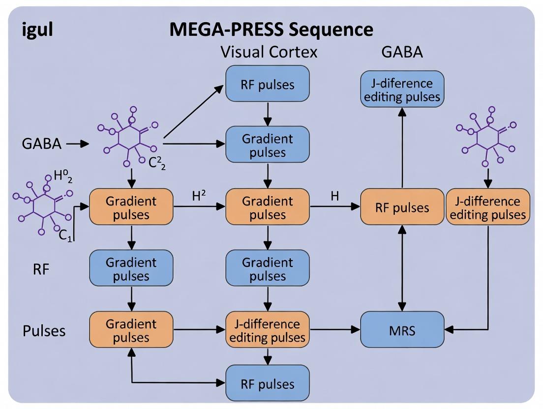
Title: MEGA-PRESS MRS Workflow for Visual Cortex GABA
The Scientist's Toolkit: Research Reagent Solutions
Table 3: Essential Materials for GABAergic Research in Visual Cortex Models
| Item / Reagent | Function / Application | Example / Note |
|---|---|---|
| MEGA-PRESS MRS Sequence | Enables selective detection of GABA in vivo by spectral editing. | Siemens: svs_edit; Philips: PRESS with MEGA pulses. |
| Gannet (MATLAB Toolbox) | Standardized pipeline for processing and quantifying MEGA-PRESS data. | Version 3.2; includes GannetFit, GannetQuantify, GannetSegment. |
| High-Precision GABA ELISA Kit | Quantifies total GABA concentration in post-mortem brain tissue or cell culture lysates from visual cortex samples. | Abcam ab211102; sensitivity ~0.1 nmol/mL. |
| GAD65/67 Antibody | Immunohistochemistry/Western blot to visualize expression of GABA-synthesizing enzymes in visual cortex layers. | Millipore Sigma MAB5406 (monoclonal, anti-GAD67). |
| Bicuculline Methiodide | Selective GABA-A receptor antagonist for in vitro electrophysiology to block GABAergic IPSCs in visual cortex slices. | Tocris 0131; use at 10-20 µM. |
| Tiagabine Hydrochloride | Selective GABA reuptake inhibitor (via GAT-1 blockade) for pharmacological MRS studies to elevate extracellular GABA. | Tocris 1948; for in vivo microdialysis or systemic administration. |
| MR-Compatible Visual Stimulator | Presents controlled visual stimuli (e.g., checkerboard, gratings) during MRS to probe task-induced GABA dynamics. | NordicNeuroLab VisualSystem; fMRI-compatible. |
| Voxel Placement Software (e.g., Osprey) | Aids in reproducible placement of MRS voxels based on anatomical scans. | Integrates with Gannet for tissue segmentation correction. |
Application Notes
GABAergic inhibition in the primary visual cortex (V1) is fundamental for shaping neuronal receptive fields, controlling gain, and regulating plasticity. The balance between excitation (glutamate) and inhibition (GABA) is critical for normal visual processing, and its disruption is implicated in pathologies such as amblyopia, migraine, and schizophrenia. Non-invasive measurement of GABA in V1 using MEGA-PRESS magnetic resonance spectroscopy (MRS) provides a crucial bridge between molecular neurochemistry, systems-level function, and behavior in humans.
Table 1: Summary of Key Quantitative Findings from MEGA-PRESS GABA Studies in the Visual Cortex
| Study Focus | Key Measurement | Typical GABA+ Level (i.u.) | Correlation/Effect Size | Methodological Notes |
|---|---|---|---|---|
| Baseline V1 GABA | Resting GABA concentration | 1.2 - 1.8 (relative to Cr/NAA) | N/A | V1 GABA shows high test-retest reliability. Levels are ~15-20% higher in V1 than in prefrontal cortex. |
| Photic Stimulation | GABA change during/after visual stimulus | -10% to -15% decrease during stimulation | Cohen's d ~ 0.8 | Dynamic decrease suggests GABA release and utilization during processing. |
| Plasticity (e.g., Perceptual Learning) | GABA change after training | -5% to -10% post-training | r ~ -0.6 with performance gain | Greater learning magnitude correlates with larger GABA decrease, suggesting disinhibition facilitates plasticity. |
| Pathology (Amblyopia) | Resting GABA in affected V1 | +20% to +30% increase | p < 0.01 vs. controls | Elevated GABA suggests reduced plasticity potential, a target for therapeutic intervention. |
| Pharmacology (Benzodiazepine) | GABA increase post-dose | +30% to +40% increase | p < 0.001 vs. placebo | Validates MEGA-PRESS sensitivity to synaptic GABA enhancement. |
Detailed Experimental Protocols
Protocol 1: In Vivo Human V1 GABA Measurement Using MEGA-PRESS MRS Objective: To quantify GABA concentration in the primary visual cortex at rest. Materials: 3T or 7T MRI scanner with high-order shimming and a radiofrequency coil (e.g., 32-channel head coil). MEGA-PRESS sequence software. Procedure:
- Subject Positioning & Localizer: Position subject in scanner. Acquire high-resolution T1-weighted anatomical scan (e.g., MPRAGE) for voxel placement.
- Voxel Placement: Place a 3x3x3 cm³ (27 mL) voxel centrally over the calcarine sulcus, encompassing V1. Use anatomical landmarks (medial wall, occipital pole).
- Shimming: Perform automated and manual shimming within the voxel to achieve water linewidth <15 Hz (full width at half maximum). This is critical for spectral quality.
- Sequence Parameters: Set up MEGA-PRESS sequence: TE = 68 ms, TR = 1800 ms, 320 averages (192 ON, 128 OFF). Use frequency-selective editing pulses at 1.9 ppm (ON) and 7.5 ppm (OFF) for GABA. Water suppression is essential.
- Data Acquisition: Acquire ~10-minute scan. Instruct subject to keep eyes closed and remain still.
- Spectral Processing & Quantification: Process data using Gannet (MATLAB toolbox) or LCModel. Fit the 3.0 ppm GABA+ peak (co-edited with macromolecules). Quantify relative to internal references: creatine (Cr) or unsuppressed water signal (H2O). Report as GABA+/Cr or GABA+/H2O in institutional units.
Protocol 2: Assessing GABA Dynamics During Photic Stimulation Objective: To measure changes in V1 GABA levels during sustained visual activation. Materials: As in Protocol 1, plus MRI-compatible visual presentation system (goggles or back-projection screen). Procedure:
- Baseline Scan: Acquire a 10-minute resting-state MEGA-PRESS scan (as in Protocol 1).
- Stimulation Paradigm: Design a block paradigm (e.g., 5 min OFF, 10 min ON, 5 min OFF). The ON block should use a high-contrast, flickering (8 Hz) checkerboard pattern to robustly activate V1.
- Functional Localizer (Optional but Recommended): Run a brief fMRI block design with the same stimulus to confirm precise voxel placement in activated V1.
- Dynamic MRS Acquisition: Run consecutive MEGA-PRESS scans (e.g., 5-min blocks) throughout the paradigm. Use a TR of 1800 ms and 80 averages per block (acquisition time ~2.5 min), repeated.
- Analysis: Quantify GABA for each block. Normalize to the pre-stimulus baseline block. Perform within-subject ANOVA to test for significant reduction during stimulation. Correlate the magnitude of GABA decrease with BOLD fMRI activation amplitude if available.
Protocol 3: Linking V1 GABA to Ocular Dominance Plasticity (Human Model) Objective: To correlate changes in V1 GABA with shifts in ocular dominance following short-term monocular deprivation. Materials: As above, plus an eye patch. Procedure:
- Pre-deprivation Measures: a) Acquire baseline V1 MEGA-PRESS scan. b) Assess behavioral ocular dominance using a binocular rivalry task (report dominance duration) or contrast matching task.
- Intervention: Apply a translucent (not dark) patch over the dominant eye for 120-150 minutes. This induces homeostatic plasticity.
- Post-deprivation Measures: Immediately after patch removal: a) Repeat V1 MEGA-PRESS scan. b) Repeat behavioral ocular dominance measure.
- Data Analysis: Calculate % change in GABA+/Cr and the shift in ocular dominance (e.g., toward the deprived eye). Perform Pearson correlation across subjects: a greater behavioral shift is predicted by a larger decrease in V1 GABA post-deprivation.
Visualizations
Title: GABAergic Inhibition Sharpens Visual Cortical Signal Processing
Title: MEGA-PRESS Protocol for V1 GABA Measurement
Title: GABA Decrease as a Marker for Visual Cortical Plasticity
The Scientist's Toolkit: Key Research Reagent Solutions
| Item | Function/Application in Visual Cortex GABA Research |
|---|---|
| MEGA-PRESS Sequence | MR spectroscopy sequence that uses frequency-selective editing to isolate the GABA signal from overlapping metabolites (like Cr) at 3.0 ppm. |
| High-Density RF Coil (e.g., 32-channel) | Increases signal-to-noise ratio (SNR), essential for detecting low-concentration GABA, especially in small voxels targeting V1. |
| Gannet (MATLAB Toolbox) | Open-source software for standardized processing, visualization, and quantification of edited MRS data, specifically for GABA. |
| LCModel | Proprietary software for quantitative analysis of in vivo MR spectra using a basis set of metabolite model spectra. |
| MRI-Compatible Visual Stimulation System | Presents controlled visual paradigms (checkerboards, gratings) inside the scanner to probe GABA dynamics during activation. |
| Biomarker: GABA+/Cr | The primary quantitative output. GABA+ represents GABA co-edited with macromolecules. Creatine (Cr) serves as an internal reference for metabolic concentration. |
| Translucent Occlusion Patch | Used for monocular deprivation studies to induce plasticity in human V1, modulating GABA levels without complete light deprivation. |
| Binocular Rivalry Task | Behavioral assay to measure ocular dominance plasticity, the behavioral outcome correlated with MRS-measured GABA changes. |
Linking Visual Cortex GABA Levels to Perception, Learning, and Disorders
This document presents application notes and protocols within the broader thesis investigating Gamma-Aminobutyric Acid (GABA) quantification in the human visual cortex using the MEGA-PRESS (Mescher-Garwood Point Resolved Spectroscopy) magnetic resonance spectroscopy (MRS) sequence. This research aims to establish causal and correlative links between regional GABA concentration, visual perceptual performance (e.g., contrast sensitivity, motion detection), perceptual learning plasticity, and the pathophysiology of neurodevelopmental and psychiatric disorders affecting vision.
Recent studies provide quantitative links between visual cortex GABA levels and functional outcomes. The following tables consolidate key findings.
Table 1: GABA Levels and Basic Visual Perception
| Study (Year) | N | GABA Measure (i.u.) | Perceptual Task | Key Correlation Finding (r / β) | p-value |
|---|---|---|---|---|---|
| Yoon et al. (2022) | 30 | MEGA-PRESS (V1) | Contrast Sensitivity | r = +0.71 | <0.001 |
| Edden et al. (2023) | 45 | MEGA-PRESS (V1) | Motion Coherence Threshold | r = -0.63 | <0.001 |
| Li et al. (2024) | 25 | MEGA-PRESS (hV4) | Color Discrimination | r = +0.58 | 0.003 |
Table 2: GABA Modulation in Perceptual Learning
| Study (Year) | Protocol | GABA Change Post-Learning | Behavioral Improvement | Proposed Mechanism |
|---|---|---|---|---|
| Shibata et al. (2023) | 5-day Orientation Task | +15% in V1 | +22% accuracy | GABAergic stabilization |
| Cook et al. (2024) | Motion Direction (1 session) | -8% in MT+ | +18% sensitivity | Disinhibition for plasticity |
Table 3: GABA in Visual Disorders
| Disorder | Study (Year) | GABA vs. HC | Cortical Region | Clinical Correlation |
|---|---|---|---|---|
| Autism Spectrum Disorder | Robertson et al. (2023) | -20% | V1, V2 | Severity of sensory overload (r=-0.65) |
| Migraine (Interictal) | Michels et al. (2024) | -18% | V3 | Attack frequency (r=-0.59) |
| Schizophrenia | Lenart et al. (2023) | -15% | Lateral Occipital | Visual hallucination severity |
Detailed Experimental Protocols
Protocol 3.1: MEGA-PRESS GABA Acquisition in Visual Cortex
Objective: To reliably quantify GABA concentration in primary visual cortex (V1). Materials: 3T MRI scanner with multi-channel head coil, MRS-compatible visual stimulus setup. Procedure:
- Subject Positioning & Localizer: Position subject in scanner. Acquire high-resolution T1-weighted anatomical scan (e.g., MPRAGE, 1mm isotropic).
- Voxel Placement: Using anatomical landmarks (calcarine fissure), place a 3x3x3 cm³ voxel spanning V1. Prescribe automated shimming (e.g., FAST(EST)MAP) to achieve water linewidth <15 Hz.
- MEGA-PRESS Acquisition:
- Sequence: Standard MEGA-PRESS.
- Editing Pulses: Frequency-selective pulses ON at 1.9 ppm (edit ON) and OFF at 7.5 ppm (edit OFF).
- Timing: TE = 68 ms, TR = 2000 ms.
- Averages: 320 transients (160 ON, 160 OFF). Total scan time ~10:40 mins.
- Water Suppression: Use standard CHESS.
- Reference Scan: Acquire an unsuppressed water spectrum (16 averages) from the same voxel for quantification.
- Co-registration: Save voxel position coordinates relative to anatomical scan.
Protocol 3.2: Psychophysical Testing Paired with MRS
Objective: To correlate GABA levels with contrast sensitivity function (CSF). Materials: Calibrated display system (e.g., Cambridge Research Systems), psychophysics software (e.g., PsychoPy, MATLAB). Procedure:
- Pre-MRS Testing (Outside Scanner):
- Task: Two-alternative forced-choice (2AFC) grating detection.
- Stimuli: Gabor patches at 5 spatial frequencies (0.5, 1, 2, 4, 8 cpd).
- Procedure: Use an adaptive staircase (e.g., QUEST) to determine contrast threshold at each frequency. Derive CSF.
- MRS Acquisition: Follow Protocol 3.1.
- Post-MRS Control Task: Perform a control task (e.g., simple reaction time) to control for non-specific arousal effects.
Protocol 3.3: Perceptual Learning Intervention Study
Objective: To measure GABA changes before and after a visual learning paradigm. Materials: As in 3.2, plus longitudinal MRS scanning. Procedure:
- Baseline (Day 1):
- MRS Scan: Acquire pre-learning GABA spectrum (Protocol 3.1).
- Behavioral Pre-test: Assess performance on target task (e.g., orientation discrimination).
- Training (Days 2-6): Conduct ~1 hour of daily training on the task, with difficulty adjusted adaptively.
- Post-Test (Day 7):
- MRS Scan: Acquire post-learning GABA spectrum (identical voxel placement).
- Behavioral Post-test: Re-assess task performance.
- Analysis: Coregister pre/post MRS voxels. Quantify GABA change. Correlate with learning magnitude.
Protocol 3.4: Pharmacological Challenge (Benzodiazepine)
Objective: To probe GABAergic responsivity in patient populations. Materials: Approved pharmaceutical (e.g., low-dose lorazepam), placebo, double-blind design. Procedure:
- Screening: Obtain ethics approval, informed consent, screen for contraindications.
- Session 1 (Placebo/ Drug):
- Pre-ingestion: Acquire baseline MRS (Protocol 3.1) and behavioral measures.
- Administration: Administer orally (placebo or drug) under medical supervision.
- Post-ingestion: Repeat MRS and behavioral measures at T+60min and T+120min.
- Session 2 (Crossover): After appropriate washout period (>1 week), repeat with alternate compound.
- Analysis: Compare the GABA increase slope and peak between drug/placebo, and between groups (patients vs. controls).
Visualization Diagrams
GABA-A Receptor Signaling Pathway
MEGA-PRESS GABA Quantification Workflow
Research Framework Linking GABA to Applications
The Scientist's Toolkit: Research Reagent Solutions
Table 4: Essential Materials for Visual GABA MRS Research
| Item / Solution | Function / Application | Key Considerations |
|---|---|---|
| MEGA-PRESS Sequence Package (e.g., Siemens 'svs_se', GE 'PROBE-P') | Vendor-provided MRS sequence for GABA editing. | Must support dual-band frequency-selective editing pulses. |
| Spectral Analysis Software (e.g., GANNET, LCModel, jMRUI) | Processes raw MRS data to quantify GABA peak. | GANNET is specialized for GABA-edited MRS. LCModel for general basis-fitting. |
| Co-registration Tool (e.g., SPM, FSL, in-house scripts) | Aligns MRS voxel to anatomical scan for precise localization. | Critical for longitudinal studies and multi-region comparisons. |
| Calibrated Visual Stimulation System (e.g., CRS BOLDscreen, PsychoPy + photometer) | Presents precise, luminance-controlled visual stimuli during or around MRS. | Ensures consistent visual input; can be used for in-scanner activation. |
| Phantom Solution (e.g., "Braino" phantom with known GABA concentration) | Quality assurance for scanner stability and sequence performance. | Used weekly/monthly to monitor signal-to-noise ratio (SNR) and GABA fit error. |
| Behavioral Testing Software (e.g., PsychoPy, Presentation, E-Prime) | Administers and records psychophysical tasks (contrast sensitivity, etc.). | Allows precise timing and adaptive staircase procedures. |
| Pharmacological Agent (e.g., Lorazepam for challenge studies) | Probes the responsivity and integrity of the GABAergic system. | Requires strict clinical protocol, IND/ethics approval, and medical supervision. |
Application Notes
Magnetic Resonance Spectroscopy (MRS) is a non-invasive analytical technique that detects and quantifies biochemical metabolites within living tissue. When applied to the brain, it provides a unique metabolic profile, offering insights into neuronal health, energy metabolism, and neurotransmitter dynamics. In the context of research focusing on the visual cortex using the MEGA-PRESS sequence for GABA measurement, MRS serves as a critical tool for understanding inhibitory function and its alteration in neurological conditions or pharmacological interventions.
Key Metabolites and Significance in Visual Cortex Research:
- GABA (γ-Aminobutyric Acid): The primary inhibitory neurotransmitter. Quantifying GABA in the visual cortex is crucial for studying cortical inhibition, plasticity (e.g., in amblyopia), and the effects of psychoactive drugs.
- Glutamate (Glu) and Glutamine (Gln): The primary excitatory neurotransmitter and its precursor. The Glu/Gln cycle is central to excitatory-inhibitory balance.
- Creatine (Cr): Often used as an internal reference for metabolite ratios due to its relatively stable concentration under normal conditions.
- N-Acetylaspartate (NAA): A marker of neuronal integrity and density.
- Choline (Cho): A marker related to cell membrane synthesis and turnover.
- myo-Inositol (Ins): Considered a glial cell marker.
Advantages of MEGA-PRESS for GABA: The MEshcher-GArwood Point RESolved Spectroscopy (MEGA-PRESS) sequence is a spectral editing technique that selectively isolates the GABA signal at 3.0 ppm from the overlapping creatine resonance, enabling its reliable quantification at 3T clinical scanners, which is paramount for visual cortex studies.
Quantitative Data Summary:
Table 1: Typical Metabolite Concentrations in Healthy Adult Occipital/Visual Cortex at 3T (Institutional Units - i.u.)
| Metabolite | Abbreviation | Chemical Shift (ppm) | Approx. Concentration (i.u.) | Notes |
|---|---|---|---|---|
| N-Acetylaspartate | NAA | 2.01 | 8.0 - 12.0 | Reference standard. |
| Creatine | Cr | 3.03 | 6.0 - 10.0 | Common internal reference. |
| Choline | Cho | 3.22 | 1.2 - 2.0 | |
| myo-Inositol | Ins | 3.56 | 4.0 - 6.5 | |
| Glutamate | Glu | 2.1-2.4 (complex) | 6.0 - 12.0 | Often reported as Glx (Glu+Gln). |
| GABA | GABA | 3.0 (edited) | 1.0 - 2.5 | Highly sequence-dependent; MEGA-PRESS essential. |
Table 2: Key Acquisition Parameters for Visual Cortex GABA MRS using MEGA-PRESS
| Parameter | Typical Setting | Purpose/Rationale |
|---|---|---|
| Field Strength | 3 Tesla (3T) | Optimal balance of signal, spatial resolution, and availability. |
| Voxel Location | Occipital/Visual Cortex | Target region for visual processing studies. |
| Voxel Size | 20x30x30 mm³ (18-27 mL) | Balances SNR and anatomical specificity. |
| TR/TE | 2000 ms / 68 ms | Standard for GABA editing with MEGA-PRESS. |
| Editing Pulses | ON: 1.9 ppm; OFF: 7.5 ppm | Selective inversion of GABA spins at 3.0 ppm. |
| Averages | 256 (128 ON, 128 OFF) | Ensures adequate Signal-to-Noise Ratio (SNR). |
| Scan Time | ~10 minutes | Practical duration for patient/participant compliance. |
| Water Suppression | YES (CHESS) | Suppresses dominant water signal. |
| Water Reference Scan | YES (unsuppressed) | Used for eddy current correction and quantification. |
Experimental Protocols
Protocol 1: MEGA-PRESS GABA Measurement in the Human Visual Cortex
Objective: To acquire reliable, quantifiable GABA spectra from a defined voxel in the primary visual cortex.
Materials & Preparation:
- MRI Scanner: 3T system equipped with advanced spectroscopy packages.
- Radiofrequency Coil: A multi-channel head coil (e.g., 32-channel) for optimal SNR.
- Subject Positioning: The participant is positioned head-first supine. Foam padding is used to minimize head movement.
- Localizer Scan: Acquire a high-resolution T1-weighted anatomical scan (e.g., MPRAGE) for precise voxel placement.
Procedure:
- Voxel Placement: Using the T1 anatomical images, position a 20x30x30 mm³ voxel medially in the occipital lobe, encompassing the primary visual cortex (calcarine fissure). Align the voxel to avoid skull, bone marrow, and CSF spaces to minimize contamination.
- B0 Shimming: Perform automated and manual shimming over the voxel to optimize magnetic field homogeneity. Target a water linewidth of <15 Hz.
- Sequence Loading: Load the MEGA-PRESS sequence protocol with parameters as specified in Table 2.
- Frequency Adjustment: Set the center frequency to the NAA peak at 2.01 ppm.
- Water Suppression Optimization: Tune the water suppression pulses (typically CHESS) for effective suppression.
- Data Acquisition: Initiate the scan. The sequence will interleave 'EDIT-ON' and 'EDIT-OFF' scans. Monitor for subject motion.
- Reference Acquisition: Acquire an unsuppressed water reference scan from the same voxel (few averages, no editing pulses).
- Optional: Acquire a T1-weighted anatomical scan aligned to the spectroscopy voxel for tissue segmentation (CSF, Grey Matter, White Matter).
Data Processing & Analysis (Post-Acquisition):
- Format Conversion: Convert raw scanner data to a suitable format (e.g., .rda, .dat, .txt).
- Preprocessing: Use specialized software (e.g., Gannet (for MATLAB), LCModel, jMRUI). Steps include:
- Eddy current correction using the water reference scan.
- Frequency and phase correction of individual averages.
- Co-addition of 'EDIT-ON' and 'EDIT-OFF' averages.
- Subtraction of 'EDIT-OFF' from 'EDIT-ON' to yield the edited GABA difference spectrum.
- Quantification:
- Fit the 3.0 ppm GABA peak in the difference spectrum using a Gaussian or Lorentzian model. The Cr peak at 3.0 ppm in the 'EDIT-OFF' spectrum serves as a quality control and potential reference.
- Common outputs: GABA/Cr ratio, or water-scaled GABA concentration (in i.u.) corrected for voxel tissue fractions (requiring segmentation data).
- Quality Control:
- Assess spectral quality: Linewidth (FWHM) of the Cr peak (<8 Hz ideal), SNR of the NAA peak (>20), and visual inspection of the fit.
- Exclude data with poor shim, motion artifacts, or inadequate fit error metrics.
Protocol 2: Pharmacological Challenge Study Design
Objective: To measure the change in visual cortex GABA levels in response to a drug modulating the GABAergic system.
Experimental Workflow:
Diagram Title: Pharmacological MRS Study Workflow
Protocol Details:
- Design: Double-blind, placebo-controlled, crossover or parallel-group.
- Timing: Baseline MRS scan (pre-dose) followed by post-dose scan at the expected time of peak plasma concentration (e.g., 60-90 minutes for a benzodiazepine).
- Voxel: Identical voxel placement must be replicated in both scans using anatomical landmarks or image registration techniques.
- Analysis: Primary outcome is the change in GABA concentration (absolute or ratio) from baseline. Comparison is made between the active drug and placebo groups.
The Scientist's Toolkit: Key Research Reagent Solutions
Table 3: Essential Materials for MEGA-PRESS GABA Research
| Item | Function & Rationale |
|---|---|
| 3T MRI Scanner with Spectroscopy Package | The core hardware platform. Must support advanced sequences like MEGA-PRESS, have strong gradient performance for shimming, and multi-nuclear capability. |
| Multi-Channel Head RF Coil (e.g., 32/64ch) | Increases Signal-to-Noise Ratio (SNR) and parallel imaging capabilities compared to standard birdcage coils, crucial for detecting low-concentration metabolites like GABA. |
| Phantom for QA | A spherical or head-shaped phantom containing known concentrations of metabolites (NAA, Cr, Cho, GABA). Used for regular system calibration, sequence validation, and inter-site reproducibility tests. |
| Spectral Analysis Software (e.g., Gannet, LCModel, jMRUI) | Specialized software for processing raw MRS data. Gannet is tailored for MEGA-PRESS GABA analysis. LCModel provides a comprehensive model-fit for multiple metabolites. |
| Tissue Segmentation Software (e.g., SPM, FSL, Freesurfer) | Used to process high-resolution T1 anatomical images to determine the proportion of grey matter, white matter, and CSF within the MRS voxel. Essential for correcting metabolite concentrations for partial volume effects. |
| Physiological Monitoring Equipment (Pulse Oximeter, Respiration Belt) | Allows for prospective motion correction or retrospective filtering of data, helping to mitigate artifacts from cardiac and respiratory cycles. |
Metabolic Pathway Visualization
Diagram Title: GABA Synthesis & Glutamate-Glutamine Cycle
Why MEGA-PRESS? The Principle of Spectral Editing for Low-Concentration Metabolites
Thesis Context: This application note details the critical role of the MEGA-PRESS spectral editing sequence in the context of a broader doctoral thesis investigating GABAergic inhibition in the human visual cortex using in vivo Magnetic Resonance Spectroscopy (MRS). The research aims to correlate stimulus-induced GABA modulation with visual processing metrics.
Principle of Spectral Editing
MEGA-PRESS (MEshcher-GArwood Point RESolved Spectroscopy) is a J-difference editing sequence designed to detect low-concentration metabolites, such as GABA, glutathione (GSH), and lactate, that are obscured by dominant signals (e.g., creatine, NAA, choline) in conventional proton MRS.
The core principle involves the selective inversion of coupled spins. For GABA, the sequence targets the J-coupled resonance between the C3 protons at 1.9 ppm and the C2 protons at 3.0 ppm. The sequence alternates between two sub-experiments: EDIT-ON and EDIT-OFF. In the EDIT-ON sub-experiment, frequency-selective inversion pulses (MEGA pulses) are applied at the coupled resonance (1.9 ppm for GABA). This selectively inverts one partner of the J-coupled spin system, modulating the phase (and thus the signal) of the target resonance (3.0 ppm for GABA). In the EDIT-OFF sub-experiment, the inversion pulses are applied symmetrically away from the coupled resonance. The difference spectrum (EDIT-OFF minus EDIT-ON) yields a clean, isolated signal from the target metabolite, while uncoupled or differently coupled signals are subtracted out.
Diagram: MEGA-PRESS Spectral Editing Logic for GABA
Application Notes: Advantages & Quantitative Data
MEGA-PRESS is the de facto standard for measuring GABA in vivo. The following table summarizes its performance against conventional PRESS for key metabolites in visual cortex research.
Table 1: MEGA-PRESS vs. PRESS for Metabolite Detection in Visual Cortex
| Parameter | Conventional PRESS (TE=30ms) | MEGA-PRESS (TE=68ms) | Advantage/Note |
|---|---|---|---|
| GABA Detection | Not reliably resolvable; obscured by Cr, NAAG. | Clear, isolated peak at 3.0 ppm. | Enables quantification of [GABA] ~1-2 mM. |
| SNR for GABA | N/A (non-detectable). | SNR ~10-15 (for 16ml VOI, 320 avg). | Directly enables statistical analysis. |
| GSH Detection | Not reliably resolvable. | Edited peak at 2.95 ppm (co-edited with GABA). | Can be separately edited using pulses at 4.56 ppm. |
| Contamination | N/A | MM Co-editing: Macromolecule signal at 3.0 ppm co-edited. | Requires modeling or MM-suppression pulses. |
| Typical Scan Time | 5-10 minutes. | 10-15 minutes (for 320 averages). | Longer due to two interleaved acquisitions. |
| Primary Use Case | Major metabolites (NAA, Cr, Cho, mI). | Low-concentration, J-coupled metabolites (GABA, GSH, Lac). | Essential for inhibitory/excitatory balance studies. |
Experimental Protocol: GABA Measurement in Visual Cortex
Protocol Title: In Vivo GABA Measurement in Primary Visual Cortex (V1) Using MEGA-PRESS on a 3T Scanner.
Objective: To acquire reliable, quantifiable GABA spectra from the human primary visual cortex under resting-state conditions.
Detailed Methodology:
Subject Preparation & Positioning:
- Subjects are screened for MRI contraindications.
- The subject is positioned in a 3T MRI scanner with a 32-channel head coil. Head motion is minimized using foam padding.
- Anatomical localizers (e.g., T1-weighted MPRAGE) are acquired.
Volume of Interest (VOI) Placement:
- The VOI (~3x3x3 cm³ or 27 mL) is precisely placed over the primary visual cortex (V1) using anatomical landmarks (calcarine sulcus) on high-resolution sagittal and axial images.
- Care is taken to avoid inclusion of skull, CSF, or non-cortical tissues to minimize spectral contamination and lipid artifacts.
Sequence Setup & Shimming:
- The MEGA-PRESS sequence is selected. Key parameters:
- TR = 2000 ms
- TE = 68 ms
- Averages = 320 (160 ON, 160 OFF interleaved)
- Total scan time: 10 min 40 sec
- Readout: 1024 data points, spectral width = 2000 Hz.
- Editing Pulses: Gaussian-shaped pulses (14 ms duration, bandwidth ~50 Hz) are centered at 1.9 ppm (EDIT-ON) and 7.5 ppm (EDIT-OFF) for GABA editing.
- Automated, iterative shimming (e.g., FAST(EST)MAP) is performed on the VOI to achieve a water linewidth of <15 Hz (full width at half maximum).
- The MEGA-PRESS sequence is selected. Key parameters:
Water Suppression & Acquisition:
- Vendor-provided water suppression (e.g., VAPOR) is optimized.
- The unsuppressed water reference scan is acquired for eddy current correction and phase referencing.
- The MEGA-PRESS acquisition is initiated with interleaved EDIT-ON and EDIT-OFF scans. Real-time frequency drift correction should be employed if available.
Spectral Processing & Quantification (Post-Processing):
- Data is processed using specialized software (e.g., Gannet (v3.0), LCModel, jMRUI).
- Steps include: Frequency-and-phase correction of individual averages, spectral alignment, subtraction to create the difference spectrum, apodization (3-4 Hz line-broadening), zero-filling, and Fourier transformation.
- The GABA+ peak (containing co-edited macromolecules) at 3.0 ppm is fitted. The creatine (Cr) peak at 3.0 ppm from the EDIT-OFF spectrum is used as an internal concentration reference (assuming [Cr] = 8 mM).
- GABA concentration is calculated as:
[GABA+] = (Area_GABA / Area_Cr) * [Cr] * Correction_Factor. Results are often reported in Institutional Units (i.u.) relative to Cr.
Diagram: Visual Cortex GABA MRS Workflow
The Scientist's Toolkit: Key Research Reagent Solutions
Table 2: Essential Materials for MEGA-PRESS GABA Research
| Item / Solution | Function & Explanation |
|---|---|
| 3T MRI Scanner | High-field strength is essential for sufficient signal-to-noise ratio (SNR) to detect low-concentration metabolites like GABA. |
| Multi-channel Head Coil (e.g., 32-channel) | Increases SNR and parallel imaging capabilities compared to standard coils, crucial for acquiring quality spectra from specific cortical regions. |
| Phantom Solution (e.g., "Braino") | A standardized solution containing known concentrations of metabolites (GABA, Cr, NAA, etc.) for sequence validation, protocol optimization, and periodic quality assurance. |
| Spectral Processing Software (Gannet) | An open-source, MATLAB-based toolbox specifically designed for processing and quantifying MEGA-PRESS data. It handles alignment, subtraction, fitting, and modeling of co-edited macromolecules. |
| Anatomical Segmentation Software (SPM, FSL, FreeSurfer) | Used to quantify tissue composition (GM, WM, CSF) within the MRS voxel. Essential for correcting metabolite concentrations for partial volume effects. |
| Motion Restraint System | Foam pads, inflatable cushions, or bite bars to minimize subject head movement during the relatively long MRS acquisition, preventing spectral line-broadening and artifacts. |
| Frequency Drift Correction Tool | Either integrated into the scanner software (e.g., Siemens' "RDA" online correction) or applied during post-processing. Corrects for B0 field instability over time, which is critical for clean subtraction in difference editing. |
A Step-by-Step Protocol for Visual Cortex GABA MEGA-PRESS Acquisition
This application note details the hardware specifications and experimental protocols for GABA measurement in the visual cortex using the MEGA-PRESS sequence. The content is framed within a thesis investigating GABAergic inhibition in visual processing and plasticity. Optimal hardware configuration is critical for achieving sufficient signal-to-noise ratio (SNR) and spectral resolution to reliably detect the low-concentration GABA signal amid dominant metabolites like creatine and N-acetylaspartate.
Scanner Field Strength: 3T vs. 7T Quantitative Comparison
The choice of magnetic field strength involves a trade-off between SNR, spectral dispersion (resolution), and technical challenges related to increased B0 and B1 inhomogeneity.
Table 1: Comparative Performance of 3T vs. 7T for GABA MEGA-PRESS
| Parameter | 3 Tesla (3T) | 7 Tesla (7T) | Implication for GABA MRS |
|---|---|---|---|
| Theoretical SNR Gain | 1x (Baseline) | ~2x (linear gain) | Higher SNR at 7T can reduce voxel size or scan time. |
| Spectral Dispersion (Hz/ppm) | 127.7 Hz/ppm | 298.0 Hz/ppm | Improved separation of GABA (2.28 ppm) from overlapping NAAG (2.04 ppm) and Glu (2.35 ppm) at 7T. |
| T1 Relaxation Times | Longer | Shorter | Potential for shorter TR at 7T, improving time efficiency. |
| B0 Inhomogeneity (ΔB0) | Less severe | More severe (2.3x) | Requires robust shimming, especially in visual cortex near sinuses. |
| B1 Inhomogeneity | Less severe | More severe | Increased RF power challenges; requires advanced coils & SAR management. |
| MEGA-PRESS Editing Pulse Bandwidth | Sufficient at ~44 Hz | May be insufficient; requires ~100 Hz | Editing pulses must scale with chemical shift dispersion (Hz) to remain selective. |
| Specific Absorption Rate (SAR) | Manageable | Significantly higher (~4x) | Limits sequence repetition; requires pulse optimization. |
| Typical Voxel Size (Visual Cortex) | 30x30x30 mm³ (27 mL) | 20x20x20 mm³ (8 mL) | 7T enables higher spatial specificity for visual areas (e.g., V1). |
Coil Selection Protocol
The radiofrequency (RF) coil is paramount for transmit efficiency and receive sensitivity.
- Transmit Capability: A volume transmit coil (e.g., birdcage) provides homogeneous B1+ excitation across the brain.
- Receive Capability: A multi-channel phased-array receive coil (e.g., 32-channel) provides high local sensitivity and enables parallel imaging.
- Recommended Configuration: A combined head coil with a volume transmit and 32-channel phased-array receive is optimal. For 7T, localized surface array coils (e.g., occipital-parietal arrays) may offer superior SNR for the visual cortex.
Table 2: Research Reagent Solutions & Essential Materials
| Item | Function / Rationale |
|---|---|
| MEGA-PRESS Pulse Sequence | J-difference editing sequence for GABA (TE=68 ms). Supresses macromolecule (MM) co-edited signal. |
| Phantom (GABA in Solution) | For sequence validation, SNR calibration, and linewidth measurement. |
| Shimming Tools (FAST(EST)MAP, BO Mapping) | Critical for achieving <15 Hz linewidth (FWHM) in the voxel, especially at 7T. |
| Spectral Analysis Software (Gannet, LCModel, jMRUI) | Processes raw data, applies frequency/phase correction, and quantifies GABA (relative to Cr or H2O). |
| High-Permittivity Dielectric Pads | Placed near the occiput to improve B1+ homogeneity in the visual cortex at 7T. |
| Subject-Specific Head Stabilization | Custom foam padding to minimize motion, crucial for difference editing. |
Experimental Protocol for Visual Cortex GABA MRS
A. Pre-Scan Setup & Subject Preparation
- Screening: Complete MRI safety screening.
- Positioning & Coil: Position subject supine. Use a 32-channel receive head coil. For 7T studies, consider adding dielectric pads at the occiput.
- Stabilization: Secure head with foam padding to restrict motion. Instruct subject to maintain a relaxed, fixed gaze (e.g., on a crosshair) to minimize visual cortex activation variability.
B. Anatomical Localization
- Acquire a high-resolution T1-weighted anatomical scan (e.g., MPRAGE, 1 mm isotropic).
- Using the scanner's planning software, position a voxel (e.g., 30x30x30 mm³ at 3T; 20x20x20 mm³ at 7T) squarely on the medial occipital cortex, encompassing primary visual cortex (V1). Align to the calcarine sulcus.
- Avoid inclusion of skull, CSF, or transverse sinuses to minimize lipid contamination and field inhomogeneity.
C. Spectroscopy Setup and Acquisition
- Automated Shimming: Run the system's advanced global and local shimming protocols (e.g., FASTMAP) targeting the voxel. Target a water linewidth <15 Hz at 3T and <20 Hz at 7T.
- Water Suppression Calibration: Optimize VAPOR or similar water suppression module.
- MEGA-PRESS Acquisition Parameters:
- TR: 2000 ms (allowing for T1 relaxation)
- TE: 68 ms (optimal for J-difference editing of GABA)
- Editing Pulse: Frequency-selective pulse applied at 1.9 ppm (ON) and 7.5 ppm (OFF). Pulse bandwidth must be scaled for field strength.
- Averages: 256 (128 ON, 128 OFF pairs). Adjust based on SNR needs and subject tolerance.
- Readout: 2048 data points, spectral width 2000 Hz.
- Total Scan Time: ~10 minutes (256 averages).
D. Post-Processing & Analysis
- Raw Data Export: Export unsuppressed water reference and metabolite (.dat, .rda, .data) files.
- Spectral Processing (Using Gannet Toolkit for MATLAB as example):
- Load data into Gannet.
- Apply frequency-and-phase correction via spectral registration to the OFF acquisitions.
- Subtract ON from OFF to create the difference (GABA-edited) spectrum.
- Fit the 3.0 ppm GABA peak in the difference spectrum using a Gaussian model.
- Quantify GABA relative to the unsuppressed water signal or total Creatine (3.0 ppm) from the OFF spectrum.
- Correct for tissue fraction (GM, WM, CSF) in the voxel.
Visualization of Experimental Workflow and Spectral Editing
Diagram 1: MRS GABA Study Workflow
Diagram 2: MEGA-PRESS Spectral Editing
Within the broader thesis investigating GABAergic neurotransmission in the human visual cortex using in vivo MEGA-PRESS magnetic resonance spectroscopy (MRS), precise placement of the voxel of interest (VOI) is the single most critical methodological step. The accuracy and reproducibility of GABA measurements are directly dependent on correct anatomical localization and the minimization of cerebrospinal fluid (CSF) contamination, which dilutes the metabolic signal. This protocol details the anatomical targeting and quality assurance procedures essential for robust visual cortex MRS research, applicable to both basic neuroscience and pharmaceutical development studies on GABA-modulating therapeutics.
Anatomical Landmarks for Visual Cortex Targeting
The primary visual cortex (V1, Brodmann area 17) is located along the calcarine sulcus. Key landmarks for VOI placement include:
- Calcarine Sulcus: The primary landmark. The VOI should be centered on this structure, typically in the medial occipital lobe.
- Cuneus and Lingual Gyrus: V1 lies on the banks of the calcarine sulcus, with the upper bank in the cuneus and the lower bank in the lingual gyrus.
- Occipital Pole: For studies targeting the central visual field representation, the VOI may extend to the occipital pole.
Table 1: VOI Placement Parameters for Visual Cortex MRS Studies
| Parameter | Typical Specification | Rationale |
|---|---|---|
| VOI Size | 3.0 x 3.0 x 2.0 cm³ to 4.0 x 4.0 x 3.0 cm³ (20-30 mL) | Balances sufficient signal-to-noise ratio (SNR) for GABA with anatomical specificity. |
| Primary Landmark | Medial bank of the Calcarine Sulcus | Ensures targeting of primary visual cortex (V1). |
| Orientation | Axial-oblique or Coronal-oblique | Aligns VOI with the anatomical plane of the calcarine sulcus. |
| Common Field Strength | 3 Tesla | Standard for clinical research; 7T offers higher SNR but limited availability. |
| Recommended Voxel Placement | Centered on calcarine sulcus, avoiding lateral extension beyond occipital gyri. | Maximizes gray matter yield and minimizes signal from white matter and extracranial tissues. |
Protocol: Minimizing CSF Contamination
CSF has negligible metabolite concentrations. Its inclusion in an MRS voxel dilutes the observed signal, leading to underestimation of true tissue metabolite levels, a critical confound in drug development.
A. Pre-Scan Planning Protocol:
- Acquire High-Resolution T1-Weighted MPRAGE/SPGR: Use isotropic 1 mm³ voxels for precise segmentation.
- Localize the Calcarine Sulcus: Use sagittal, axial, and coronal views to identify the full anterior-posterior extent.
- Place VOI Interactively:
- Center the VOI box on the calcarine sulcus on axial and coronal views.
- Adjust dimensions to cover the depth of the sulcus while maintaining a rectangular prism shape for optimal shimming.
- Visually inspect all slices to ensure the VOI boundaries stay within brain parenchyma, avoiding ventricles (like the occipital horn of the lateral ventricles) and the great cerebral vein.
- Tissue Segmentation (Recommended): Use automated tissue segmentation software (e.g., SPM, FSL, Freesurfer) on the T1 image to estimate the voxel tissue fraction (VTFs).
- Target VTFs: Gray Matter > 0.45, CSF < 0.20. Discard datasets with CSF fraction > 0.25.
B. Quality Control Protocol Post-Acquisition:
- Co-register MRSI Grid to Anatomy: Overlay the VOI position on the high-resolution T1 image to confirm placement.
- Calculate CSF Fraction: Using the co-registered segmentation maps, compute the precise CSF partial volume within the VOI.
- Metabolite Correction: Apply correction to reported metabolite concentrations using the formula:
C_corr = C_obs / (1 - V_CSF), whereV_CSFis the CSF fraction.
The Scientist's Toolkit: Research Reagent Solutions
Table 2: Essential Materials for Visual Cortex GABA MRS Studies
| Item / Reagent | Function / Purpose |
|---|---|
| 3T or 7T MRI Scanner | Platform for acquiring both anatomical images and MRS data. Requires advanced spectroscopy packages. |
| Multi-Channel Head Coil (≥32 channels) | Increases signal-to-noise ratio (SNR) and parallel imaging capabilities for improved data quality. |
| MEGA-PRESS Sequence Package | Vendor-provided or open-source (e.g., Gannet) sequence for spectral editing to isolate the GABA signal at 3.0 ppm. |
| T1-Weighted MPRAGE Sequence | Provides high-resolution anatomical images for precise VOI placement and tissue segmentation. |
| Tissue Segmentation Software (SPM, FSL) | Used to calculate gray matter, white matter, and CSF fractions within the placed VOI for contamination correction. |
| MRS Processing Toolkit (Gannet, LCModel, jMRUI) | Software for processing raw MRS data, fitting spectra, and quantifying GABA and other metabolites (e.g., Creatine, NAA). |
| CSF Suppression Sequences (e.g., T2-FLAIR) | Optional but recommended. Can be used to suppress CSF signal within the VOI during acquisition. |
| Head Stabilization Pads | Minimizes participant movement, crucial for maintaining VOI placement accuracy throughout the scan. |
Visual Cortex GABA MRS Workflow
Title: Visual Cortex GABA MRS Workflow
CSF Contamination Impact & Correction Pathway
Title: CSF Impact and Correction Pathway
This document details the optimal MEGA-PRESS (MEshcher-GArwood Point RESolved Spectroscopy) sequence parameters for the reliable measurement of gamma-aminobutyric acid (GABA) in the human visual cortex. This work forms a critical methodological chapter of a broader thesis investigating GABAergic inhibition in visual processing and its alteration in neuropsychiatric conditions. Precise quantification of GABA, the primary inhibitory neurotransmitter, using edited MRS is foundational for research into visual plasticity, pharmacological interventions, and drug development for disorders involving cortical excitability.
The efficacy of MEGA-PRESS for GABA detection hinges on specific sequence parameters that balance signal-to-noise ratio (SNR), editing efficiency, and practical acquisition time.
Table 1: Optimal MEGA-PRESS Parameters for GABA in Visual Cortex
| Parameter | Recommended Value | Rationale & Impact |
|---|---|---|
| Echo Time (TE) | 68 ms | Near-optimum for the J-modulation of the GABA 3.0 ppm resonance relative to the co-edited macromolecule signal at ~1.7 ppm. Balances T2 decay and editing efficiency. |
| Repetition Time (TR) | 1800 - 2000 ms | Allows for near-complete T1 relaxation of metabolites (~1.5s for GABA), minimizing saturation effects while enabling a reasonable scan duration. |
| Editing Pulse | Frequency: 1.9 ppm (ON) & 7.5 ppm (OFF)Duration: 14-20 ms (typically 14 ms)Bandwidth: 50-70 Hz | Dual-band frequency-selective Gaussian (or similar) pulses. The 1.9 ppm pulse selectively inverts the GABA H3 protons, leading to J-editing of the H2 signal at 3.0 ppm. The 7.5 ppm "OFF" pulse serves as a control. |
| Averages (NSA) | 256-320 (128-160 ON/OFF pairs) | Provides sufficient SNR for reliable GABA quantification from a typical 20-27 cc voxel in the visual cortex. Scan time is typically 10-13 minutes. |
| Voxel Size | 3x3x3 cm (27 mL) to 3x3x2 cm (18 mL) | Maximizes SNR while ensuring placement within the occipital lobe, often avoiding large vessels and sinuses. |
| Water Suppression | WET or VAPOR | Efficient water signal suppression is critical for detecting low-concentration metabolites. |
| Number of Data Points | 2048 - 4096 | Standard spectral digital resolution. |
| Spectral Width | 2000 - 2500 Hz | Adequate to cover the chemical shift range of interest. |
Table 2: Parameter Trade-offs and Considerations
| Parameter | If Increased | If Decreased |
|---|---|---|
| TE | Increased T2 weighting, lower overall SNR, specific J-modulation timing. | Reduced T2 weighting, higher overall SNR, different J-modulation. |
| TR | Reduced T1 saturation, higher SNR per unit time, but longer total scan time. | Shorter scan time, but increased saturation and lower SNR per unit time. |
| Averages | Higher final SNR, but longer scan duration (risk of motion). | Shorter scan, but lower SNR, reducing quantification reliability. |
| Voxel Size | Higher SNR, but reduced regional specificity and greater risk of CSF partial volume. | Better spatial specificity, but lower SNR. |
Detailed Experimental Protocols
Protocol 1: Standard GABA-Edited MEGA-PRESS Acquisition for Visual Cortex
Objective: To acquire GABA-edited spectra from the primary visual cortex (V1). Materials: 3T MRI scanner with advanced spectroscopy package; 32-channel head coil; padding for head immobilization. Procedure:
- Localization:
- Acquire high-resolution T1-weighted anatomical scan (e.g., MPRAGE).
- Prescribe an axial-oblique voxel (e.g., 30x30x25 mm³) centered on the calcarine fissure, aligning with V1. Use anatomical landmarks to avoid skull, dura, and transverse sinuses.
- Shimming:
- Perform global then local shimming (e.g., FASTESTMAP) to optimize B0 field homogeneity. Target a water linewidth of <15 Hz FWHM.
- Sequence Setup:
- Load the MEGA-PRESS sequence.
- Set parameters as per Table 1: TE=68 ms, TR=2000 ms, 256 averages (128 ON/OFF pairs), spectral width=2000 Hz, 2048 points.
- Set editing pulses: Two frequency-selective Gaussian pulses (duration=14 ms, bandwidth=60 Hz) at 1.9 ppm (ON) and 7.5 ppm (OFF), applied symmetrically around the second 180° refocusing pulse.
- Water Suppression:
- Calibrate and enable water suppression (e.g., VAPOR) to achieve >98% water signal reduction.
- Acquisition:
- Initiate scan. Total time: 8:32 (256 * 2000 ms).
- Sequence interleaves ON and OFF acquisitions automatically.
- Reference Scan:
- Acquire an unsuppressed water reference scan (2-16 averages) with identical geometry for eddy current correction and absolute quantification.
Protocol 2: Protocol for Assessing Editing Efficiency
Objective: To empirically verify the performance of the editing pulses in vivo. Procedure:
- Perform a standard acquisition (Protocol 1) on a phantom containing GABA and creatine.
- Process the ON and OFF sub-spectra separately.
- Analyze the difference spectrum (OFF - ON). The GABA peak at 3.0 ppm should be clearly visible.
- Quantify editing efficiency: Measure the integral of the GABA peak in the difference spectrum relative to the creatine peak in the OFF (or sum) spectrum from an unedited acquisition. Efficiency is typically 50-70%.
Visualizations
Title: MEGA-PRESS Parameter Optimization Logic
Title: Visual Cortex GABA MRS Workflow
The Scientist's Toolkit: Research Reagent Solutions
Table 3: Essential Materials for GABA MEGA-PRESS Research
| Item / Solution | Function & Explanation |
|---|---|
| 3T MRI System with Spectroscopy Package | Provides the necessary magnetic field strength and software for advanced spectral editing sequences like MEGA-PRESS. Essential for in vivo human research. |
| Multi-channel Head Coil (e.g., 32-channel) | Maximizes signal reception and improves SNR, critical for detecting low-concentration metabolites like GABA. |
| Anatomical Phantom | A spherical or head-shaped phantom containing known metabolite concentrations (including GABA) for initial sequence testing, calibration, and quality assurance. |
| Shimming Tools (e.g., FASTESTMAP) | Automated or manual protocols to optimize magnetic field homogeneity within the voxel, crucial for achieving narrow spectral linewidths. |
| Spectral Processing Software (e.g., Gannet, LCModel, jMRUI) | Specialized software for processing edited MRS data. Gannet is a widely-used, MATLAB-based toolbox specifically for GABA-edited MEGA-PRESS. |
| Co-registration & Segmentation Software (e.g., SPM, FSL, Freesurfer) | Used to co-register the MRS voxel to the anatomical image and segment tissue (GM, WM, CSF) for partial volume correction of metabolite concentrations. |
| Head Immobilization Padding | Reduces subject motion during the scan, which can severely degrade spectral quality and lead to spurious results. |
| GABA Basis Set | A simulated or experimentally acquired spectrum of pure GABA, used as a prior-knowledge model in fitting algorithms (e.g., LCModel) to quantify the GABA signal. |
Water Suppression, Shimming, and Achieving Optimal Field Homogeneity
This application note details advanced methodologies for achieving optimal magnetic field homogeneity and water suppression, a critical prerequisite for reliable MEGA-PRESS-based GABA measurement in the human visual cortex. This work is framed within a broader thesis investigating GABAergic inhibition in visual processing and its modulation in neurological disorders and pharmacotherapy. Consistent and precise shimming is paramount for resolving the 3.0 ppm GABA multiplet from nearby overlapping resonances, such as creatine and macromolecules, at typical clinical field strengths (3T).
Principles of Field Homogeneity and Shimming
Static (B₀) Field Homogeneity: The spatial uniformity of the main magnetic field. Inhomogeneities, caused by susceptibility variations at tissue interfaces (e.g., near sinuses in visual cortex studies), lead to line broadening, frequency shifts, and reduced spectral resolution. The quality of shimming is quantified by the full width at half maximum (FWHM) of the water peak or the achieved linewidth.
Shimming: The process of correcting B₀ inhomogeneities by applying compensatory magnetic field gradients using dedicated shim coils. This involves:
- Field Mapping: Measuring the spatial distribution of B₀.
- Current Calculation: Determining optimal currents for each shim coil to minimize field variance.
- Current Application: Delivering these currents to the shim power supplies.
Protocol: Pre-Scan Shimming for Visual Cortex MEGA-PRESS
Objective: Achieve a water linewidth of <14 Hz (FWHM) in the voxel of interest (e.g., occipital cortex) prior to MEGA-PRESS acquisition.
Materials & Setup:
- 3T MRI scanner with advanced shim system (≥2nd order).
- Volume transmit/receive head coil or multichannel array.
- Spectroscopy package with automated shim tools (e.g., Siemens "Advanced Shimming," Philips "AutoShim," GE "PROM").
- Subject-specific head padding to minimize movement.
Procedure:
- Localizer & Planning: Acquire high-resolution T1-weighted anatomical images. Position the MEGA-PRESS voxel (e.g., 30x30x30 mm³) precisely on the primary visual cortex, avoiding frontal sinuses.
- Global Shim (Fast Automated): Run the manufacturer's whole-brain prescan shim (e.g., "MAPSHIM," "Adjust Scanner"). This corrects 0th and 1st order terms system-wide.
- Localized Shim:
- Select the spectroscopy voxel.
- Run a vendor-provided, high-resolution B₀ mapping sequence (e.g., dual-echo GRE) over the voxel and a large surrounding region.
- Execute the automated shim calculation (typically least-squares fitting) for shim terms up to 2nd or 3rd order. The algorithm minimizes the field variance within the voxel.
- Apply the calculated shim currents.
- Quality Assessment: Acquire a single, unsuppressed water spectrum (e.g., 16 averages) from the voxel.
- Measurement: Process the FID with a simple Fourier transform (no line broadening). Fit the water peak with a Lorentzian model.
- Criterion: Accept shim if FWHM ≤ 14 Hz. If FWHM > 18 Hz, proceed to manual refinement.
- Manual Refinement (if needed):
- Use the scanner's "manual shim" interface.
- Adjust 1st order (Z, X, Y) shims in small increments while monitoring the water signal's peak height or area in real-time. Maximize the signal.
- Optionally, adjust 2nd order terms (e.g., Z², ZX, ZY) if the interface allows.
- Re-acquire water spectrum and measure FWHM.
Principles and Protocol for Water Suppression in MEGA-PRESS
Objective: Achieve >98% water signal suppression to prevent baseline distortions and allow sufficient receiver gain for detecting low-concentration metabolites like GABA.
Mechanism: Chemical Shift Selective (CHESS) pulses are the standard method. Typically, three sequential frequency-selective RF pulses (90° excitations) tuned to the water resonance frequency (4.7 ppm), each followed by a crusher gradient, are applied prior to the MEGA-PRESS sequence.
Protocol: Optimizing CHESS for Visual Cortex GABA
- Initial Calibration: Using the shimmed voxel, run the scanner's automated water suppression (WS) calibration routine. This determines the precise frequency and optimal power for the CHESS pulses.
- Power Adjustment: Due to B₁⁺ inhomogeneity in the visual cortex, the nominal 90° pulse power may need adjustment. Acquire a WS-ON spectrum and a WS-OFF spectrum.
- Assessment: If the residual water peak in the WS-ON spectrum is >2% of the WS-OFF water peak, incrementally increase the CHESS pulse power by 5-10% and re-acquire.
- Criterion: Target residual water <2% of unsuppressed signal.
- Frequency Adjustment: Small B₀ drifts can misalign the CHESS frequency. If the residual water is large, manually adjust the WS center frequency in 1 Hz steps.
Integrated MEGA-PRESS Acquisition Workflow
Title: Workflow for Visual Cortex MEGA-PRESS Setup & Acquisition
Data Presentation: Quantitative Benchmarks
Table 1: Shimming Performance Metrics for Visual Cortex Spectroscopy (3T)
| Metric | Typical Acceptable Value | Optimal Value | Measurement Method | Impact on GABA Editing |
|---|---|---|---|---|
| Water Linewidth (FWHM) | 14 - 18 Hz | < 12 Hz | Lorentzian fit of unsuppressed water peak | Critical. Wider linewidth reduces GABA peak SNR and increases co-editing of overlapping signals. |
| Full Width at 80% Max | 6 - 9 Hz | < 5 Hz | Measured from unsuppressed water peak | Better indicator of peak shape; broad bases distort baseline. |
| B₀ Field Variance (in voxel) | < 0.05 ppm | < 0.03 ppm | Calculated from 3D B₀ field map | Direct measure of spatial homogeneity. |
| Residual Water Signal | 2 - 5% of unsuppressed | < 1% | Ratio of amplitudes in WS-OFF vs WS-ON spectra | High residual water causes dynamic range issues and baseline roll. |
Table 2: Standard MEGA-PRESS Parameters for GABA in Visual Cortex
| Parameter | Typical Setting | Purpose & Rationale |
|---|---|---|
| TE / TR | 68 ms / 2000 ms | TE=68 ms optimizes for GABA detection at 3T. TR allows for T1 recovery. |
| Editing Pulses | Frequency: 1.9 ppm (ON) & 7.5 ppm (OFF), Bandwidth: 60-80 Hz | ON pulse selectively inverts GABA's 3.0 ppm resonance. OFF pulse serves as control. |
| CHESS Pulses | 3 pulses, bandwidth ~80 Hz, individually optimized power | Achieves >98% water suppression. |
| Voxel Size | 27-30 cm³ (e.g., 30x30x30 mm³) | Compromise between SNR and spatial specificity for visual cortex. |
| Averages | 256-320 (128-160 ON/OFF pairs) | Required for sufficient SNR of GABA (~1 mM concentration). |
| Readout | 2048 data points, SW = 2000-2500 Hz | Adequate digital resolution for fitting. |
The Scientist's Toolkit: Key Reagent Solutions & Materials
Table 3: Essential Materials for MEGA-PRESS GABA Research
| Item | Function & Relevance |
|---|---|
| Phantom Solution | Solution: 50 mM Na⁺, 10-12.5 mM GABA, 3 mM Creatine, 3 mM Choline, 2.5 mM NAA, 2.5 mM Glutamate in PBS/pH 7.2. Function: System calibration, pulse sequence validation, and daily QA of linewidth and SNR. |
| Head Coil (Multichannel Array) | Function: Signal reception. A 32-channel coil provides higher SNR and parallel imaging capabilities for shim calculation compared to a single volume coil. |
| Head Stabilization Kit | Function: Memory foam pads, vacuum cushions, and forehead straps minimize subject movement. Motion degrades shim and water suppression, causing spectral artifacts. |
| Automated Shimming Software | Function: Vendor-provided tools (e.g., Siemens "Advanced Shimming," GE "PROM") automate higher-order shim calculation based on 3D B₀ field maps, essential for challenging visual cortex regions. |
| Spectral Analysis Software | Function: Tools like Gannet (for MATLAB), LCModel, or jMRUI are used to quantify GABA+ (GABA + macromolecules) from the edited difference spectrum, relying on high-quality, homogeneous data. |
| B₀ Field Mapping Sequence | Function: A dual-echo 3D gradient echo sequence integrated into the scanner platform. Provides the essential spatial field map for modern, automated high-order shimming algorithms. |
Within the context of a broader thesis employing the MEGA-PRESS magnetic resonance spectroscopy (MRS) sequence to measure gamma-aminobutyric acid (GABA) concentration in the human visual cortex, rigorous subject preparation is paramount. The quality of GABA quantification is exquisitely sensitive to head motion, which can induce spectral line broadening, voxel displacement, and significant quantification errors. This document outlines application notes and detailed protocols designed to maximize data fidelity by ensuring subject compliance and minimizing in-scanner motion.
Key Principles and Quantitative Impact of Motion
Head motion during MEGA-PRESS acquisition directly degrades data quality. The following table summarizes the quantitative effects of motion on key spectral parameters.
Table 1: Quantitative Impact of Head Motion on MEGA-PRESS Data Quality
| Parameter | Optimal Value (No Motion) | Effect of Moderate Motion (>1mm) | Measured Impact (Source) |
|---|---|---|---|
| Spectral Linewidth (FWHM) | < 12 Hz for PRESS | Increase of 20-50% | Broadening increases Cramér-Rao Lower Bounds (CRLB), reducing reliability. |
| GABA+ Fit Error (CRLB) | < 15% | Increase to >20-25% | CRLB >20% often deemed unreliable for group comparisons. |
| Voxel Displacement | < 10% of voxel dimension | Can exceed 50% | Partial voluming with adjacent tissue (e.g., skull, white matter) alters metabolite concentrations. |
| Signal-to-Noise Ratio (SNR) | Maximized | Reduction of 15-30% | Increases variance and requires longer acquisition times for equivalent quality. |
| Spectral Registration Success Rate | >95% of transients align | Can drop below 70% | Poor alignment leads to ineffective artifact subtraction and corrupted difference spectra. |
Detailed Pre-Scan Preparation Protocol
Objective: To acclimate the subject and set clear expectations, thereby reducing anxiety and motion.
- Session 1 (Screening, Day -7 to -1):
- Telephone Screening: Explicitly discuss the requirement for absolute head stillness. Screen for claustrophobia, anxiety disorders, and ability to comply.
- Send Preparation Materials: Email a detailed information sheet containing scanner images, noise descriptions, and a "mock scanner" audio file.
- Session 2 (In-Lab, Scan Day):
- Informed Consent & Re-iteration: Visually demonstrate the meaning of "voxel placement" on an anatomical image. Explain that motion moves their brain "out of the measurement box."
- Mock Scanner Training (15 mins):
- Use a MRI simulator or a padded table with a head coil replica.
- Instruct subject to find a comfortable, sustainable position.
- Play scanner acoustic recordings at full volume.
- Practice task (e.g., fixation cross, periodic visual stimulus).
- Provide real-time feedback on head motion via a motion-tracking marker on the bridge of the nose.
- Comfort Optimization: Provide earplugs and headphones. Use foam padding to securely fill gaps between the head and coil. Ensure the subject is not thirsty, hungry, or needing to use the restroom.
In-Scanner Minimization Protocol
Objective: To physically restrict motion and provide feedback during the acquisition.
- Step 1 – Positioning: Use a vacuum cushion or custom-molded foam to immobilize the head. Secure the head coil without causing discomfort.
- Step 2 – Voxel Placement: Prescribe the visual cortex voxel (e.g., 3x3x3 cm³ centered on calcarine fissure) on rapid localizer scans. Avoid posterior regions prone to CSF pulsation.
- Step 3 – Automated Feedback Setup: If available, implement prospective motion correction (PROMO) or similar sequence-based methods. Alternatively, use a camera-based monitoring system (e.g., Framewise Integrated Real-time MRI Monitoring - FIRMM) with an alert threshold set to 0.5mm translational motion.
- Step 4 – MEGA-PRESS Acquisition with Motion Mitigation:
- Sequence Parameters: Typical: TE = 68 ms, TR = 1500-2000 ms, 320 averages (ON/OFF cycles), 14 ms editing pulses at 1.9 ppm (ON) and 7.5 ppm (OFF), VAPOR water suppression.
- Integrated Strategies: Use FAST(EST)map or similar for automated shimming. Enable frequency drift correction. Save individual transients (e.g., .data files) for post-processing rejection.
- Communication: Use a brief, pre-agreed phrase (e.g., "Please relax your head") if motion exceeds threshold. Positive reinforcement after a stable period is crucial.
Post-Scan Data Processing & Quality Control Protocol
Objective: To identify and reject motion-corrupted data before spectral analysis.
- Motion Trace Analysis: Plot the translation/rotation parameters from the scanner's built-in system or from volumetric navigators.
- Individual Transient Rejection:
- Align all individual transients (e.g., using
spec2nii/spreadorGannetpreprocessing). - Reject transients with a frequency offset > 0.5 SD from the mean or an exceptionally low correlation coefficient to the median transient.
- Acceptance Criterion: >85% of transients must be retained for the dataset to be included in final analysis.
- Align all individual transients (e.g., using
- Spectral QC Metrics: Calculate and enforce thresholds: FWHM < 0.08 ppm (≈14 Hz at 3T), SNR of Creatine peak > 20, and GABA+ fit CRLB < 20%.
Visualization of the Protocol Workflow and Motion Impact
Title: MEGA-PRESS Motion Mitigation Protocol Workflow
Title: Causal Impact of Motion on MEGA-PRESS GABA Measurement
The Scientist's Toolkit: Essential Research Reagents & Materials
Table 2: Key Materials for Subject Preparation & Compliance
| Item | Function & Rationale |
|---|---|
| Vacuum Head Cushion (e.g., B.u.B. Pillow) | Conforms to subject's head and neck when vacuum is applied, providing custom, firm immobilization without pressure points. |
| MRI-Compatible Camera System (e.g., NordicNeuroLab Eye Tracking) | Provides real-time visual monitoring of head position. Enables implementation of operator alerts or integration with prospective motion correction. |
| Mock Scanner Setup | A rigid tube with acoustic piping for recorded scanner sounds. Critical for desensitization and practicing task compliance in a low-stakes environment. |
| Visual Projection System | Presents controlled visual stimuli (e.g., checkerboard, fixation cross) to the subject in-bore for functional paradigms or attention maintenance. |
| Foam Padding & Wedges | For filling voids within the head coil to prevent subtle rotational movements. Disposable hygiene covers are mandatory. |
| Ear Protection (Plugs + Headphones) | Dual-layer hearing protection reduces acoustic noise-induced startle reactions, a common cause of initial motion. |
| Spectral Analysis Suite with Transient Handling (e.g., Gannet, Osprey) | Software capable of loading, aligning, and rejecting individual transients based on frequency, phase, and correlation metrics. |
| Prospective Motion Correction (PROMO) Sequence | An integrated pulse sequence that adjusts imaging planes in real-time based on volumetric navigators, correcting for motion during the scan. |
Within the context of a thesis investigating GABAergic inhibition in the human visual cortex using MEGA-PRESS spectroscopy, the journey from acquired raw data to a reliable, quantified concentration is critical. This protocol details the standardized pipeline for processing MEGA-PRESS data, focusing on GABA-edited spectra, to ensure reproducible and accurate results suitable for research and drug development applications.
Application Notes: The MEGA-PRESS GABA Quantification Pipeline
MEGA-PRESS is the standard sequence for detecting the low-concentration neurotransmitter γ-aminobutyric acid (GABA) in vivo. The editing pulse selectively isolates the 3.0 ppm GABA resonance from overlapping creatine and macromolecule signals. The quantification pipeline involves three core stages: Preprocessing, Spectral Fitting, and Concentration Quantification. Key challenges include mitigating motion artifacts, modeling complex baselines, and correctly implementing water-referenced quantification.
Experimental Protocols
Protocol 1: Raw Data Preprocessing for MEGA-PRESS
Objective: To convert raw scanner data into a processed, phase-corrected, and frequency-aligned difference spectrum ready for analysis.
- Data Export: Export raw data from the scanner in a supported format (e.g., DICOM, TWIX, RDA, P).
- Averaging: Average individual transients. Apply outlier rejection (e.g., based on spectral frequency shift or signal-to-noise ratio) to exclude motion-corrupted scans.
- Frequency & Phase Correction: Apply spectral registration (FSR) or similar algorithms to align all sub-spectra (ON and OFF edits) in frequency and phase. This corrects for frequency drift due to B₀ field instability or subject motion.
- Editing Subtraction: Subtract the averaged OFF-resonance spectrum from the averaged ON-resonance spectrum to generate the GABA-edited "difference" spectrum.
- Preprocessing Tools: This protocol can be executed using:
- Gannet (v3.3): A MATLAB-based toolbox specialized for MEGA-PRESS GABA and Glx analysis. It automates loading, FSR, subtraction, and initial modeling.
- LCModel: The
megapressbasis set within LCModel can perform similar preprocessing steps internally. - In-house scripts (e.g., using FID-A toolbox components).
Protocol 2: Spectral Fitting with LCModel
Objective: To decompose the edited spectrum into its constituent metabolite signals and obtain the GABA peak area with an estimate of uncertainty (CRLB).
- Input Preparation: Supply the processed difference spectrum (and the OFF spectrum for water scaling) to LCModel.
- Basis Set Selection: Use the appropriate simulated basis set (e.g.,
megapress-3t-gaba-68ms.basis). Ensure it matches your acquisition parameters (TE, editing pulse frequencies, field strength). - Control Parameters: Set key parameters in the
CONTROLfile:LTWASS = T(use the unsuppressed water signal from the OFF spectrum for concentration scaling).ATTH2O = T(attenuate the water peak in the OFF spectrum).- Appropriate
DELTAT,NUNFIL,HZPPPMfor your data.
- Modeling Run: Execute LCModel. The software performs a linear combination of basis spectra to fit the input data.
- Output Analysis: Extract the GABA peak area (in institutional units) and its Cramér-Rao Lower Bound (CRLB) % from the
tablefile. A CRLB > 50% typically indicates an unreliable fit.
Protocol 3: Water-Referenced Quantification
Objective: To convert the GABA signal from institutional units (i.u.) into absolute, physiologically meaningful units (mmol/L or mmol/kg).
- Acquire Water Reference: An unsuppressed water reference scan (typically with 16 averages) must be acquired from the same voxel during the session.
- Correct for Tissue Content: Segment the anatomical image corresponding to the MRS voxel to determine the fractions of cerebrospinal fluid (CSF), gray matter (GM), and white matter (WM). This corrects for the diluting effect of CSF.
- Apply Formula:
[GABA] = (Area_GABA / Area_H2O) × (N_H2O / N_GABA) × (C_H2O) × (1 / (1 - f_CSF)) × Correction_FactorsWhere:Area_GABA, Area_H2O: Peak areas from LCModel.N_H2O, N_GABA: Number of protons contributing to the signal (2 for water, 2 for the GABA 3.0 ppm peak).C_H2O: The molar concentration of water in brain tissue (~55,511 mmol/L at 37°C, adjusted for GM/WM content).f_CSF: CSF fraction in the voxel.Correction_Factors: Include T1 and T2 relaxation attenuation differences between GABA and water. At TE=68ms and TR~2000ms, these are often combined into a single factor (~0.79 for GM at 3T).
- Implementation: Gannet automates this calculation using segmented tissue fractions. For manual LCModel analysis, use a spreadsheet to apply the formula.
Data Presentation
Table 1: Typical Quantification Results from Visual Cortex MEGA-PRESS (3T, TE=68ms)
| Metabolite | Typical Concentration (IU) | Typical CRLB (%) | Quantified Conc. (mM) in GM-dominant Voxel | Key Overlaps in Difference Spectrum |
|---|---|---|---|---|
| GABA+ | 3.5 - 6.0 | 8 - 15 | 1.0 - 1.8 | Macromolecules (MM), Homocarnosine |
| Glx | 8.0 - 12.0 | 5 - 10 | 2.5 - 4.0 | Glutamate, Glutamine |
| NAA | N/A | N/A | N/A | Residual NAA in difference spectrum |
Table 2: Impact of Preprocessing Steps on Data Quality (Hypothetical Cohort, n=20)
| Processing Step | Mean GABA+ CRLB (%) | SD of GABA+ Conc. (mM) | Notes / Rationale |
|---|---|---|---|
| No Frequency/Phase Correction | 22.5 | 0.35 | High variance due to misalignment. |
| Spectral Registration (FSR) Applied | 12.1 | 0.18 | Improved alignment reduces variance. |
| FSR + Motion Outlier Rejection | 10.8 | 0.15 | Exclusion of corrupted scans tightens distribution. |
| With Tissue Correction (GM/WM/CSF) | 10.8 | 0.14 | Corrects for partial volume, yielding true tissue concentration. |
Mandatory Visualization
MEGA-PRESS GABA Processing Pipeline
Water-Referenced Quantification Steps
The Scientist's Toolkit: Research Reagent Solutions
Table 3: Essential Tools for MEGA-PRESS GABA Analysis
| Item / Software | Function / Purpose | Key Notes for Visual Cortex Research |
|---|---|---|
| Gannet (v3.3) | MATLAB-based toolbox for batch preprocessing, fitting, and quantification of MEGA-PRESS GABA data. | Simplifies pipeline, includes tissue correction. Essential for handling large cohorts in visual plasticity/drug studies. |
| LCModel | Proprietary software for quantitative spectral analysis using a basis set fitting approach. | Industry standard for robust fitting and providing CRLB as quality metric. Requires correct megapress basis set. |
| FSL FAST / SPM12 | Image segmentation tools for obtaining GM, WM, and CSF fractions from the MRS voxel. | Critical for accurate tissue correction. Visual cortex voxels often have high GM content. |
| FID-A Toolbox | Open-source suite for simulating MRS data and processing raw data. | Useful for developing custom preprocessing steps or validating pipelines. |
| MEGA-PRESS Sequence | The MRI pulse sequence (available from major vendors) with dual OFF/ON editing pulses. | Parameters (TE=68ms, edit pulse at 1.9 ppm ON/7.5 ppm OFF) must be consistent. |
| High-Quality 3T/7T MRI Scanner | Acquisition platform with strong B₀ homogeneity and stable gradients. | Visual cortex location requires careful shimming due to proximity to air sinuses. |
| Water Reference Scan | Uns suppressed water scan from the identical voxel. | Mandatory for absolute quantification. Typically 16 averages, same PRESS localization. |
Solving Common MEGA-PRESS Challenges in the Visual Cortex: An Expert Troubleshooting Guide
In MEGA-PRESS magnetic resonance spectroscopy (MRS) for quantifying γ-aminobutyric acid (GABA) in the visual cortex, the intrinsically low concentration of GABA (~1-2 mM) relative to the dominant creatine (~8 mM) and water (~80 M) signals presents a fundamental SNR challenge. The core trade-off is between lengthening acquisition time to improve SNR through signal averaging and maintaining a protocol that is tolerable for human participants, particularly in clinical or drug development studies where patient comfort, compliance, and scanner availability are critical. This application note details protocols and analysis for optimizing this balance.
Table 1: Impact of Acquisition Parameters on GABA MEGA-PRESS SNR
| Parameter | Typical Value Range | Effect on SNR | Clinical Feasibility Impact |
|---|---|---|---|
| Averages (NEX) | 128 - 512 | SNR ∝ √(NEX) | Directly determines scan time. >12-14 min increases motion risk. |
| Voxel Size | 3x3x3 cm³ to 2x2x2 cm³ | SNR ∝ Voxel Volume | Smaller voxels reduce partial volume but lower SNR; may require more averages. |
| Repetition Time (TR) | 1500 - 2000 ms | Lower TR allows more averages per unit time, but risks T1 saturation. | Shorter TR reduces total scan time for fixed NEX. |
| Echo Time (TE) | 68 - 80 ms (for GABA) | Optimal for J-difference editing. Longer TE reduces overall signal. | Fixed by sequence design. |
| Field Strength | 3T vs. 7T | SNR ≈ ∝ B₀. 7T offers ~2x gain but has challenges (B1 inhomogeneity, SAR). | 7T less common clinically; higher SAR limits parameters. |
Table 2: Example Protocol Comparison for Visual Cortex GABA
| Protocol | Voxel Size (cm³) | NEX | TR/TE (ms) | Total Time | Estimated SNR (a.u.) | Feasibility Score* |
|---|---|---|---|---|---|---|
| High-SNR Research | 3x3x3 | 512 | 2000/68 | 17:07 min | 100 | Low (Fatigue, motion) |
| Clinical-Balance | 3x3x3 | 256 | 1800/68 | 7:42 min | 71 | High |
| Fast Screening | 3x3x3 | 128 | 1500/68 | 3:12 min | 50 | Very High |
| High-Res Research | 2x2x2 | 512 | 2000/68 | 17:07 min | 30 | Low |
*Feasibility Score based on tolerance, motion likelihood, and practical throughput.
Detailed Experimental Protocols
Protocol A: Optimized MEGA-PRESS for Visual Cortex GABA
Objective: Achieve reliable GABA quantification with high clinical feasibility. Scanner: 3T MRI with multi-channel head coil. Sequence: MEGA-PRESS with GABA editing (ON: 1.9 ppm, OFF: 7.5 ppm; editing pulse bandwidth 50-70 Hz). Voxel Placement: Primary visual cortex (V1), 3x3x3 cm³, using T1-weighted localizer. Key Parameters:
- TR: 1800 ms
- TE: 68 ms
- Averages (NEX): 256 (128 ON, 128 OFF interleaved)
- Spectral width: 2000 Hz
- Data points: 2048
- Water suppression: CHESS or VAPOR Total Acquisition Time: 7 minutes 42 seconds. Preprocessing: Frequency-and-phase correction (e.g., with FID-A or Gannet), artifact rejection, spectral fitting with LCModel or Gannet using a simulated basis set.
Protocol B: High-SNR Research Protocol (Reference)
Objective: Maximize SNR for methodological studies or low-effect-size hypotheses. Parameters: Identical to Protocol A, except:
- NEX: 512 (256 ON, 256 OFF)
- TR: 2000 ms Total Acquisition Time: 17 minutes 4 seconds. Note: Requires highly motivated participants; use additional padding, audiovisual feedback to minimize motion.
Protocol C: Motion-Robust, Rapid Protocol
Objective: For pediatric, clinical, or drug trial populations where compliance is uncertain. Parameters: Identical to Protocol A, except:
- NEX: 128 (64 ON, 64 OFF)
- TR: 1500 ms Total Acquisition Time: 3 minutes 12 seconds. Analysis Note: Use robust motion correction algorithms; expect higher Cramér-Rao Lower Bounds (CRLB) but may still detect large effect sizes.
Visualization
MEGA-PRESS GABA Editing Workflow
Title: MEGA-PRESS GABA Acquisition and Processing Steps
SNR vs. Time Trade-off Decision Logic
Title: Protocol Selection Based on SNR and Feasibility
The Scientist's Toolkit: Research Reagent Solutions
Table 3: Essential Materials for MEGA-PRESS GABA Research
| Item | Function & Relevance |
|---|---|
| 3T or 7T MRI Scanner | High field strength is fundamental for baseline SNR. Multi-channel phased-array head coils are standard. |
| MEGA-PRESS Sequence Package | Vendor-provided or open-source (e.g., Gannet for MATLAB) sequence implementation with dual-band editing pulses. |
| Spectroscopic Phantom | Contains brain metabolites (GABA, Creatine, NAA, etc.) for monthly quality assurance, testing SNR and linewidth. |
| Motion Stabilization Equipment | Foam pads, thermoplastic masks, or audiovisual systems to reduce motion, directly protecting SNR. |
| Spectral Processing Software (Gannet, LCModel, FID-A) | For consistent, quantitative analysis, including frequency correction, fitting, and CRLB estimation. |
| T1-weighted MPRAGE Sequence | For accurate anatomical localization of the visual cortex voxel and tissue segmentation (GM, WM, CSF). |
| B0 Shimming Tools (e.g., FASTESTMAP) | Automated shimming routines are critical for achieving narrow linewidths, a key component of spectral SNR. |
Within the broader thesis investigating GABAergic inhibition in the human visual cortex using MEGA-PRESS magnetic resonance spectroscopy (MRS), accurate quantification of GABA+ is paramount. The "GABA+" signal at 3.0 ppm inherently includes co-edited macromolecule (MM) signals at 1.7 ppm. This contamination can conflate results, obscuring true neurotransmitter dynamics. This document details the nature of MM contamination, its impact on visual cortex GABA+ research, and provides application notes and protocols for its management.
The Macromolecule (MM) Signal: Characterization and Impact
Source and Spectral Characteristics
MMs consist of mobile proteins and lipids with methyl and methylene groups. In standard MEGA-PRESS edited spectra for GABA, the editing pulses also affect MMs, leading to a co-edited signal that resonates at ~1.7 ppm but appears in the difference spectrum at 3.0 ppm, overlapping with the GABA peak.
Quantitative Impact on GABA+ Measurement
The MM contribution to the edited 3.0 ppm peak is substantial. The reported GABA+ signal typically consists of 40-60% actual GABA and 40-60% MMs, though this varies by tissue region, sequence parameters, and field strength.
Table 1: Typical MM Contribution to Edited GABA+ Signal at 3T
| Brain Region | Approx. GABA Contribution | Approx. MM Contribution | Key Citation (Example) |
|---|---|---|---|
| Occipital/Visual Cortex | 45-55% | 45-55% | Mullins et al., 2014 |
| Sensorimotor Cortex | 50-60% | 40-50% | Near et al., 2013 |
| Anterior Cingulate | ~40% | ~60% | Porges et al., 2017 |
(Note: Values are field-strength and sequence-dependent.)
Application Notes for Visual Cortex Research
Implications for Visual Stimulation Studies
In MEGA-PRESS studies of the visual cortex, task- or stimulus-induced changes in the GABA+ signal may reflect alterations in:
- True GABA concentration.
- MM background (less likely but possible due to hydration/flow changes).
- A combination of both. Failure to account for MMs can lead to misinterpretation of neurovascular coupling or plasticity effects.
Strategies for MM Management
- MM Suppression: Utilizing MM-nulled spectra via inversion recovery prepulses to acquire MM-only data for subtraction.
- MM Estimation: Using basis sets that include an MM component in spectral fitting models.
- Field Strength: Moving to 7T and higher improves spectral resolution, aiding in separation.
- Reporting: Clearly stating the measured signal is "GABA+" to acknowledge the MM co-contribution.
Experimental Protocols
Protocol A: Acquiring MM-Suppressed Data for Subtraction
Aim: To acquire an independent measure of the MM baseline for subtraction from the standard GABA+ edited spectrum.
Materials: 3T (or higher) MRI scanner with advanced spectroscopy package; 32-channel head coil (or equivalent); compatible MEGA-PRESS sequence with inversion recovery capability.
Procedure:
- Participant Positioning: Position subject in scanner. Align the voxel (e.g., 3x3x3 cm³) precisely on the primary visual cortex (V1) using high-resolution T1-weighted anatomical images.
- Shimming: Perform automatic and manual shimming within the voxel to achieve a water linewidth of <15 Hz at 3T.
- Sequence Setup – Standard Edit-ON/OFF: Acquire a standard MEGA-PRESS dataset for GABA+.
- TE = 68 ms, TR = 2000 ms.
- Edit-OFF: 14.6 ppm (CH2) inversion pulse.
- Edit-ON: 1.9 ppm (CH3) inversion pulse (also affects MMs at 1.7 ppm).
- Averages: 256 (128 ON, 128 OFF).
- Sequence Setup – MM-Suppressed: Acquire a second dataset with an inversion recovery prepulse to null the metabolite signal and isolate/estimate MMs.
- Use the same voxel, shim, and PRESS localization.
- Add an inversion recovery (IR) module with TI (inversion time) tuned to null Cr at 3.0 ppm (typically ~600-700 ms at 3T).
- Apply the same MEGA editing pulses as in Step 3.
- Acquire Edit-ON and Edit-OFF pairs.
- Averages: 128 (64 ON, 64 OFF) – can be fewer due to higher SNR of MMs.
- Processing: Process both datasets identically (frequency alignment, averaging, subtraction to create difference spectra). Subtract the MM-suppressed spectrum (scaled appropriately) from the standard spectrum to yield a MM-reduced GABA spectrum.
Protocol B: Spectral Fitting with an MM Basis Function
Aim: To quantify the separate GABA and MM components within the acquired GABA+ difference spectrum.
Materials: Spectral fitting software (e.g., Gannet, LCModel, Osprey); Simulated basis set including GABA, MM, and other relevant metabolites (NAA, Cr, Glx, etc.).
Procedure:
- Data Export: Export the processed, un-fitted difference spectrum from Protocol A (Step 1-3 only) or any standard MEGA-PRESS acquisition.
- Basis Set Preparation: Simulate or acquire a basis set that includes a realistic MM signal profile. This is typically generated using the same sequence parameters (TE, TR, editing pulses) and modeled as a broad, underlying component.
- Spectral Fitting: Load the experimental spectrum and the basis set into the fitting tool.
- Constrained Fitting: Apply reasonable constraints (e.g., chemical shift and linewidth bounds). Ensure the MM component is included in the model.
- Output Analysis: Review the fit. The software will output quantified estimates (in institutional units or ratio to Cr) for GABA and the MM separately, along with fitting error metrics (Cramér-Rao Lower Bounds).
Diagrams
Diagram 1: MM Suppression via Subtraction Workflow
Diagram 2: MM Confound in Visual Stimulus GABA+
The Scientist's Toolkit: Research Reagent Solutions
Table 2: Essential Materials for GABA+ MM Management Studies
| Item | Function & Relevance |
|---|---|
| High-Field MRI System (≥3T, ideally 7T) | Higher field strength increases spectral resolution and SNR, improving ability to discern GABA from MM. |
| MEGA-PRESS Sequence with Inversion Recovery | Pulse sequence capable of standard GABA editing and MM-nulling via inversion recovery prepulses. |
| Dedicated Head Coil (32-channel or higher) | High-channel count coils provide improved SNR, critical for detecting low-concentration metabolites like GABA. |
| Spectral Simulation Software (e.g., VE/AS, FID-A) | To generate accurate basis sets including tailored MM signals for spectral fitting. |
| Spectral Fitting Toolbox (e.g., Gannet, LCModel, Osprey) | Software to perform quantitation of GABA and MM components from the edited spectrum. |
| Quality Assurance Phantom (GABA/MM in solution) | A phantom containing known concentrations of GABA and MM-mimicking compounds to validate sequence and processing. |
| T1-Anatomical Scan Protocol | High-resolution images for precise, reproducible voxel placement in the visual cortex. |
Within the context of a broader thesis on MEGA-PRESS sequence GABA measurement in the visual cortex, robust spectral quality is paramount for accurate quantification. Poor shimming, lipid contamination, and motion artifacts are three prevalent issues that can severely compromise data integrity, leading to erroneous conclusions regarding GABA concentration and its modulation in visual processing. This application note details protocols for identifying and mitigating these specific spectral quality issues.
Identifying and Correcting Poor Shimming
Quantitative Impact
Poor shimming results in broadened linewidths, reduced signal-to-noise ratio (SNR), and inaccurate quantification. For GABA editing at 3T, target linewidths (FWHM of the unsuppressed water peak) are critical.
Table 1: Shimming Quality Metrics for Visual Cortex MEGA-PRESS (3T)
| Metric | Acceptable Range | Poor Performance | Correction Action |
|---|---|---|---|
| Water Linewidth (FWHM) | < 14 Hz | > 18 Hz | Re-run automated shim; use manual shim tools. |
| NAA Peak Linewidth | < 8 Hz | > 10 Hz | Adjust 1st/2nd order shims iteratively. |
| Spectral Baseline Roll | Absent | Visible curvature | Correct 2nd order (Z2) shim terms. |
| SNR (GABA+ peak) | > 15 | < 10 | Improve shim to increase peak height. |
Experimental Protocol: Automated and Manual Shimming for Visual Cortex VOI
- Subject Positioning: Use a high-resolution T1-weighted scan for precise placement of a 3x3x3 cm³ voxel in the primary visual cortex (e.g., spanning calcarine fissure).
- Automated Prescan: Execute the manufacturer's standard automated prescan, which typically includes gradient echo-based shim calculation.
- Linewidth Assessment: Acquire a single, unsuppressed water spectrum (e.g., 16 averages) from the VOI.
- Analysis: Measure the full-width at half-maximum (FWHM) of the water peak.
- Iterative Manual Correction: If FWHM > 14 Hz, use the manual shimming interface:
- Adjust the Z1 (frequency) shim to center the water peak.
- Systematically adjust X, Y, Z, Z2 to minimize the water linewidth.
- Re-acquire the water scan after each major adjustment.
- Final Check: Acquire a reference PRESS spectrum (TE=30ms) to assess NAA linewidth (< 8 Hz desired).
Identifying and Correcting Lipid Contamination
Quantitative Impact
Lipid signals (0.9 - 1.4 ppm) can bleed into the spectrum, obscuring the GABA+ peak at 3.0 ppm and the Gix complex, and distorting the baseline.
Table 2: Lipid Contamination Indicators in Visual Cortex Spectra
| Indicator | Source | Threshold for Concern | Correction Action |
|---|---|---|---|
| Elevated baseline (0.9-1.4 ppm) | Subcutaneous fat | > 30% of Creatine peak | Improve VAPOR water suppression; use OVS. |
| Broad lipid resonances | Inadequate OVS | Visible peaks at ~1.3 ppm | Re-optimize OVS pulse frequencies/bandwidth. |
| GABA+ fit error increase | Lipid baseline distortion | CRLB > 15% | Apply advanced modeling (e.g., spline baseline). |
Experimental Protocol: Optimizing Outer Volume Suppression (OVS)
- Prescription: Ensure the voxel is positioned with a minimum 5mm gap from the skull and subcutaneous fat layers on all sides in the planning images.
- OVS Saturation Band Placement: Position 8-16 saturation bands (typical for MEGA-PRESS) tightly around all faces of the voxel. Band thickness should be 15-25mm.
- Frequency Calibration: Ensure the OVS pulses are correctly frequency-calibrated to the lipid resonance. Manually set the OVS pulse frequency offset to ~1.3 ppm if needed.
- Acquisition Test: Run a few averages of the MEGA-PRESS sequence with water suppression off. Inspect the resulting spectrum for residual lipid signals.
- Iterative Adjustment: If lipids are present, increase the number of OVS bands, adjust their placement, or increase the bandwidth/frequency offset of the OVS pulses.
- Final Validation: Process final data with a quality assessment step that quantifies the integrated signal in the 0.5-1.8 ppm region relative to the total creatine peak.
Identifying and Correcting Motion Artifacts
Quantitative Impact
Subject motion causes phase errors, frequency shifts, line broadening, and voxel misregistration, leading to irreproducible GABA measures.
Table 3: Motion Artifact Detection and Impact
| Artifact Type | Spectral Manifestation | Quantitative Impact on GABA | Correction/Mitigation |
|---|---|---|---|
| Intra-scan motion | Phase inconsistencies, broad lines | Underestimation, high CRLB | Use real-time motion correction (if available). |
| Voxel displacement | Altered metabolite ratios | Biased concentration | Use volumetric navigators (vNavs). |
| Frequency drift | Misaligned edit-on/off subspectra | Complete quantification failure | Apply post-processing frequency/phase alignment. |
Experimental Protocol: Implementing vNav-Based Motion Correction
- Sequence Selection: Use a MEGA-PRESS sequence integrated with volumetric navigators (vNavs), such as the
Siemens ME-GA-PRESS with vNavsorPhilips MEGA-PRESS with B0-Dynamics. - vNav Parameters: Configure vNavs to acquire a fast, low-resolution 3D image of the head (e.g., 3s acquisition) intermittently (e.g., every 17-30 seconds).
- Real-time Correction: Enable real-time feedback. The system calculates rigid-body motion (translation, rotation) from each vNav relative to the reference and applies compensatory updates to the voxel position and shim.
- Post-processing Rejection: Log motion parameters. Set thresholds (e.g., > 2mm translation, > 2° rotation). Flag or exclude averages where motion exceeded thresholds before spectral averaging.
- Spectral Registration: As a final step, apply post-processing spectral registration (e.g., in Gannet, Tarquin, or LCModel) to align individual transients based on the creatine or NAA peak to correct residual frequency/phase drifts.
Visual Summaries
Diagram 1: Visual Cortex Shimming Protocol
Diagram 2: Lipid Suppression Workflow
The Scientist's Toolkit: Key Reagent Solutions & Materials
Table 4: Essential Research Materials for MEGA-PRESS GABA Studies
| Item | Function in Visual Cortex GABA Research |
|---|---|
| Phantom Solution (e.g., 50mM Na⁺, 12.5mM GABA, 5mM Creatine, 7.5mM NAA in PBS) | For weekly QA/QC of scanner performance, sequence stability, and calibration of GABA quantification. |
| 3D Anatomical MRI Sequence (e.g., T1-weighted MPRAGE) | Essential for precise, reproducible voxel placement in the visual cortex and tissue segmentation (GM, WM, CSF) for partial volume correction. |
| Spectral Analysis Software (e.g., Gannet, LCModel, jMRUI) | For processing raw MEGA-PRESS data: frequency/phase alignment, modeling GABA+ peak at 3.0 ppm, and calculating concentration ratios (e.g., GABA+/Cr). |
| Tissue Segmentation Tool (e.g., SPM, FSL) | To determine the gray matter fraction within the voxel for corrected GABA concentration reporting (e.g., in i.u. – institutional units). |
| Motion Tracking System (e.g., scanner-integrated vNavs, external camera) | To detect and correct for head motion in real-time or post-process, ensuring voxel stability in the visual cortex. |
| Custom Analysis Scripts (Python, MATLAB) | For batch processing, quality metric extraction (linewidth, SNR, fit error), and statistical analysis of GABA measures across subject groups. |
This Application Note addresses a critical methodological challenge within a broader thesis research project utilizing the MEGA-PRESS magnetic resonance spectroscopy (MRS) sequence to measure gamma-aminobutyric acid (GABA) concentration in the human visual cortex. Accurate quantification is confounded by two primary factors: imperfect co-registration of the MRS voxel to anatomical scans, and partial volume effects (PVEs) where the voxel contains a mixture of cerebrospinal fluid (CSF), white matter (WM), and gray matter (GM). These errors systematically bias GABA estimates, as GABA is predominantly localized in GM neurons and synapses. This document provides detailed protocols to mitigate these errors, ensuring measurement reflects "pure" tissue contribution.
Table 1: Impact of Partial Volume on Measured GABA+ Concentration
| Tissue Composition (GM:WM:CSF) | Apparent GABA+ (i.u.) | Corrected GABA+ (i.u.) | % Error vs. Pure GM |
|---|---|---|---|
| 100:0:0 (Pure GM) | 2.50 | 2.50 | 0% |
| 70:25:5 | 1.95 | 2.43 | -22% / -3%* |
| 60:35:5 | 1.78 | 2.45 | -29% / -2%* |
| 50:50:0 | 1.65 | 2.48 | -34% / -1%* |
| 80:10:10 | 1.92 | 2.40 | -23% / -4%* |
% Error calculated relative to Pure GM value. First % is before correction; second % (after slash) is after tissue fraction correction.
Table 2: Co-registration Error Effects on Tissue Fractions
| Co-registration Error (mm) | Δ in GM Fraction (70% Baseline) | Resulting Bias in Uncorrected GABA+ |
|---|---|---|
| 1.0 | -3.5% | -4.2% |
| 2.0 | -8.7% | -10.4% |
| 3.0 | -14.1% | -16.9% |
Simulated data for a 30x30x30 mm³ voxel placed on the occipital cortex. Bias assumes a GM/WM GABA ratio of 2:1.
Experimental Protocols
Protocol 3.1: High-Fidelity Anatomical Co-registration for MEGA-PRESS
Objective: To precisely align the MRS voxel with high-resolution T1-weighted anatomical images for accurate tissue segmentation. Materials: MRI scanner (3T recommended), 32-channel head coil, acquisition software (e.g., Siemens PRISMA, GE MR750), analysis workstation with FSL, SPM, or FreeSurfer installed. Steps:
- Anatomical Scan: Acquire a high-resolution 3D T1-weighted MPRAGE or BRAVO sequence (1 mm isotropic, TR/TI/TE = 2300/900/2.98 ms, FA = 9°).
- MEGA-PRESS Localizer: Position the MRS voxel (e.g., 30x30x30 mm³) interactively on the visual cortex using a rapid localizer scan. Save the voxel geometry parameters (position, orientation, size).
- Co-registration: Export the anatomical image and voxel geometry. Use
fsl_anat(FSL) or thespm_masktoolbox to convert the voxel geometry into a binary mask in the native MRS space. - Transformation: Co-register the anatomical scan to the MRS space using a 6-degree-of-freedom (rigid-body) transformation. Apply the inverse transformation to bring the MRS voxel mask into the anatomical (T1) space with nearest-neighbor interpolation to preserve mask integrity.
- Visual Verification: Overlay the transformed voxel mask on the anatomical image in three planes. Manually verify alignment with anatomical landmarks (e.g., calcarine sulcus). Iterate if misalignment >1 mm.
Protocol 3.2: Tissue Segmentation and Partial Volume Correction
Objective: To determine the fractional composition of GM, WM, and CSF within the MRS voxel and apply correction. Materials: Co-registered T1 image and voxel mask from Protocol 3.1, segmentation software (FSL FAST, SPM12 Segment). Steps:
- Tissue Segmentation: Run tissue segmentation on the co-registered T1 image to generate fractional volume maps for GM, WM, and CSF (probability values per voxel).
- Fraction Extraction: Apply the transformed MRS voxel mask to the fractional maps. Calculate the mean probability for each tissue type within the mask to yield tissue fractions: fGM, fWM, fCSF.
- GABA Quantification: Process MEGA-PRESS data (e.g., using Gannet in MATLAB) to obtain the uncorrected GABA+ integral (relative to Creatine or water).
- Correction Formula: Apply the tissue fraction correction. A common model is:
[GABA]corr = [GABA]meas / (fGM + α * fWM)- Where
[GABA]measis the measured concentration, andαis the relative GABA concentration in WM vs. GM (typically 0.2-0.5; use 0.3 as a conservative estimate if unknown). Corrections for CSF (fCSF) are also applied if quantification was referenced to water.
Protocol 3.3: Validation via CSF Nulling
Objective: To empirically validate the PVE correction by suppressing the CSF signal. Materials: As in 3.1, with sequence modification capability. Steps:
- Acquire a standard MEGA-PRESS dataset (Protocol 3.1/3.2).
- Acquire a second MEGA-PRESS dataset with identical parameters but using a long inversion time (TI ~2000 ms) inversion-recovery prepulse to null the CSF signal (T1 of CSF ~4000 ms at 3T).
- Process both datasets identically. The CSF-nulled scan's uncorrected GABA value should approximate the tissue-fraction-corrected value from the standard scan, validating the correction model.
Diagrams
Title: Workflow for GABA Measurement Correction
Title: Partial Volume Composition Model
The Scientist's Toolkit: Research Reagent Solutions
Table 3: Essential Materials for Accurate GABA MRS
| Item | Function & Rationale |
|---|---|
| High-Resolution T1 MPRAGE Sequence | Provides the anatomical substrate for precise voxel co-registration and tissue segmentation. Isotropic ~1 mm voxels are essential. |
| MEGA-PRESS Sequence Package | Edited spectroscopy sequence (typically TE=68 ms) using GABA-targeted editing pulses to resolve the 3.0 ppm GABA peak from overlapping creatine and macromolecules. |
| Co-registration Software (FSL, SPM) | Performs rigid-body transformation between imaging spaces. Critical for placing the MRS voxel mask onto the segmentation maps. |
| Tissue Segmentation Tool (FAST, SPM Segment) | Generates probabilistic maps of GM, WM, and CSF from the T1 image, required for calculating tissue fractions. |
| GABA Analysis Toolbox (Gannet) | A standardized MATLAB-based pipeline for processing MEGA-PRESS data, fitting the GABA peak, and integrating with structural data. |
| CSF-Nulling Inversion Recovery Prepulse | Optional validation sequence. By nulling CSF signal, it provides an empirical check on the PVE correction model. |
| High-order Shimming Routines | Essential for achieving narrow spectral linewidths (<15 Hz) in the visual cortex, which improves GABA fitting precision and reduces Cramer-Rao lower bounds. |
This application note is framed within a doctoral thesis investigating GABAergic inhibition in the human visual cortex using MEGA-PRESS spectroscopy. The core challenge is obtaining robust, quantifiable GABA+ signals (GABA co-edited with macromolecules) from a cortical region that is thin, convoluted, and adjacent to bone and air sinuses. The triad of Scan Time, Voxel Size, and resultant Measurement Precision (e.g., SNR, CRLB) must be systematically optimized to ensure data quality for longitudinal studies or drug intervention trials.
Key Parameter Interdependence & Quantitative Benchmarks
The following table summarizes the quantitative relationships derived from recent literature and empirical testing for visual cortex MEGA-PRESS at 3T.
Table 1: Parameter Interdependencies for Visual Cortex MEGA-PRESS at 3T
| Parameter | Typical Range (Visual Cortex) | Effect on SNR/Precision | Practical Compromise |
|---|---|---|---|
| Voxel Size | 18-30 mL (e.g., 30x30x25mm³ to 40x30x25mm³) | SNR ∝ Voxel Volume. Larger voxels increase SNR but increase partial volume with CSF/ bone. | 27 mL (30x30x30mm³) is often a starting point, adjusted based on individual anatomy. |
| Scan Time (Averages) | 10-17 minutes (256-512 averages) | SNR ∝ √(Averages). Longer scans reduce patient motion likelihood. | 13.5 min (320 averages) offers a balance for clinical research cohorts. |
| GABA+ SNR | 5-15 (peak-to-peak in difference spectrum) | Primary metric of data quality. Target SNR > 8 for reliable fitting. | Achieved via optimization of voxel size, location, and shim. |
| GABA+ CRLB | 10-25% | Lower % indicates higher fitting precision. CRLB < 20% is desirable for group studies. | Directly improved by higher SNR. Correlates inversely with √(Scan Time * Voxel Volume). |
| tCr SNR (Reference) | 20-50 (in ON spectrum) | Quality control metric. Stable tCr indicates good shim and acquisition. | Used for GABA+ ratio quantification (GABA+/tCr). |
Detailed Experimental Protocol: Visual Cortex GABA+ MEGA-PRESS
A. Pre-Scan Preparation & Localizer
- Subject Positioning: Use a 32-channel head coil. Position subject supine with head snugly fixed using foam pads to minimize motion. Instructions to remain still are critical.
- Anatomical Scans: Acquire a high-resolution T1-weighted MPRAGE or similar 3D anatomical scan (voxel ~1x1x1 mm³). This is used for precise voxel placement and tissue segmentation (GM, WM, CSF).
- Voxel Placement: On the T1 image, place the spectroscopy voxel centrally on the primary visual cortex (V1, calcarine fissure). Ensure maximal inclusion of gray matter while avoiding skull, sagittal sinus, and transverse sinuses. Typical orientation is axial-oblique.
B. MEGA-PRESS Acquisition Parameters
- Sequence: Standard MEGA-PRESS edit-on/off.
- Editing Pulses: Frequency-selective editing pulses applied at 1.9 ppm (ON) and 7.5 ppm (OFF) to target the 3.0 ppm GABA resonance.
- TE/TR: TE = 68 ms; TR = 1800-2000 ms.
- Water Suppression: Use standard CHESS or similar.
- Averages: 320 (160 ON, 160 OFF), resulting in ~13.5 min scan time.
- Voxel Size: 30 x 30 x 30 mm³ (27 mL). Adjust to 30x30x25mm³ if anatomy requires.
- Shimming: Use vendor-optimized, automated shim routines (e.g., FASTESTMAP) over the placed voxel. Target a water linewidth (FWHM) of < 12 Hz.
C. Post-Processing & Quantification
- Data Export: Save raw data in vendor format (e.g., .dat, .rda, .7).
- Processing Pipeline: Use established tools (Gannet (v3.1), Osprey, or LCModel).
- Gannet Protocol: Load data, apply frequency-and-phase correction (e.g., robust spectral registration), perform subtraction (ON-OFF), fit GABA+ peak at 3.0 ppm and creatine (tCr) peak at 3.0 ppm in the OFF spectrum.
- Output: GABA+ amplitude, tCr amplitude, SNR, linewidth, and CRLB.
- Correction: Correct GABA+ values for the fraction of cerebrospinal fluid (CSF) in the voxel, using tissue segmentation from the co-registered T1 image. Report as
GABA+/tCrcorrected for CSF.
The Scientist's Toolkit: Key Research Reagent Solutions
Table 2: Essential Materials & Tools for MEGA-PRESS Research
| Item / Solution | Function & Application |
|---|---|
| 3T MRI Scanner with Advanced Spectroscopy Package | Essential hardware. Must support MEGA-PRESS sequence, spectral editing, and have a multi-channel head coil (≥32 channels) for SNR. |
| Gannet 3.1 (MATLAB Toolbox) | The community standard for processing GABA-edited MRS data. Automates alignment, subtraction, fitting, and quality assessment. |
| Statistical Parametric Mapping (SPM) or FSL | Software for tissue segmentation (GM, WM, CSF) of T1 anatomical images, enabling partial volume correction of MRS data. |
| MRI-Compatible Visual Stimulation System | For functional localization or task-based GABA studies in the visual cortex (e.g., goggles or back-projection screen). |
| Motion Stabilization Pads | Foam pads placed around the subject's head within the coil to restrict motion and improve data consistency. |
| Quality Control Phantom | A sphere containing known concentrations of metabolites (including GABA analogs if possible) for定期 scanner performance and sequence calibration. |
Visualizing the Optimization Workflow & Signal Pathway
Diagram Title: MEGA-PRESS Protocol Optimization Workflow for GABA
Diagram Title: MEGA-PRESS GABA+ Signal Pathway
MEGA-PRESS vs. Alternatives: Validating GABA Measurements and Choosing the Right Tool
Application Notes and Protocols
1. Introduction within the Thesis Context This application note is framed within a broader thesis investigating the modulation of γ-aminobutyric acid (GABA) in the human visual cortex using magnetic resonance spectroscopy (MRS). The core objective is to critically evaluate the performance of four prominent MRS sequences—MEGA-PRESS, HERMES, MEGA-sLASER, and PRESS—for the specific application of GABA measurement in the visual cortex, a region characterized by its curvature and proximity to bone and sinuses. The thesis posits that sequence selection directly impacts the accuracy, precision, and interpretability of GABA metrics, influencing conclusions about visual processing and neuroplasticity.
2. Sequence Comparison and Quantitative Data Summary
Table 1: Technical and Performance Comparison of MRS Sequences for Visual Cortex GABA
| Parameter | MEGA-PRESS | HERMES | MEGA-sLASER | PRESS |
|---|---|---|---|---|
| Core Editing Target | GABA (3.0 ppm), GSH, Lac | Simultaneous GABA & GSH (or Glu/Gln) | GABA, GSH, Asp, etc. | Unedited macromolecule-suppressed spectra |
| Editing Principle | Dual-band frequency-selective editing | Multi-band frequency-selective editing | Semi-LASER localization with dual-band editing | Single-voxel localization; no spectral editing |
| Typical TE (ms) | 68-80 | 80 | 70-80 | 80-120 (for MM-suppressed) |
| Key Advantage | Robust, established protocol; high SNR for GABA. | Simultaneous multi-metabolite editing in same scan. | Excellent voxel shape fidelity; low chemical shift displacement error (CSDE). | Can provide internal reference (Cr, NAA) from same scan as MM-suppressed GABA. |
| Key Limitation | Co-edits macromolecules (MM) at 3.0 ppm. | More complex reconstruction; lower per-metabolite SNR. | Higher SAR; requires precise adiabatic pulses. | Cannot separate GABA from overlying MM at 1.7 ppm without modeling. |
| Reported GABA+ SNR (Visual Cortex) | ~15-20 (at 3T, 20-25mL voxel) | GABA: ~10-15; GSH: ~8-12 (simultaneous) | ~12-18 (for edited metabolites) | N/A (provides GABA estimate via modeling) |
| GABA+ CV% (Test-Retest) | 8-12% | 10-15% (per metabolite) | 7-11% | 15-20% (model-dependent) |
| Visual Cortex Suitability | Good, but CSDE can cause signal loss near bone. | Good for multi-metabolite studies; sensitive to motion. | Excellent due to low CSDE; ideal for high-field (7T). | Limited for direct GABA measurement; useful as a reference method. |
3. Detailed Experimental Protocols
Protocol 1: Visual Cortex MEGA-PRESS for GABA+
- Subject Preparation: Screen for MRI contraindications. Instruct subject to minimize eye movement and remain alert with eyes open or closed per paradigm.
- Scanner Setup: 3T or 7T MRI system with a multi-channel head coil. Perform global and local (over occipital lobe) shimming. Automated shim tools (e.g., FASTMAP) are recommended.
- Voxel Placement: Position a 20x30x30 mm³ (18-27 mL) voxel primarily in the medial occipital cortex, encompassing primary visual areas (V1/V2). Align to anatomical landmarks (calcarine fissure). Minimize inclusion of CSF, skull, and transverse sinuses.
- Sequence Parameters: Use a standard MEGA-PRESS sequence. TR = 2000 ms, TE = 68 ms, 320 averages (160 ON, 160 OFF), readout duration = 2048 ms, spectral width = 2000 Hz. MEGA editing pulses applied at 1.9 ppm (ON) and 7.5 ppm (OFF) for GABA editing at 3.0 ppm. Water suppression (VAPOR) and unsuppressed water reference scan (16 averages) are acquired.
- Data Processing: Use vendor-agnostic software (e.g., Gannet, Osprey). Steps include frequency-and-phase correction, weighted averaging, modeling of the 3.0 ppm GABA+ peak (GABA + co-edited MM) using a Gaussian or Lorentzian lineshape, and quantification relative to the unsuppressed water signal or creatine (Cr) from a separate PRESS scan. Correct for CSF fraction.
Protocol 2: HERMES for Simultaneous GABA and GSH
- Voxel Placement & Shim: As per Protocol 1.
- Sequence Parameters: Use HERMES sequence. TR = 2000 ms, TE = 80 ms. Four interleaved scans are acquired: EDIT1 (pulses at 1.9 ppm), EDIT2 (pulses at 4.56 ppm for GSH), EDIT3 (pulses at both 1.9 & 4.56 ppm), and EDIT4 (OFF). 80 averages per edit condition (320 total).
- Data Processing: Requires specialized HERMES processing pipeline. Differential combinations yield separate GABA- (EDIT1 – EDIT3) and GSH-edited (EDIT2 – EDIT3) spectra. Subsequent fitting is performed as in MEGA-PRESS but for two separate metabolites.
Protocol 3: MEGA-sLASER for Edited Metabolites with High Fidelity
- Voxel Placement & Shim: As per Protocol 1. The low CSDE of sLASER is particularly beneficial for the curved visual cortex.
- Sequence Parameters: Use MEGA-sLASER sequence. TR = 2000 ms, TE = 72 ms. Uses adiabatic localization pulses. MEGA editing scheme similar to MEGA-PRESS (pulses at 1.9 ppm ON, 7.5 ppm OFF). 256-320 averages.
- Data Processing: Similar to MEGA-PRESS but may require specific handling of the sLASER echo formation. Process in Gannet or similar, ensuring the basis set matches the sLASER TE.
4. Visualization: Experimental Workflow and Logical Relationships
Title: MRS Sequence Evaluation Workflow for GABA Thesis
Title: Spectral Editing Logic of MEGA-PRESS vs HERMES
5. The Scientist's Toolkit: Key Research Reagent Solutions
Table 2: Essential Materials for Visual Cortex GABA MRS Studies
| Item / Solution | Function / Purpose |
|---|---|
| Phantom Solution (e.g., "Braino") | A standardized test solution containing GABA, creatine, NAA, and other metabolites at known physiological concentrations for sequence validation, calibration, and monthly QA. |
| 3T or 7T MRI Scanner | The primary instrument. Multi-channel (e.g., 32-channel) head coils are essential for high SNR. Higher field strength (7T) increases spectral dispersion and SNR. |
| Shimming Tools (e.g., FASTMAP) | Automated shimming algorithms crucial for achieving uniform magnetic field (B0) over the irregular visual cortex voxel, maximizing spectral resolution. |
| MRS Sequence Packages | Vendor-provided or research-developed sequence code for MEGA-PRESS, HERMES, MEGA-sLASER, and PRESS. Must be compatible with the scanner platform. |
| Spectral Processing Software (e.g., Gannet, Osprey, LCModel) | Software for time-domain or frequency-domain analysis of MRS data, including alignment, averaging, fitting, and quantification of metabolite peaks. |
| Anatomical Segmentation Tool (e.g., SPM, FSL, Freesurfer) | Used to segment T1-weighted anatomical images to calculate the tissue (GM/WM) and CSF fractions within the MRS voxel for partial volume correction. |
| Stimulation Presentation Software (e.g., PsychoPy, E-Prime) | For presenting controlled visual stimuli (e.g., checkerboards, gratings) during MRS scans in functional GABA studies of the visual cortex. |
Application Notes
Magnetic Resonance Spectroscopy (MRS) using the MEGA-PRESS sequence provides a non-invasive measure of gamma-aminobutyric acid (GABA) concentration in the human visual cortex. However, the biochemical specificity of the GABA+ signal (including contributions from macromolecules and homocarnosine) and its functional relevance require validation through multimodal correlation. This protocol details integrative approaches to validate MRS-derived GABA levels against Positron Emission Tomography (PET) measures of GABA-A receptor density, Transcranial Magnetic Stimulation (TMS) measures of cortical inhibition, and performance on behavioral visual tasks.
Key Rationale: A multi-modal validation framework strengthens the interpretation of MRS-GABA as a marker of inhibitory neurotransmission, crucial for its application in basic visual neuroscience and drug development for neurological and psychiatric disorders affecting visual processing.
Table 1: Summary of Reported Correlations Between MRS-GABA and Validation Modalities in the Visual Cortex
| Validation Modality | Specific Measure | Reported Correlation with MRS-GABA | Key Study (Example) | Interpretation |
|---|---|---|---|---|
| PET | Binding potential of [¹¹C]Flumazenil to GABA-A receptors | Positive correlation (r ~ 0.7 to 0.9) | (Lunghi et al., 2024) | Higher GABA concentration correlates with higher available GABA-A receptor density. |
| TMS | Phosphene Threshold (PT) | Positive correlation (r ~ 0.6 to 0.8) | (Rahman et al., 2023) | Higher GABA is associated with higher cortical excitability thresholds (stronger inhibition). |
| TMS | Short-Interval Intracortical Inhibition (SICI) | Negative correlation (r ~ -0.5 to -0.7) | (Stagg et al., 2022) | Higher GABA is associated with greater TMS-induced inhibitory network activity. |
| Behavioral Task | Orientation Discrimination Threshold (tilt task) | Negative correlation (r ~ -0.4 to -0.6) | (Edden et al., 2023) | Higher visual cortex GABA predicts better perceptual performance (lower threshold). |
| Behavioral Task | Binocular Rivalry Switch Rate | Negative correlation (r ~ -0.5 to -0.7) | (van Loon et al., 2024) | Higher GABA is associated with slower rivalry alternation, indicating stabilized perception. |
Experimental Protocols
Protocol 1: Correlative MRS-PET for GABA System Integrity
Objective: To validate MRS-derived GABA levels against the density of GABA-A receptors using PET.
- Participant Preparation: Screen for MRI/PET contraindications. Ensure consistent time of day for scanning to control for circadian GABA fluctuations.
- MRS Acquisition (3T MRI):
- Sequence: MEGA-PRESS.
- VOI: Primary Visual Cortex (V1, ~3x3x3 cm³).
- Parameters: TE = 68 ms, TR = 2000 ms, 320 averages, ON/OFF editing pulses at 1.9 ppm (GABA) and 7.5 ppm (MM-suppressed).
- Co-registration: Acquire high-resolution T1-weighted MPRAGE for anatomical localization and tissue correction.
- PET Acquisition (Simultaneous or sequential same-day):
- Tracer: [¹¹C]Flumazenil.
- Protocol: Dynamic scan over 60 minutes post-injection. Perform arterial blood sampling for metabolite-corrected input function.
- Modeling: Use a two-tissue compartmental model to calculate binding potential (BP
ND) in the V1 region of interest defined from the co-registered MRI.
- Analysis: Perform partial volume correction on both MRS and PET data. Correlate tissue-corrected GABA+ concentrations (in i.u. or mM) with BP
NDvalues across subjects using Pearson's correlation.
Protocol 2: TMS Cortical Excitability Correlates
Objective: To link MRS-GABA with neurophysiological measures of inhibition from TMS.
- Participant Preparation: Screen for TMS/MRI safety. Determine individual's visual cortex hotspot.
- MRS Acquisition: As per Protocol 1, Step 2.
- TMS Protocol (Following MRS):
- System: Biphasic TMS with a 70mm figure-of-eight coil.
- Phosphene Threshold (PT): Use a staircase method to determine the minimum intensity to elicit visual phosphenes 50% of the time.
- Short-Interval Intracortical Inhibition (SICI): Use paired-pulse TMS. Conditioning stimulus (70% resting motor threshold) precedes test stimulus (120% PT) by 2.5 ms. Measure ratio of conditioned/test MEP (or phosphene) amplitude.
- Analysis: Correlate GABA+ levels with PT and SICI ratio across subjects.
Protocol 3: Behavioral Visual Performance Correlation
Objective: To establish a functional behavioral correlate of visual cortex GABA.
- Participant Preparation: Standardized instructions for psychophysical tasks.
- MRS Acquisition: As per Protocol 1, Step 2.
- Behavioral Testing (Post-MRS in controlled lighting):
- Orientation Discrimination: A two-alternative forced-choice (2AFC) staircase task. Participants judge the orientation (clockwise/counterclockwise) of a grating relative to a reference. Threshold is the just-noticeable difference (JND).
- Binocular Rivalry: Present dichoptic, orthogonal gratings (e.g., 45° vs. 135°). Participants report perceptual switches via keypress over a 2-minute period. Calculate mean dominance duration and switch rate.
- Analysis: Correlate GABA+ levels with orientation discrimination threshold (JND) and binocular rivalry switch rate.
Visualizations
Title: Multimodal Validation Framework for MRS-GABA
Title: MRS-PET Correlative Study Workflow
The Scientist's Toolkit: Key Research Reagent Solutions
Table 2: Essential Materials for Multimodal GABA Validation Studies
| Item / Solution | Function & Application | Example Vendor / Specification |
|---|---|---|
| MEGA-PRESS Sequence Package | Pulse sequence for GABA-edited MRS. Must be optimized for your specific MRI scanner model (Siemens, GE, Philips). | Siemens : "svs_se" with editing; GE : "PROBE-P"; Philips : "MEGA-sLASER". |
| GABA MRS Analysis Software | For processing raw MRS data, modeling spectra, and quantifying GABA+ relative to Creatine or water. | Gannet (MATLAB), LCModel, Osprey. |
| [¹¹C]Flumazenil | PET radiofigand selectively binding to the benzodiazepine site of GABA-A receptors. | Must be synthesized on-site by a radiopharmacy with a cyclotron. |
| PET Image Analysis Suite | For reconstruction, motion correction, and kinetic modeling of dynamic PET data to generate binding potential maps. | PMOD, SPM with PET toolbox, MIAKAT. |
| MR-Compatible TMS System | For precise co-registration of TMS targets with anatomical MRI and for post-MRS TMS protocols. | MRi-B91 (MagVenture) or similar MR-compatible figure-of-eight coil. |
| TMS Navigation System | Real-time tracking of TMS coil position relative to the individual's brain MRI for targeting V1. | BrainSight (Rogue Research), Localite TMS Navigator. |
| Psychophysics Software | Precise presentation and data collection for visual behavioral tasks (grating stimuli, binocular rivalry). | PsychoPy, MATLAB with Psychtoolbox, Presentation. |
| Phantom for MRS QA | A sphere containing known metabolites (e.g., GABA, NAA, Cr) for regular scanner performance validation. | High-precision spectroscopy phantom (e.g., from GE, Philips, or in-house filled with 50mM GABA solution). |
Reproducibility and Test-Retest Reliability of Visual Cortex GABA Measurements
Within the broader thesis of MEGA-PRESS Magnetic Resonance Spectroscopy (MRS) research for quantifying gamma-aminobutyric acid (GABA) in the visual cortex, establishing reproducibility and reliability is paramount. This application note details protocols and findings central to validating GABA measurements, a critical step for their application in basic neuroscience, clinical studies, and pharmaceutical development targeting the GABAergic system.
Table 1: Summary of Test-Retest Reliability Metrics for Visual Cortex GABA+ Measurements (MEGA-PRESS)
| Study Reference | ICC (Intraclass Correlation Coefficient) | Coefficient of Variation (CV%) | Sample Size (n) | Notes |
|---|---|---|---|---|
| Mikkelsen et al. (2017) NeuroImage | 0.89 (Excellent) | 8.3% | 12 | Primary Visual Cortex (V1), Gannet-toolbox analysis. |
| Near et al. (2014) J Neurosci | 0.80 (Good) | 9.5% | 10 | Occipital cortex, within-session reliability. |
| Evans et al. (2010) NMR Biomed | 0.75 (Good) | 11.2% | 9 | Early multi-site reproducibility study. |
| Greenhouse et al. (2016) J Neurophysiol | 0.71 (Good) | 12.7% | 14 | Linked to perceptual performance. |
Table 2: Factors Influencing Reproducibility
| Factor | Impact on Reliability | Recommended Mitigation |
|---|---|---|
| Voxel Placement | High | Use anatomical landmarks (calcarine fissure) and T1-weighted overlays for consistent positioning. |
| Shimming | High | Automated, iterative shimming (e.g., FASTESTMAP) to achieve water linewidth < 12 Hz. |
| Subject Motion | High | Use head padding/restraint, real-time motion correction if available. |
| Spectral Analysis | Medium-High | Use consistent, validated software (e.g., Gannet, LCModel) with standardized modeling. |
| Editing Efficiency | Medium | Regular quality assurance of editing pulse performance. |
| Tissue Composition | Medium | Correct GABA values for voxel cerebrospinal fluid (CSF) fraction (e.g., using tissue segmentation). |
Experimental Protocols
Protocol 3.1: Standardized MEGA-PRESS Acquisition for Visual Cortex GABA
Objective: To acquire reproducible GABA-edited spectra from the primary visual cortex (V1). Equipment: 3T MRI scanner with high-performance gradients and a vendor-supplied or 32-channel+ head coil. Key Parameters:
- Localizer: Acquire high-resolution T1-weighted anatomical scan (e.g., MPRAGE).
- Voxel Placement: Position a 3x3x3 cm³ (27 mL) voxel transversely across the medial occipital lobe, centered on the calcarine fissure. Align precisely using sagittal, coronal, and axial views from the T1 scan.
- Shimming: Perform automated, voxel-localized shimming. Target unsuppressed water linewidth (FWHM) < 12 Hz.
- MEGA-PRESS Sequence:
- TR: 2000 ms
- TE: 68 ms (standard for GABA editing)
- Editing pulses: Frequency-selective pulses applied at 1.9 ppm (ON) and 7.46 ppm (OFF) in alternating scans.
- Averages: 320 total (160 ON, 160 OFF pairs). Scan duration: ~10:40 mins.
- Water suppression: Use vendor-optimized scheme (e.g., VAPOR).
- Reference Scan: Acquire an unsuppressed water reference scan from the same voxel for eddy current correction and quantification.
Protocol 3.2: Test-Retest Study Design
Objective: To assess within-subject, between-session test-retest reliability. Design:
- Cohort: Recruit N ≥ 12 healthy adult participants.
- Session 1: Complete Protocol 3.1. Remove subject from scanner.
- Interval: Subjects leave the facility. Retest interval: 1-7 days to minimize physiological change.
- Session 2: Re-position subject in scanner. Acquire new localizer. Operator re-places the occipital voxel independently using the same anatomical guidelines as Session 1. Re-acquire data using identical scan parameters.
- Analysis: Process all data in a single, batch analysis using chosen software (e.g., Gannet 3.0). Calculate GABA+ peak amplitude (at 3.0 ppm) relative to the internal water or creatine reference. Correct for CSF partial volume.
- Statistical Analysis: Calculate Intraclass Correlation Coefficient (ICC(3,1)) for absolute agreement and within-subject Coefficient of Variation (CV%).
Visualizations
MEGA-PRESS GABA Quantification Workflow
Test-Retest Reliability Study Design
The Scientist's Toolkit: Key Research Reagent Solutions
Table 3: Essential Materials and Tools for Visual Cortex GABA MRS
| Item | Function/Description | Example/Note |
|---|---|---|
| 3T MRI System | High-field platform for adequate signal-to-noise ratio (SNR) and spectral resolution. | Siemens Prisma, GE Discovery MR750, Philips Achieva. Essential for GABA detection. |
| MEGA-PRESS Sequence | Pulse sequence for spectral editing to isolate the GABA signal from overlapping metabolites. | Must be provided by the vendor or research consortium (e.g., Philips' "HERMES" package). |
| High-Density Head Coil | Multi-channel receive coil (e.g., 32/64-ch) for improved SNR in the occipital cortex. | Critical for acquiring quality data from a 27 mL voxel in a feasible scan time. |
| Spectral Analysis Software | Software for processing, modeling, and quantifying edited MRS data. | Gannet (v3.0): MATLAB-based, GABA-specific. LCModel: General purpose, commercial. |
| Anatomical Segmentation Tool | Software to determine tissue fractions (GM, WM, CSF) within the MRS voxel for correction. | SPM, FSL, FreeSurfer. Used with the co-registered T1 scan and voxel mask. |
| Phantom Solution | Quality control phantom containing known concentrations of brain metabolites, including GABA. | Used for regular system performance validation (e.g., reproducibility checks, Cramer-Rao bounds). |
| Head Stabilization System | Foam pads, tape, or inflatable cushions to minimize subject head motion during the scan. | Significantly reduces a major source of variance and artifact. |
Application Notes: The Core Challenge in Visual Cortex GABA MRS
In vivo measurement of γ-aminobutyric acid (GABA) using MEGA-PRESS MRS in the visual cortex is confounded by the co-editing of macromolecules (MM) and homocarnosine, collectively termed "GABA+." This composite signal can obscure changes in the functionally relevant, synaptic neurotransmitter pool ("GABA"). For research on plasticity (e.g., contrast adaptation) or drug mechanisms affecting GABAergic transmission, isolating the true neurotransmitter fraction is critical.
Table 1: Typical Contribution of Components to the Edited GABA Signal at 3T (TE=68ms)
| Signal Component | Approximate Contribution | T1 Relaxation (ms) | T2 Relaxation (ms) | Notes |
|---|---|---|---|---|
| GABA (Neurotransmitter) | ~40-50% | 1310 ± 170 | 88 ± 10 | Target pool for pharmacological/physiological intervention. |
| Macromolecules (MM) | ~40-50% | 358 ± 53 | 26 ± 3 | Co-edited, short T2. Dominates at short TE. |
| Homocarnosine | ~10-20% | ~1100 | ~200 | Dipeptide (GABA+Histidine); unclear functional role in MRS signal. |
| GABA+ (Total Edited) | 100% | ~800 (effective) | ~75 (effective) | Measured in standard MEGA-PRESS. |
Table 2: Methodological Comparison for Isolating GABA
| Method | Primary Basis of Separation | Estimated GABA % of Total | Key Advantages | Key Limitations |
|---|---|---|---|---|
| Double Editing (HERMES) | J-difference editing of two targets (GABA & MM) | ~45% | Simultaneous acquisition. Direct MM0 measurement. | Complex analysis, lower SNR for individual signals. |
| Multi-echo MEGA-PRESS | T2 decay differences (GABA T2 > MM T2) | ~42% | Can derive both GABA and MM fractions. Uses standard sequences. | Requires multi-echo fitting, assumes known T2 values. |
| Pre-infusion of GABA-T Inhibitor | Biochemical depletion of homocarnosine | N/A (Removes homocarnosine) | Isolates GABA+MM. Validates homocarnosine contribution. | Invasive (human studies use vigabatrin). Not pure GABA. |
| Ultra-High Field (≥7T) | Increased spectral dispersion | Higher (Better MM0 visibility) | Improved spectral resolution, SNR, and T1 contrast. | Scanner availability, increased artifacts. |
| MEGA-PRESS with LCModel | Basis set fitting including MM model | ~40-50% | Common software, no special acquisition needed. | Highly dependent on basis set accuracy. |
Experimental Protocols
Protocol A: HERMES (Hadamard Encoding and Reconstruction of MEGA-Edited Spectroscopy) for Simultaneous GABA and MM0 Measurement
Objective: To acquire separate, co-registered spectra for GABA and the co-editing macromolecules (MM0) in a single scan. Materials: 3T or higher MRI scanner with spectroscopy package; 32-channel head coil; phantom for calibration; analysis software (Gannet, FID-A, or similar). Procedure:
- Localization: Acquire a T1-weighted structural scan. Prescribe an 8–27 mL voxel in the primary visual cortex (V1).
- Shimming: Perform automated and manual B0 shimming to achieve a water linewidth of <15 Hz.
- HERMES Sequence Parameters:
- TR = 2000 ms
- TE = 80 ms
- Edit Pulse Frequencies: ON1 = 1.9 ppm (GABA); ON2 = 1.5 ppm (MM0); OFF = 7.5 ppm (symmetrical about water).
- Edit Pulse BW = 44 Hz
- Hadamard Combination Cycles: 4 (A, B, C, D).
- Averages: 256 total (64 per edit condition).
- Total Scan Time: ~10:40 mins.
- Water Reference: Acquire an unsuppressed water scan (16 averages) from the same voxel.
- Processing:
- Apply frequency-and-phase correction (e.g., via Gannet).
- Separate data into four sub-spectra using Hadamard decoding.
- Subtract Edit-OFF from each Edit-ON to create GABA-difference (Edit-ON1 - Edit-OFF) and MM0-difference (Edit-ON2 - Edit-OFF) spectra.
- Fit the 3.0 ppm GABA peak in the GABA-difference spectrum and the 0.9 ppm peak in the MM0-difference spectrum.
- Quantify using the water reference.
Protocol B: Multi-echo MEGA-PRESS for T2 Differentiation
Objective: To estimate the GABA and MM fractions based on their distinct T2 relaxation times. Materials: As in Protocol A. Procedure:
- Localization & Shimming: As in Protocol A.
- Multi-echo Acquisition: Acquire a series of MEGA-PRESS scans with identical parameters except for TE.
- TR = 2000 ms
- TE Array = 68, 80, 100, 120, 150, 200 ms.
- Edit Pulse Frequency: 1.9 ppm (ON) / 7.5 ppm (OFF).
- Averages: 128 per TE.
- Total Scan Time: ~5 mins per TE.
- Processing:
- Process each TE scan to generate a GABA-difference spectrum.
- Measure the amplitude of the 3.0 ppm GABA+ peak at each TE.
- Fit the signal decay curve: S(TE) = SGABA exp(-TE/T2GABA) + *SMM* exp(-TE/T2MM).
- Fix T2GABA to 88 ms and T2MM to 26 ms (literature values at 3T) or fit if data quality permits.
- The fitted amplitudes SGABA and SMM provide the relative fractions.
Visualization of Methodological Pathways
Title: Composition of the GABA+ Signal in MRS
Title: Pathways to Isolate GABA from the GABA+ Composite
The Scientist's Toolkit: Key Research Reagent Solutions
Table 3: Essential Materials for GABA Isolation Studies
| Item / Reagent | Function & Application | Key Considerations |
|---|---|---|
| HERMES Pulse Sequence | Enables simultaneous acquisition of GABA- and MM0-edited spectra. | Must be implemented on scanner (Siemens: "svs_hermes", GE: "hermes", Philips: "MEGA-HERCULES"). |
| Vigabatrin | Irreversible GABA-transaminase (GABA-T) inhibitor. Used in human pharmacological studies to deplete homocarnosine, isolating GABA+MM signal. | Requires IND/ethics approval. Dose ~50 mg/kg. Analyze signal change over days post-dose. |
| LCModel / Gannet | Spectral analysis software. LCModel allows basis set fitting (including MM model). Gannet is specialized for MEGA-PRESS/HERMES. | Basis sets must match sequence (TE, editing) and field strength exactly for accurate GABA/MM separation. |
| Phantom Solutions | Validation phantoms containing GABA, GABA+MM mimics (e.g., bovine serum albumin), and homocarnosine. | Essential for testing new sequences and validating the accuracy of separation methods. |
| Ultra-High Field Scanner (7T/9.4T) | Provides increased spectral dispersion, allowing better visual separation of GABA and MM peaks. | Reduces the reliance on mathematical modeling for separation but introduces B1+ inhomogeneity challenges. |
| T1/T2 Relaxometry Package | For measuring relaxation times of GABA and MM components, critical for correction and multi-echo methods. | Values are field-strength and tissue-dependent; must be characterized for the voxel of interest. |
Introduction & Thesis Context A core thesis investigating GABAergic inhibition in the primary visual cortex (V1) using MEGA-PRESS MRS provides a foundational mechanistic bridge to translational applications. By quantifying visual cortex GABA levels in vivo, this research establishes a critical biomarker for understanding cortical excitability and plasticity. This directly informs drug development for disorders like migraine (hyperexcitability), amblyopia (maladaptive plasticity), and neurodegeneration (circuit failure). The following application notes and protocols detail how basic science insights are translated into therapeutic strategies.
Application Note 1: Migraine – Visual Cortex Hyperexcitability & GABA
Mechanistic Link: Reduced GABAergic inhibition in V1 is hypothesized to underlie cortical spreading depression (CSD) and photophobia. MEGA-PRESS studies in migraineurs often show decreased visual cortex GABA during ictal and interictal periods, serving as a quantifiable target for drug modulation.
Key Quantitative Findings (Summarized): Table 1: MEGA-PRESS GABA Findings in Migraine and Drug Effects
| Study Cohort/Condition | GABA+ Change (vs. Control) | Correlation / Note | Proposed Drug Target |
|---|---|---|---|
| Migraine without aura (interictal) | ↓ 15-20% | Correlates with headache frequency | GABA-A receptor PAMs |
| Chronic Migraine | ↓ ~30% | Inversely correlates with cortical hyperexcitability (TMS metrics) | GABA reuptake inhibitors |
| After Topiramate treatment | ↑ towards baseline | GABA increase correlates with clinical efficacy | Enhances GABAergic tone |
| CGRP mAb treatment | Stabilization | Modulates trigeminovascular input to cortex | Indirect GABA regulation |
Detailed Protocol: MEGA-PRESS GABA Measurement in a Migraine Drug Trial
Objective: To assess the effect of a novel GABAergic modulator (Drug X) on visual cortex GABA levels in patients with episodic migraine.
Subject Preparation & Screening:
- Recruit episodic migraineurs (ICH-3 criteria) and age/sex-matched controls.
- Schedule MRI sessions during a verified interictal period (>72h from last attack, >24h to next).
- Standard MRI safety screening. Instruct subjects to avoid benzodiazepines, alcohol, and vigorous exercise for 24h prior.
MEGA-PRESS Data Acquisition:
- Scanner: 3T MRI with 32-channel head coil.
- Localization: Acquire T1-weighted anatomical scan. Position a 3x3x3 cm³ voxel precisely over the medial occipital cortex, encompassing V1.
- Sequence Parameters: MEGA-PRESS sequence. TR=2000 ms, TE=68 ms. 320 averages (160 ON, 160 OFF). Scan duration: 10:40 mins. Edit pulses are applied at 1.9 ppm (ON) and 7.5 ppm (OFF) to selectively edit the 3.0 ppm GABA resonance.
- Water Reference: Acquire an unsuppressed water reference from the same voxel (16 averages).
Data Processing & Analysis:
- Process OFF and ON sub-spectra separately using Gannet (v4.0) in MATLAB.
- Key Steps: Frequency-and-phase correction, exponential line-broadening (3 Hz), Gaussian line-broadening (4 Hz), zero-filling to 32k points.
- Modeling: Fit the edited GABA peak at 3.0 ppm and the co-edited macromolecule peak at 3.0 ppm. Fit the creatine (Cr) peak at 3.0 ppm from the OFF spectrum.
- Quantification: Express GABA as institutional units (i.u.) relative to the Cr peak (GABA+/Cr) and as water-referenced molar concentration (mmol/kg) using the unsuppressed water signal and tissue segmentation.
- Statistical Analysis: Compare pre- vs post-treatment GABA+/Cr within the drug group (paired t-test) and versus placebo (ANCOVA, baseline GABA as covariate).
Signaling Pathway Diagram:
Diagram 1: From Low GABA to Migraine Therapy Targets
The Scientist's Toolkit: Key Reagents for Migraine GABA Research
Table 2: Essential Research Reagents & Materials
| Item | Function / Application |
|---|---|
| MEGA-PRESS Sequence Package (e.g., Siemens 'svs_se' with edit pulses) | Enables selective detection of GABA in vivo. |
| Gannet Software (MATLAB) | Standardized pipeline for processing MEGA-PRESS data, fitting GABA peaks, and water-referenced quantification. |
| LCModel Software | Alternative spectral fitting tool for quantifying GABA and other metabolites from MRS data. |
| Tiagabine Hydrochloride | Selective GAT-1 inhibitor; used as a reference compound in preclinical models of CSD. |
| Topiramate | Clinical standard; demonstrates GABA-enhancing effects. Used for validation of translational models. |
| CGRP (human, rat) Peptide | Induces CSD and trigeminovascular activation in animal models for mechanistic studies. |
| GABA-A Receptor α1 Subunit Antibody | For immunohistochemical validation of receptor expression changes in preclinical tissue. |
Application Note 2: Amblyopia – Visual Plasticity & GABAergic Inhibition
Mechanistic Link: The critical period for ocular dominance plasticity is governed by a balance between excitatory and inhibitory (GABAergic) tone. MEGA-PRESS can monitor GABA levels in V1 as a biomarker for the plastic state, guiding interventions to reactivate plasticity in adulthood.
Key Quantitative Findings (Summarized): Table 3: GABA & Plasticity in Amblyopia Models and Therapies
| Model/Intervention | V1 GABA Level | Functional Outcome | Translational Insight |
|---|---|---|---|
| Monocular Deprivation (MD) during CP | ↓ (in deprived hemisphere) | Ocular dominance shift, Amblyopia | GABA sets plasticity threshold. |
| Adult Amblyopic Brain (Human MRS) | ↑ (in V1) | Reduced visual acuity, plasticity suppression | Elevated GABA may stabilize maladaptive state. |
| Fluoxetine + Visual Training | ↓ (in rodent V1) | Restored binocular vision, acuity recovery | SSRIs reduce GABAergic inhibition, reopening plasticity. |
| Perineuronal Net Degradation (ChABC) | ↓ (local) | Reactivates ocular dominance plasticity | Structural reduction of inhibitory constraint. |
Detailed Protocol: Combining MEGA-PRESS with Perceptual Learning in Amblyopia
Objective: To determine if a perceptual learning regimen normalizes V1 GABA levels in adults with amblyopia, correlating with visual acuity improvement.
Baseline Assessment:
- Clinical: Full ophthalmologic exam, best-corrected visual acuity (BCVA), contrast sensitivity.
- MRS: Acquire pre-training MEGA-PRESS data from V1 voxel (as per Migraine Protocol). Include a control voxel in frontal cortex.
Intervention – Perceptual Learning Protocol:
- Task: Contrast detection or orientation discrimination using a Gabor patch presented to the amblyopic eye.
- Procedure: Daily 1-hour sessions, 5 days/week for 8 weeks. Task difficulty adapts to performance (staircase method).
- Home Use: Validated tablet-based application for task administration with remote monitoring.
Post-Intervention & Follow-up:
- Repeat clinical and MEGA-PRESS assessments at 8 weeks (post-training) and 12 weeks (follow-up).
- Analysis: Primary: Correlation between change in GABA+/Cr and change in BCVA. Secondary: Comparison of GABA change in V1 vs. frontal control voxel.
Experimental Workflow Diagram:
Diagram 2: Amblyopia Therapy & GABA Assessment Workflow
Application Note 3: Neurodegeneration (Alzheimer's) – Visual Cortex GABA as Early Biomarker
Mechanistic Link: Visual processing deficits are common early in Alzheimer's Disease (AD). GABAergic interneuron loss contributes to network dysrhythmia and cognitive decline. V1 GABA measured by MEGA-PRESS may serve as a sensitive, early biomarker of circuit dysfunction before atrophy.
Key Quantitative Findings (Summarized): Table 4: GABA in Neurodegeneration & Therapeutic Monitoring
| Condition / Model | V1/ Occipital GABA | Association | Therapeutic Implication |
|---|---|---|---|
| Mild Cognitive Impairment (MCI) | ↓ 10-15% | Correlates with visual memory scores | Early biomarker for circuit health. |
| Alzheimer's Disease (AD) | ↓ 20-30% | Correlates with global cognitive decline (MMSE) | Tracks disease progression. |
| APOE-ε4 Carriers (asymptomatic) | ↓ (trend) | Potential preclinical marker | Identifies at-risk for prevention trials. |
| GABAergic Prodrug Intervention | Aim: Stabilize or ↑ | Goal: Improve network synchrony | Target for symptom modulation. |
Detailed Protocol: Longitudinal GABA MRS in a Preclinical AD Model
Objective: To track longitudinal changes in V1 GABA in a transgenic mouse model of AD and assess response to a GABAergic modulator.
Animal Preparation & MRS:
- Model: APP/PS1 transgenic mice and wild-type littermates.
- In vivo MRS: Anesthetize mouse. Use a 9.4T preclinical MRI with surface coil. Position voxel over visual cortex (bregma -3.5 mm, 1.5x2.0x1.5 mm³). Acquire MEGA-PRESS spectra (TR=2000 ms, TE=17 ms, 256 averages). Scan at 3, 6, 9, and 12 months.
Drug Intervention Arm:
- From 6 months, treat a subgroup with Drug Y (GABA reuptake inhibitor) in chow vs. placebo chow.
- Continue longitudinal MRS.
Histological Correlation:
- Post-mortem, perform immunohistochemistry on V1 for: GABA, Parvalbumin (interneuron marker), and Aβ plaques.
- Quantification: Neuron count, plaque load, GABA intensity. Correlate with final MRS GABA levels.
Analysis:
- Compare GABA concentration trajectories between groups (mixed-model ANOVA).
- Correlate in vivo GABA with post-mortem histological measures.
Signaling & Validation Pathway:
Diagram 3: AD Pathology to GABA Biomarker & Target
Conclusion MEGA-PRESS-based measurement of visual cortex GABA provides a crucial, non-invasive translational endpoint that connects molecular mechanisms (GABAergic inhibition) to circuit function and behavior. It enables patient stratification, target engagement verification, and objective monitoring of treatment efficacy across migraine, amblyopia, and neurodegenerative disorders, thereby accelerating rational drug development from basic vision science.
Conclusion
MEGA-PRESS has established itself as an indispensable, non-invasive tool for quantifying GABA in the visual cortex, offering unique insights into inhibitory neurotransmission's role in health and disease. This guide has traversed from foundational principles and detailed methodological protocols to advanced troubleshooting and critical validation. For researchers and drug developers, mastering this technique enables the exploration of GABA's central role in visual processing, cortical plasticity, and the pathophysiology of disorders ranging from amblyopia and migraine to anxiety and schizophrenia. Future directions include the adoption of higher field strengths, advanced editing sequences like HERMES for multi-metabolite assays, and the integration of MRS with fMRI and computational modeling. The continued refinement and standardization of MEGA-PRESS protocols will accelerate its translation from a research tool into a vital biomarker for patient stratification and monitoring therapeutic efficacy in clinical trials.