Real-Time Neurochemical Monitoring: Advanced Electrochemical Techniques for Neuroscience Research and Drug Development
This comprehensive review explores cutting-edge electrochemical techniques revolutionizing real-time neurochemical monitoring for neuroscience research and pharmaceutical development.
Real-Time Neurochemical Monitoring: Advanced Electrochemical Techniques for Neuroscience Research and Drug Development
Abstract
This comprehensive review explores cutting-edge electrochemical techniques revolutionizing real-time neurochemical monitoring for neuroscience research and pharmaceutical development. It addresses the critical limitations of traditional methods like microdialysis and establishes the foundational principles of voltammetric approaches that enable simultaneous measurement of tonic and phasic neurotransmitter dynamics. The article systematically examines innovative platforms such as the Multifunctional Apparatus for Voltammetry, Electrophysiology, and Neuromodulation (MAVEN), advanced sensor materials including carbon-based nanocomposites, and emerging methodologies for seamless integration into clinical workflows. Through comparative analysis of validation frameworks and optimization strategies incorporating artificial intelligence, this work provides researchers and drug development professionals with essential insights for implementing robust, high-fidelity neurochemical sensing systems. The convergence of electrochemical innovation with clinical neuroscience promises to accelerate biomarker discovery, therapeutic optimization, and personalized neuromodulation strategies for neurological and psychiatric disorders.
Fundamental Principles and Limitations in Neurochemical Monitoring Technologies
The Critical Need for Real-Time Neurochemical Monitoring in Neuroscience and Drug Development
Neurotransmitters are fundamental chemical messengers that regulate a vast array of physiological and psychological functions, including mood, memory, muscle movement, and appetite [1]. The concentrations of these neurochemicals, such as dopamine, serotonin, and glutamate, are directly linked to the pathogenesis of numerous neurological and psychiatric disorders, including Parkinson's disease, Alzheimer's disease, depression, and schizophrenia [1] [2]. Imbalances in these systems often underlie the symptoms of these conditions, making the accurate tracking of neurochemical dynamics a critical component of both neuroscience research and the development of new therapeutics.
Traditional methods for detecting neurotransmitters, including high-performance liquid chromatography (HPLC) and mass spectrometry, are often expensive, time-consuming, and labor-intensive, requiring complex sample pretreatment [1]. Crucially, they generally provide only single time-point measurements, failing to capture the rapid, dynamic fluctuations that are characteristic of neurochemical signaling in the living brain [2]. This represents a significant blind spot in our understanding of brain function and the mechanisms of drug action.
Electrochemical techniques for real-time neurochemical monitoring offer a powerful solution to this challenge. These sensors provide rapid response, high sensitivity and selectivity, and the capacity for miniaturization, making them exceptionally well-suited for continuous, real-time monitoring both in laboratory settings and, increasingly, in wearable and implantable formats [1] [2]. This application note details the critical role of real-time neurochemical monitoring, provides structured comparisons of available technologies, and offers detailed experimental protocols to advance research in this field.
The In Vivo Monitoring Challenge and Technological Imperative
Direct in vivo measurement of neurochemicals presents a unique set of challenges that sensor design must overcome. The ideal sensor must be highly sensitive and selective, must respond on a behaviorally-relevant timescale, and must exhibit long-term stability and biocompatibility within the harsh neurological environment [2].
Key Performance Challenges for In Vivo Sensors
- Quantitative Analysis: The brain environment is chemically complex, with thousands of potentially interfering compounds. Target analytes like dopamine exist at basal levels as low as 1–200 nM but can surge to several µM during stimulated release or in drug-treated states, demanding a sensor with a low limit of detection and a wide dynamic range [2]. Furthermore, the timescales of neurochemical events are highly variable, from sub-second transients caused by neuronal firing to tonic releases over seconds to minutes [2].
- The Inflammatory Response: The brain's natural defense mechanism triggers a foreign body response upon implantation. This includes microglial activation, protein adsorption, and the release of reactive oxygen species, which can degrade the sensor, foul its surface, and alter local neurochemistry, thereby compromising the accuracy and longevity of measurements [2].
Advantages of Electrochemical Sensing
Electrochemical techniques stand out due to their rapid response, high sensitivity and selectivity, cost-effectiveness, and ease of operation [1]. Their capability for miniaturization is a key advantage, enabling integration into flexible substrates and minimally invasive probes for chronic implantation [1]. Common electrochemical techniques include amperometry (AMP), differential pulse voltammetry (DPV), square wave voltammetry (SWV), and fast-scan cyclic voltammetry (FSCV) [1].
Table 1: Performance Metrics of Electrochemical Neurotransmitter Sensors
| Neurotransmitter | Detection Technique | Limit of Detection (LOD) | Linear Range | Key Sensor Material / Configuration | Application Context |
|---|---|---|---|---|---|
| Dopamine (DA) | Multimodal Voltammetry with ML | 5 nM [3] | Not specified | Nafion-coated Laser-Induced Graphene (LIG) [3] | Urine analysis |
| Dopamine (DA) | Not specified | 1–200 nM (basal); µM (stimulated) [2] | nM to µM [2] | Microarray electrode [2] | In vivo brain measurement |
| Serotonin (SER) | Multimodal Voltammetry with ML | 5 nM [3] | Not specified | Nafion-coated Laser-Induced Graphene (LIG) [3] | Urine analysis |
| γ-Aminobutyric Acid (GABA) | Non-enzymatic electrochemical sensor | Not specified | Not specified | Ligand-based graphene oxide modified electrode [1] | In vitro buffer measurement |
| Glutamate (Glu) | Polymer-based microsensor | Not specified | Not specified | Flexible dual glutamate and GABA microsensor [1] | In vivo brain sensing |
Table 2: Comparison of Electrochemical Detection Techniques
| Technique | Temporal Resolution | Sensitivity | Selectivity Strategy | Primary Use Case |
|---|---|---|---|---|
| Amperometry (AMP) | Very High (sub-second) | High | Primarily from applied potential | Tracking real-time release events (e.g., vesicular exocytosis) |
| Fast-Scan Cyclic Voltammetry (FSCV) | High (sub-second to seconds) | Very High | "Chemical fingerprint" from voltammogram | Distinguishing closely related analytes (e.g., DA vs. DOPAC) in vivo |
| Differential Pulse Voltammetry (DPV) | Moderate | Very High | Applied potential & pulse waveform | Quantitative, high-precision measurement in complex media |
| Square Wave Voltammetry (SWV) | Moderate to High | Very High | Applied potential & square waveform | High-sensitivity detection and quantification |
Experimental Protocols for Real-Time Neurochemical Sensing
The following protocols provide detailed methodologies for key experiments in the field of real-time neurochemical monitoring.
Protocol 1: In Vivo Dopamine Transient Monitoring using Fast-Scan Cyclic Voltammetry (FSCV)
Objective: To measure real-time, stimulus-evoked changes in extracellular dopamine concentration in the striatum of an anesthetized rodent model.
The Scientist's Toolkit: Research Reagent Solutions
| Item | Function / Explanation |
|---|---|
| Carbon-Fiber Microelectrode | The working electrode; a single carbon fiber provides a small, sensitive surface for dopamine oxidation and is biocompatible for implantation. |
| Ag/AgCl Reference Electrode | Provides a stable and reproducible reference potential against which the working electrode's voltage is controlled. |
| FSCV Potentiostat | The core instrument that applies the triangular waveform to the electrode and measures the resulting current with high temporal fidelity. |
| Stimulating Electrode | A bipolar electrode used to deliver a precise, brief electrical pulse to the dopamine-producing neurons in the midbrain to evoke release. |
| Guide Cannula & Micromanipulator | Allows for the precise, stereotaxically guided implantation of the carbon-fiber electrode into the target brain region. |
| Artificial Cerebrospinal Fluid (aCSF) | A buffered salt solution that mimics the ionic composition of natural CSF, used to keep the brain tissue hydrated and healthy during surgery. |
| Phosphate Buffered Saline (PBS) | Used for calibration of the carbon-fiber electrode in a solution of known dopamine concentration before and after the experiment. |
Methodology:
- Electrode Preparation and Calibration:
- Pull a single carbon fiber (diameter ~7 µm) to a length of 50-100 µm to construct the microelectrode.
- Soak the new electrode in isopropyl alcohol for 10 minutes, followed by a rinse in deionized water.
- Calibrate the electrode by recording FSCV signals in a standard PBS solution, then in PBS containing known concentrations of dopamine (e.g., 1 µM). This establishes the relationship between current and dopamine concentration for that specific electrode.
Surgical Preparation:
- Anesthetize the rodent and secure it in a stereotaxic frame.
- Perform a craniotomy to expose the brain surface.
- Stereotaxically implant the guide cannula above the target region (e.g., striatum).
In Vivo Measurement:
- Lower the carbon-fiber microelectrode through the guide cannula into the striatum.
- Lower the stimulating electrode into the ventral tegmental area or substantia nigra.
- Apply the FSCV triangular waveform (e.g., -0.4 V to +1.3 V and back, 400 V/s, 10 Hz) to the working electrode.
- Deliver a brief electrical stimulus (e.g., 60 Hz, 2 ms pulse width, for 2 seconds) via the stimulating electrode.
- Record the faradaic current in real-time. The primary dopamine oxidation peak typically appears at approximately +0.6 V.
Data Analysis:
- Use principal component analysis (PCA) or other background subtraction techniques to isolate the dopamine-specific current from the background charging current.
- Convert the background-subtracted current to dopamine concentration using the calibration factor obtained in Step 1.
Troubleshooting:
- Low Signal-to-Noise Ratio: Ensure the carbon fiber is freshly cut and clean. Check electrical connections for interference.
- Poor Selectivity: Use chemometric analysis (PCA) to distinguish dopamine from other electroactive species like pH changes or ascorbic acid.
- Signal Drift: This can be caused by protein fouling. Using Nafion-coated electrodes can significantly improve stability by repelling large, anionic interferents [3].
Protocol 2: Fabrication of a Laser-Induced Graphene (LIG) Sensor for Multiplexed Detection
Objective: To fabricate a flexible, Nafion-coated LIG electrochemical sensor for the simultaneous detection of dopamine and serotonin in a biological fluid, optimized with machine learning for data analysis.
Methodology:
- LIG Electrode Fabrication:
- Use a CO2 laser cutter to convert a polyimide film substrate into porous graphene.
- Optimize the laser power and speed to achieve a high-quality graphene structure. Studies show that two laser passes improve sensor performance compared to a single pass [3].
- Define the working, counter, and reference electrode areas through the laser patterning process.
Sensor Modification for Selectivity:
- Drop-cast a solution of Nafion (e.g., 0.5-1.5% in aliphatic alcohols) onto the LIG working electrode surface.
- Allow the solvent to evaporate, forming a thin, uniform film. This cation-exchange membrane repels anionic interferents like ascorbic acid and uric acid, while attracting cationic analytes like dopamine and serotonin, enhancing selectivity by over 88% relative to interferents [3].
Electrochemical Measurement and ML Integration:
- Immerse the sensor in a solution containing dopamine and serotonin, or in a filtered, undiluted urine sample.
- Collect multimodal voltammetry data (e.g., combining DPV and SWV) to create a rich electrochemical "fingerprint" for each analyte.
- Train a machine learning model (e.g., a support vector machine or convolutional neural network) on a dataset of these fingerprints from known concentrations of DA and SER.
- Use the trained model to deconvolute the overlapping signals in unknown samples, achieving detection limits as low as 5 nM, a significant improvement over single-mode voltammetry [3].
Validation:
- Validate sensor performance against the gold-standard method, HPLC, demonstrating less than 10% relative error [3].
- Perform spike-and-recovery tests in the biological matrix, with acceptable recovery rates of 91%–108% as per FDA guidelines [3].
Visualization of Experimental Workflows
In Vivo FSCV Experimental Workflow
LIG Sensor Fabrication and Analysis Workflow
Real-time neurochemical monitoring via advanced electrochemical sensors is an indispensable tool for bridging the gap between neuronal activity, biochemical signaling, and behavior. The protocols and data summarized here provide a framework for researchers to investigate neurochemical dynamics with the high spatial and temporal resolution required to understand brain function and dysfunction.
The future of this field lies in the continued development of highly selective, robust, and biocompatible materials to mitigate the inflammatory response and enable chronic, stable recordings [1] [2]. Furthermore, the integration of machine learning for data analysis, as demonstrated in the LIG sensor protocol, will be crucial for deconvoluting complex signals in real biological environments [3]. Finally, the trend toward miniaturization and flexible substrates will accelerate the translation of these technologies from benchtop research tools to implantable and wearable devices for both clinical diagnostics and personalized medicine [1].
This application note provides a structured comparison of three foundational techniques in neuroscience research—microdialysis, functional magnetic resonance imaging (fMRI), and positron emission tomography (PET) imaging. Within the context of advancing electrochemical sensing platforms for real-time neurochemical monitoring, we detail the specific methodological limitations of each traditional approach, supported by quantitative data and standardized experimental protocols. The content is designed to assist researchers and drug development professionals in selecting appropriate methods and in understanding the compelling rationale for the development of next-generation electrochemical biosensors.
Understanding brain function and the dynamics of neurochemicals is paramount in neuroscience and neuropharmacology. For decades, techniques such as microdialysis, fMRI, and PET have been cornerstones of this research, providing invaluable insights [4] [5] [6]. However, the drive towards understanding neural communication at its fundamental temporal and spatial scales has revealed significant constraints in these conventional methods. This document systematically outlines these limitations, framing them within the growing need for technologies that offer direct, real-time measurement of neurochemical activity, a niche increasingly filled by advanced electrochemical techniques.
Comprehensive Method Limitations and Quantitative Comparison
The following sections and comparative table summarize the critical limitations of each technology.
Microdialysis
Microdialysis is a minimally-invasive sampling technique for measuring unbound analyte concentrations in extracellular fluid [7] [8]. A probe with a semipermeable membrane is implanted into the tissue and perfused with a physiological solution. Analytes diffuse across the membrane and are collected for off-line analysis.
Key Limitations:
- Poor Temporal Resolution: Typical sample collection intervals are 5-20 minutes, which is insufficient to track the rapid dynamics of neurotransmitter release, often occurring on the millisecond to second timescale [4] [9] [7].
- Tissue Damage and Foreign Body Response: Probe implantation (probes typically range from 200-300 µm in diameter) causes significant tissue damage, blood-brain barrier compromise, and a robust inflammatory response including gliosis. This can confound data interpretation by sampling from a compromised microenvironment [7].
- Limited Spatial Resolution: The spatial resolution is limited to 100–200 µm. While smaller probes can be fabricated, they face challenges with reduced surface area and sample volume [7].
- Variable and Low Analytic Recovery: The recovery of analytes across the membrane is often low and variable, influenced by flow rate, probe design, and tissue properties, complicating accurate quantification [7] [8].
Functional Magnetic Resonance Imaging (fMRI)
fMRI is a non-invasive imaging technique that maps brain activity by detecting changes in blood oxygenation and flow related to neural activity, known as the Blood-Oxygen-Level-Dependent (BOLD) signal [5] [10].
Key Limitations:
- Indirect Measure of Neural Activity: The BOLD signal reflects hemodynamic changes (blood flow and oxygenation), which are consequences of neural activity, not the electrical or chemical activity itself. This introduces a lag of several seconds [5].
- Poor Temporal Resolution vs. Neural Events: While faster than microdialysis, fMRI data acquisition occurs on a scale of seconds, which is too slow to track individual neuronal firing or rapid neurochemical transmission [5] [10].
- Ambiguity in Signal Interpretation: An increase in BOLD signal can be correlated with both excitatory and inhibitory activity, making its physiological interpretation ambiguous [5]. Furthermore, the signal can be influenced by large vessels, meaning the maximal signal may originate from draining veins rather than the actual site of neural activation [5].
Positron Emission Tomography (PET)
PET imaging uses radiolabeled molecules (radiotracers) to visualize and quantify physiological processes, such as glucose metabolism or receptor binding, based on the detection of gamma rays emitted by positron-emitting isotopes [6] [11].
Key Limitations:
- Use of Ionizing Radiation: The requirement for a radioactive tracer limits the number of repeated scans permissible in a single subject, especially in vulnerable populations and longitudinal studies [6].
- Low Spatial Resolution: The spatial resolution of PET is fundamentally lower than that of MRI, limiting its ability to resolve small brain structures [6] [11].
- Long Data Acquisition Times: Data acquisition can take from minutes to hours, limiting its utility for capturing rapid neurochemical events and increasing susceptibility to motion artifacts [6] [11].
- Complex Logistics and Cost: The need for a nearby cyclotron to produce short-lived radioisotopes and the high cost of scanners and tracers make PET a complex and expensive technique [6].
Table 1: Quantitative Comparison of Technical Limitations
| Parameter | Microdialysis | fMRI | PET Imaging |
|---|---|---|---|
| Temporal Resolution | 5-20 minutes [7] | Seconds [10] | Minutes to hours [6] |
| Spatial Resolution | 100-200 µm [7] | Millimeter (1-3 mm) [5] | Millimeter (4-6 mm) [6] [11] |
| Invasiveness | Invasive (probe implantation) [7] | Non-invasive | Minimally invasive (IV tracer injection) |
| Directness of Measure | Direct sampling of chemicals | Indirect (hemodynamic response) [5] | Direct for tracer, indirect for endogenous processes |
| Analytical Sensitivity | Nanomolar to picomolar (analyte-dependent) [7] | N/A (relative signal change) | Picomolar (high sensitivity to tracer) [6] |
| Key Technical Constraints | Low analyte recovery, tissue damage [7] | BOLD signal interpretation, scanner noise [5] | Radiation exposure, tracer availability [6] |
Standard Experimental Protocols
Protocol: In Vivo Microdialysis for Neurotransmitter Sampling
This protocol describes a standard procedure for implanting a microdialysis probe in the brain of a rodent model to sample extracellular neurotransmitters like dopamine and glutamate.
1. Probe Preparation:
- Select a concentric-style microdialysis probe with a suitable membrane molecular weight cut-off (e.g., 20 kDa) and membrane length for the target brain region.
- Condition the probe by perfusing with 70% ethanol followed by sterile artificial cerebrospinal fluid (aCSF) at a high flow rate (e.g., 10-15 µL/min) for 20 minutes prior to implantation.
- Connect the probe to a microinfusion pump via FEP tubing.
2. Surgical Implantation:
- Anesthetize the rodent and secure it in a stereotaxic frame.
- Perform a craniotomy at the coordinates calculated for the target brain region (e.g., striatum for dopamine).
- Slowly lower the guide cannula into the brain and fix it to the skull with dental acrylic.
- Insert the microdialysis probe through the guide cannula, ensuring the membrane is positioned precisely in the target region.
- Perfuse the probe with aCSF at a low, stable flow rate (e.g., 1-2 µL/min) overnight to allow the tissue to stabilize, reducing the acute impact of implantation.
3. Sample Collection:
- The following day, begin sample collection. Continue perfusing with aCSF at a flow rate of 1-2 µL/min.
- Collect dialysate samples into microvials over set intervals (typically 5-15 minutes). Keep samples on ice or a refrigerated fraction collector to prevent analyte degradation.
- For pharmacological challenges, drugs can be administered systemically or added directly to the perfusate (retrodialysis).
4. Sample Analysis:
- Analyze samples using high-performance liquid chromatography (HPLC) coupled with electrochemical (EC) detection for catecholamines or fluorescence detection for amino acid neurotransmitters [4] [7].
Protocol: Block-Design BOLD fMRI Experiment
This protocol outlines a standard block-design fMRI experiment to localize brain activity in response to a sensorimotor task.
1. Subject Preparation and Safety Screening:
- Screen the subject for any contraindications for MRI (e.g., pacemakers, metal implants).
- Position the subject in the scanner, using foam padding to minimize head motion.
- Use a dedicated radiofrequency head coil for signal reception.
2. Data Acquisition:
- Acquire a high-resolution anatomical scan (e.g., T1-weighted).
- For the functional scan, use a T2*-weighted gradient-echo echo-planar imaging (GE-EPI) sequence sensitive to BOLD contrast.
- The experimental paradigm consists of alternating blocks of "Task" (e.g., finger tapping) and "Rest" (e.g., visual fixation), each lasting 20-30 seconds. This block structure is repeated multiple times.
3. Data Analysis (Overview):
- Preprocessing: Steps include slice-timing correction, realignment for motion correction, co-registration of functional and anatomical images, spatial normalization to a standard brain template (e.g., MNI space), and spatial smoothing.
- Statistical Analysis: Using the General Linear Model (GLM), the BOLD time series data from each voxel is fitted to a model of the expected hemodynamic response to the block design.
- Activation Mapping: Statistically significant voxels are identified and overlaid on the anatomical image to create an activation map showing brain regions responsive to the task [5] [10].
Logical Workflow and Signaling Pathways
The following diagram illustrates the fundamental signaling pathway that traditional fMRI measures indirectly, highlighting the disconnect between neural activity and the recorded signal.
The Scientist's Toolkit: Research Reagent Solutions
Table 2: Essential Materials for Featured Experiments
| Item | Function/Application | Key Characteristics |
|---|---|---|
| Artificial Cerebrospinal Fluid (aCSF) | Perfusate for microdialysis; mimics ionic composition of brain extracellular fluid [7]. | Isotonic, contains NaCl, KCl, CaCl₂, MgCl₂, NaHCO₃, NaH₂PO₄; pH ~7.4. |
| Concentric Microdialysis Probe | In vivo sampling device for extracellular fluid [4] [8]. | Features a semipermeable hollow fiber membrane (e.g., 200-300 µm diameter, 6-100 kDa MWCO) at the tip. |
| Microinfusion Pump | Drives perfusate through the microdialysis probe at a precise, constant flow rate [7]. | Capable of low flow rates (0.1 - 5 µL/min) with high accuracy. |
| Gadolinium-Based Contrast Agent | Administered intravenously to enhance contrast in some MRI/fMRI scans [10]. | Paramagnetic agent that shortens T1 relaxation time, brightening specific tissues in T1-weighted images. |
| Radiotracer (e.g., [¹⁸F]FDG) | The signaling molecule for PET imaging; its distribution reveals physiological function [6]. | [¹⁸F]FDG: A glucose analog used to measure metabolic activity. Emits positrons. |
| SCRAM | Subject Contains Radioactive Material. A container for safe handling and disposal of used radiotracers and contaminated materials [6]. | Lead-shielded, securely closable container labeled with radiation warning symbols. |
The limitations of microdialysis, fMRI, and PET imaging are not merely technical footnotes but fundamental constraints that shape neuroscientific inquiry. The poor temporal resolution of microdialysis and PET, the indirect nature of the fMRI signal, and the invasiveness or logistical complexity common to all three methods create a significant performance gap. This gap underscores the critical value of developing and deploying advanced electrochemical biosensors, which promise direct, real-time measurement of neurochemicals with high spatial and temporal resolution, thereby offering a path to a more immediate and granular understanding of brain function.
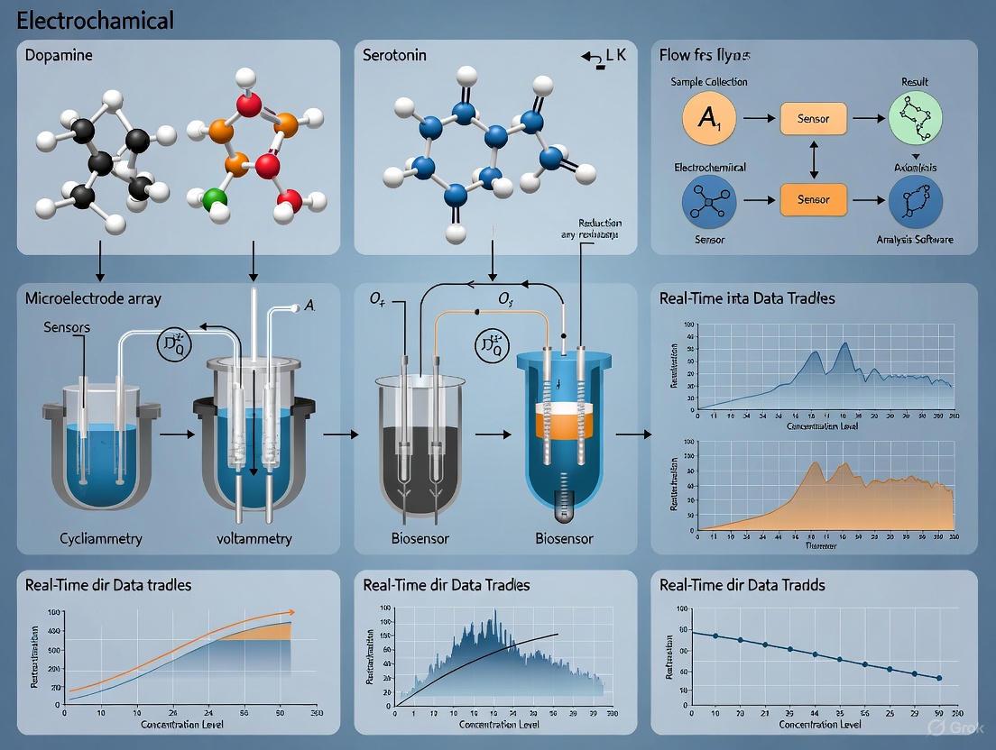
Electrochemical monitoring techniques have become an indispensable tool in neuroscience for the real-time detection and quantification of neurotransmitters. These methodologies integrate advances in sensor materials, innovative electrochemical techniques, and computational analysis to balance sensitivity, selectivity, and spatial resolution. The evolution of carbon-based sensors, fast-scan cyclic voltammetry (FSCV), and novel deep learning algorithms has enabled unprecedented insight into both rapid phasic and slower tonic neurochemical events [12]. Neurotransmitters such as dopamine (DA), serotonin (5-HT), epinephrine (EP), and glutamate (Glu) are essential chemical messengers that facilitate neuronal communication and influence various physiological functions, including mood, cognition, and motor control. Imbalances in these neurochemicals are implicated in numerous neurological disorders, making their precise measurement vital for advancing disease diagnosis and understanding pathological processes [13].
Detecting neurotransmitters in biological samples presents significant challenges due to their structural similarities, low concentrations, and the complex matrix effects of biological environments. In cell culture media, for instance, competitively adsorbing molecules can be present at concentrations up to 350,000-fold excess compared to the neurotransmitter of interest [14]. Electrochemical biosensors provide an interface between biological systems and digital technologies to monitor, measure, and analyze these biochemical processes. A typical biosensor comprises three essential components: a biological recognition element that interacts with the target analyte, a transducer that converts this interaction into a quantifiable electrical signal, and a signal processor for interpretation [13]. Among these, carbon microelectrodes (CMEs) have gained prominence for neurochemical sensing due to their unique properties, including high biocompatibility, exceptional spatiotemporal resolution, and minimal tissue damage, making them ideal for monitoring fast and dynamic biochemical changes at the cellular level [13].
Core Voltammetry Principles and Techniques
Voltammetry encompasses a family of electrochemical techniques that measure current resulting from applied potential waveforms to quantify electroactive species. The fundamental principle involves applying a controlled potential sequence to a working electrode immersed in an electrolyte solution containing the analyte and measuring the resulting current. This current-potential relationship provides qualitative and quantitative information about the analyte, including its concentration and redox properties.
Fast-Scan Cyclic Voltammetry (FSCV) has emerged as a particularly powerful technique for near real-time monitoring of neurotransmitter release. In FSCV, the electrode potential is cycled rapidly (typically at hundreds of volts per second) between two set potentials, generating characteristic oxidation and reduction currents when neurotransmitters adsorb to and react at the electrode surface [13] [12]. The high scan rates enable temporal resolution on the subsecond timescale, capturing neurotransmitter dynamics during behavioral tasks and pharmacological interventions. For the monoamine neurotransmitters like dopamine and serotonin, FSCV typically employs a triangular waveform scanning from a negative holding potential (-0.4 V to -0.2 V) to a positive switching potential (+1.0 V to +1.3 V) and back at 400-1000 V/s [15]. The electrode holding potential between scans influences analyte adsorption, while the positive switching potential cleans the electrode surface from oxidation products.
Recent Advanced Voltammetry Approaches have expanded the capabilities of traditional FSCV:
- N-shape (sawtooth) waveforms were developed specifically for serotonin detection, increasing the scan rate to 1000 V/s and altering holding potentials to improve sensitivity and reduce fouling [15].
- Fast-cyclic square-wave voltammetry superimposes triangle or N-shape waveforms on pre-patterned staircase waveforms, improving sensitivity and selectivity for both dopamine and serotonin detection [15].
- Rapid Pulse Voltammetry (RPV) utilizes short pulses (2 ms) rather than fast linear sweeps to reduce fouling and produce informative faradaic and non-faradaic currents. This approach enables multi-analyte monitoring across timescales, allowing quantification of both basal and stimulated neurotransmitter levels using the same waveform [15].
- Machine-learning-guided waveform design represents the cutting edge, using Bayesian optimization to navigate intractable waveform search spaces. This approach systematically designs analyte-specific voltammetry waveforms by tuning step potentials, lengths, order, and hold times to maximize detection performance [15].
Table 1: Comparison of Voltammetry Techniques for Neurotransmitter Detection
| Technique | Temporal Resolution | Key Analytes | Advantages | Limitations |
|---|---|---|---|---|
| Fast-Scan Cyclic Voltammetry (FSCV) | Subsecond | Dopamine, Serotonin | High temporal resolution, well-established analysis | Limited multi-analyte capability, electrode fouling issues |
| N-shape Waveform Voltammetry | Subsecond | Serotonin | Reduced fouling for serotonin | Specialized for serotonin |
| Fast-Cyclic Square-Wave Voltammetry | Subsecond | Dopamine, Serotonin | Improved sensitivity & selectivity | Complex waveform design |
| Rapid Pulse Voltammetry (RPV) | Seconds to minutes | Multiple neurotransmitters simultaneously | Multi-analyte detection, measures basal levels | Requires advanced data analysis |
| Machine-Learning-Optimized Voltammetry | Application-dependent | Tunable for specific analytes | Optimized sensitivity/selectivity, data-driven design | Complex implementation, computational requirements |
Experimental Protocols
Protocol 1: Carbon Fiber Microelectrode Fabrication for FSCV
Carbon fiber microelectrodes (CFMEs) are widely used for neurotransmitter detection due to their excellent physicochemical and electrochemical properties, microscale diameter (~7-10 microns), and minimal tissue damage [13].
Materials and Equipment:
- Polyacrylonitrile (PAN)-based carbon fibers (e.g., T-650) or pitch-based carbon fibers (e.g., Cytec Thornel P-55)
- Glass capillaries for electrode insulation
- Capillary puller
- Epoxy resin
- Vacuum system
- Scanning electron microscope (SEM) for quality control
Step-by-Step Procedure:
- Carbon Fiber Preparation: Cut carbon fibers to desired length (typically 2-5 cm). PAN-based fibers offer faster electron transfer kinetics and lower background currents, while pitch-based fibers provide higher conductivity and can handle larger currents [13].
- Glass Capillary Preparation: Use a capillary puller to create two identical tapered glass capillaries from a single glass tube.
- Fiber Aspiration: Under microscope visualization, aspirate a single carbon fiber into the tapered end of a glass capillary using a vacuum system.
- Sealing: Apply epoxy resin to seal the fiber-glass interface, ensuring only the tip of the carbon fiber is exposed.
- Curing: Allow the epoxy to cure completely according to manufacturer specifications.
- Cutting and Polishing: Precisely cut the carbon fiber to expose a clean disk electrode surface. Polish if necessary using fine abrasive materials.
- Quality Control: Verify electrode geometry and surface integrity using SEM imaging [13].
Electrochemical Pretreatment (Optional): Electrochemical treatments in alkaline solutions (e.g., KOH) can enhance performance by increasing porosity, regenerating the carbon surface, and introducing oxygen functional groups that benefit adsorption and electron transfer [13].
Protocol 2: MOF-based Aptasensor Fabrication for Multi-Neurotransmitter Detection
This protocol details the fabrication of a wearable electrochemical aptasensor utilizing a metal-organic framework (MOF) heterostructure for simultaneous detection of dopamine, serotonin, and epinephrine [16].
Materials and Reagents:
- Indium chloride (InCl₃·4H₂O) and copper nitrate (Cu(NO₃)₂)
- 2-aminoterephthalic acid (H₂BDC-NH₂) and 1,3,5-benzenetricarboxylic acid
- N,N-Dimethylformamide (DMF) and ethanol
- Gold nanoparticles (AuNPs) for electrodeposition
- Thiolated nucleic acid aptamers specific to dopamine, serotonin, and epinephrine
- 6-Mercapto-1-hexanol (MCH) for blocking non-specific binding
- Flexible screen-printed carbon electrodes (SPCEs)
Step-by-Step Fabrication:
- InMOF Synthesis:
- Combine 80 mg InCl₃·4H₂O and 40 mg H₂BDC-NH₂ in 2 mL DMF and 2 mL Milli-Q water
- Sonicate for 30 minutes until fully dissolved
- Transfer to Teflon-lined autoclave and heat at 120°C for 48 hours
- Collect precipitate by centrifugation, wash with ethanol, and vacuum-dry
CuMOF@InMOF Heterostructure Synthesis:
- Prepare Solution A: Polyvinylpyrrolidone (PVP-K30) and InMOF in DMF/ethanol mixture
- Prepare Solution B: Cu(NO₃)₂ and H₂BDC-NH₂ in DMF
- Combine Solutions A and B, sonicate for 30 minutes
- Heat at 80°C for 8 hours with continuous stirring
- Isolate product by centrifugation, rinse with ethanol, and dry
Electrode Modification:
- Prepare CuMOF@InMOF suspension (1 mg in 2 mL ultrapure water)
- Drop-cast 5 μL suspension onto working electrode surface
- Air-dry at room temperature
Gold Nanoparticle Electrodeposition:
- Using cyclic voltammetry, apply voltage from 0 to 0.8 V at 50 mV/s for 15 cycles
- Verify AuNP deposition by color change and electrochemical characterization
Aptamer Immobilization:
- Incubate AuNP-modified electrode with thiolated aptamer solution (specific to dopamine, serotonin, and epinephrine) for 12-16 hours
- Form Au-S bonds between gold nanoparticles and thiol groups of aptamers
Surface Blocking:
- Treat electrode with 1 mM MCH solution for 40 minutes to block non-specific binding sites
- Rinse with buffer to remove unbound MCH
Detection Mechanism: Upon exposure to neurotransmitters, the aptamers form specific aptamer-neurotransmitter complexes through molecular recognition and hydrogen bonding. This interaction causes conformational changes in the aptamers, leading to measurable reduction in peak current proportional to neurotransmitter concentration [16].
Protocol 3: Machine-Learning-Guided Waveform Optimization
The SeroOpt workflow utilizes Bayesian optimization to design optimized voltammetry waveforms for selective serotonin detection, representing a paradigm shift from traditional "guess-and-check" approaches [15].
Materials and Software:
- Carbon fiber microelectrodes
- Standard electrochemical setup with potentiostat
- Serotonin and dopamine solutions for calibration
- Python environment with Bayesian optimization libraries
- Custom code available at github.com/csmova/SeroOpt and github.com/csmova/SeroWare
Procedure:
- Define Optimization Parameters:
- Specify waveform variables: step potentials, pulse lengths, sequence order, hold times
- Set objective function (e.g., serotonin detection accuracy, selectivity over dopamine)
- Define constraints based on hardware limitations and physiological relevance
Initial Data Collection:
- Collect training data using a set of initial waveforms (random or heuristic-based)
- Measure sensor performance metrics for each waveform
Surrogate Modeling:
- Use Gaussian process regression to build a probabilistic model of the objective function
- The model approximates the relationship between waveform parameters and detection performance
Acquisition Function Optimization:
- Apply an acquisition function (e.g., Expected Improvement) to determine the most promising next waveform to test
- Balance exploration of unknown regions vs. exploitation of known good performers
Iterative Experimental Testing:
- Test the suggested waveform experimentally
- Update the surrogate model with new results
- Repeat steps 4-5 for multiple iterations (typically 20-50 cycles)
Waveform Validation:
- Validate optimized waveform performance using independent test data
- Compare against conventional waveforms (FSCV triangle, N-shape) and randomly designed waveforms
This data-driven approach has demonstrated superior performance compared to random and human-guided waveform designs, with the optimized waveforms enabling selective serotonin detection even in the presence of interferents like dopamine [15].
Machine Learning Waveform Optimization Workflow
Performance Metrics and Data Analysis
The performance of electrochemical sensors for neurotransmitter detection is evaluated using several key metrics, including sensitivity, selectivity, limit of detection (LOD), dynamic range, and stability. Recent advancements in sensor materials and waveform design have led to significant improvements in these parameters.
Table 2: Performance Comparison of Advanced Neurotransmitter Sensors
| Sensor Platform | Analyte | Linear Range | Limit of Detection | Selectivity Features | Reference |
|---|---|---|---|---|---|
| CuMOF@InMOF Aptasensor | Dopamine | 1 nM - 10 µM | 0.18 nM | Specific aptamers, Au-S bonding | [16] |
| CuMOF@InMOF Aptasensor | Serotonin | 1 nM - 10 µM | 0.33 nM | Specific aptamers, MOF heterostructure | [16] |
| CuMOF@InMOF Aptasensor | Epinephrine | 1 nM - 10 µM | 0.27 nM | Specific aptamers, multi-analyte detection | [16] |
| Nickel Oxide/Hydroxide Paper Sensor | Serotonin | 0.007 nM - 500 µM | 0.024 nM (low range)383.7 nM (high range) | Two linear ranges, validated in Drosophila | [17] |
| SWCNT Sensor | Dopamine/Serotonin | Nanomolar range | Nanomolar | Selective in cell culture medium | [14] |
| Machine-Learning Optimized Waveform | Serotonin | Not specified | Improved over conventional | Enhanced selectivity over dopamine | [15] |
Data Analysis Approaches: Modern electrochemical monitoring employs sophisticated data analysis techniques to interpret complex signals:
- Chemometrics: Application of mathematical and statistical techniques to interpret complex chemical data obtained from sensor outputs [12].
- Partial Least Squares Regression (PLSR): Multivariate statistical method that projects both predictors and responses to new spaces to model relationship between them.
- Deep Learning: Neural networks with multiple layers process large quantities of data to discern subtle patterns in complex datasets, differentiating between structurally similar neurotransmitters [12].
- Background Current Processing: Advanced algorithms for separating faradaic (analyte) currents from non-faradaic (background) currents, crucial for accurate quantification.
For wearable sensors, performance validation includes stability testing under flexible conditions, reproducibility across multiple electrodes, and selectivity against common interferents such as ascorbic acid, uric acid, and glucose [16].
The Scientist's Toolkit: Essential Research Reagents and Materials
Table 3: Key Research Reagent Solutions for Neurotransmitter Sensing
| Material/Reagent | Function | Example Application | Key Characteristics |
|---|---|---|---|
| Carbon Fibers (PAN-based) | Microelectrode core material | FSCV measurements | Fast electron transfer, low background currents, high tensile strength |
| Carbon Nanotubes (CNTs) | Electrode nanomaterial | SWCNT sensors for in vitro detection | High surface area, enhanced conductivity, improved sensitivity |
| Metal-Organic Frameworks (MOFs) | Porous sensing platform | CuMOF@InMOF aptasensor | Large surface area, tunable pore size, enhanced biomolecule interaction |
| Specific Aptamers | Biological recognition elements | Selective neurotransmitter binding | High binding affinity, molecular specificity, thiol modification for immobilization |
| Gold Nanoparticles (AuNPs) | Signal amplification | Electrodeposited on MOF surfaces | Enhanced electron transfer, facile aptamer immobilization via Au-S bonds |
| Bayesian Optimization Algorithms | Waveform design | SeroOpt workflow | Navigates intractable search spaces, data-driven optimization |
Applications in Neurochemical Research
Electrochemical neurotransmitter detection platforms have enabled significant advances in both basic neuroscience research and clinical applications:
In Vitro Neuropharmacology: Single-walled carbon nanotube (SWCNT) sensors have demonstrated the capability to selectively measure dopamine and serotonin at nanomolar concentrations directly from cell culture medium, despite the presence of competitively adsorbing molecules in vast excess. This enables real-time monitoring of spontaneous transient activity from dopaminergic cell cultures without altering culture conditions, providing unprecedented opportunities for drug discovery and high-throughput screening of complex neuronal models such as organoids [14].
In Vivo Monitoring: Carbon fiber microelectrodes with FSCV have been extensively used to monitor neurotransmitter dynamics in behaving animals, revealing patterns of dopamine and serotonin release during behavioral tasks, learning, and reward processing. These measurements have provided fundamental insights into the neurochemical basis of motivation, decision-making, and neurological disorders.
Wearable Neurochemical Monitoring: The development of flexible, wearable electrochemical biosensors integrated into microfluidic patches enables non-invasive monitoring of neurotransmitters in sweat during physical exercise. These platforms, incorporating advanced materials like MOF-on-MOF heterostructures, allow for continuous, real-time tracking of neurochemical biomarkers relevant to mental health conditions, opening new possibilities for personalized healthcare and depression monitoring [16].
Genetic Model Research: Electrochemical sensors have been successfully deployed in genetically engineered model organisms, such as Drosophila melanogaster, to investigate serotonin level changes under different genetic and disease conditions. The correlation of electrochemical measurements with gold-standard HPLC analysis validates these approaches for precise neurochemical phenotyping [17].
Neurotransmitter Sensing Application Pipeline
Troubleshooting and Technical Considerations
Electrode Fouling: Serotonin and its oxidation byproducts can foul electrode surfaces, reducing sensitivity over time [15]. Mitigation strategies include:
- Using optimized waveforms with positive switching potentials to renew the electrode surface [15]
- Incorporating anti-fouling coatings such as Nafion or biomimetic membranes
- Applying rapid pulse voltammetry instead of continuous sweeps to reduce adsorption [15]
Selectivity Challenges: Structurally similar neurotransmitters (dopamine, serotonin, norepinephrine) have overlapping redox potentials, creating identification challenges. Solutions include:
- Machine learning approaches to differentiate subtle signal patterns [12]
- Incorporation of highly specific bioreceptors (aptamers, enzymes) [16]
- Multi-dimensional waveforms that elicit distinct current signatures for different analytes [15]
Sensitivity in Complex Media: Biological samples contain numerous interferents that can reduce sensor sensitivity. Effective approaches include:
- Background subtraction algorithms and advanced signal processing
- Physical barriers (size-exclusion membranes) and chemical selectivity layers
- In the case of SWCNT sensors, inherent selectivity that functions even in complex cell culture media [14]
Reproducibility and Standardization: Batch-to-batch variations in electrode fabrication remain a challenge. Quality control measures include:
- SEM characterization of electrode surfaces [13]
- Standardized electrochemical activation procedures
- Performance validation with standard solutions before biological measurements
As the field advances, the integration of innovative materials, sophisticated waveform design, and machine learning analytics continues to address these challenges, pushing the boundaries of what can be detected and measured in the complex environment of the nervous system.
Distinguishing Tonic versus Phasic Neurotransmitter Signaling Dynamics
In the field of real-time neurochemical monitoring, a fundamental challenge lies in accurately capturing and distinguishing between two primary modes of neuronal communication: tonic and phasic neurotransmitter signaling. These distinct dynamics operate over different temporal scales and serve separate but complementary functional roles in regulating brain function [2]. Tonic signaling represents the steady-state, ambient neurotransmitter levels that set the overall excitability of neural circuits over seconds to minutes, providing a modulatory background that influences behavioral states [2] [18]. In contrast, phasic signaling comprises the brief, pulsatile neurotransmitter release events—typically lasting milliseconds to seconds—that are tightly coupled to neuronal firing and encode discrete information about stimuli, rewards, or actions [2] [18].
The ability to differentiate these signaling modes is not merely academic; it provides critical insights into normal brain function and the pathological mechanisms underlying neurological and psychiatric disorders. Imbalances in tonic and phasic dynamics have been implicated in Parkinson's disease, substance use disorders, depression, and other conditions [18]. For researchers and drug development professionals, mastering techniques to resolve these dynamics is essential for understanding drug mechanisms, developing diagnostic tools, and creating targeted therapies [2].
This application note situates these concepts within the broader context of electrochemical techniques for real-time neurochemical monitoring, providing both theoretical framework and practical methodologies for investigating these distinct signaling modalities.
Conceptual Framework and Neurobiological Significance
Characteristics of Tonic and Phasic Signaling
The differential characteristics of tonic and phasic neurotransmitter signaling are summarized in Table 1 below.
Table 1: Key Characteristics of Tonic vs. Phasic Neurotransmitter Signaling
| Characteristic | Tonic Signaling | Phasic Signaling |
|---|---|---|
| Temporal Scale | Seconds to minutes [2] | Milliseconds to seconds [2] [18] |
| Concentration Range | Low nanomolar (basal levels) [2] | Nanomolar to micromolar (during release events) [2] |
| Primary Function | Homeostatic regulation, setting neural circuit excitability [2] [18] | Rapid information transfer, encoding discrete stimuli [2] |
| Relationship to Neural Activity | Uncoupled from immediate firing events; reflects sustained modulatory state [2] | Tightly coupled to neuronal firing patterns, especially bursting [2] |
| Representative Measurement Techniques | Multiple Cyclic Square Wave Voltammetry (MCSWV), fluorescence lifetime photometry [18] [19] | Fast-Scan Cyclic Voltammetry (FSCV) [18] |
Functional Roles in Neural Circuit Operation
Tonic and phasic dopamine signaling in the striatum provides an excellent model for understanding the functional interplay between these dynamics. Phasic dopamine transients, often occurring at sub-second timescales during burst firing of dopaminergic neurons, carry teaching signals for reward prediction and motivated behavior [2] [19]. Conversely, tonic dopamine levels create a sustained background that modulates the gain of neural responses to phasic signals and regulates long-term behavioral states [19] [18].
Recent research using fluorescence lifetime photometry (FLIPR) has revealed that tonic dopamine levels vary significantly across striatal subregions, with higher levels observed in the tail of the striatum compared to the nucleus accumbens core [19] [20]. Furthermore, these striatal subregions display differential and dynamic responses in both phasic and tonic dopamine to appetitive and aversive stimuli [19] [20]. This spatial and temporal specialization highlights the importance of measuring both signaling modes to fully understand circuit function.
The functional relationship between these signaling modes can be visualized as follows:
Figure 1: Interplay between Tonic and Phasic Signaling Dynamics. Tonic levels set background excitability that modulates responses to phasic signals, which in turn carry discrete information.
Technical Approaches and Methodologies
Electrochemical Techniques
Electrochemical methods provide the temporal resolution necessary for resolving neurochemical dynamics, with specific techniques optimized for different signaling modes:
Fast-Scan Cyclic Voltammetry (FSCV) is the gold standard for measuring phasic neurotransmitter dynamics. By applying rapid triangular waveforms (typically 400 V/s) at 10 Hz frequency, FSCV enables millisecond resolution detection of neurotransmitter transients caused by neuronal burst firing [18]. The technique involves scanning through oxidation and reduction potentials of the target analyte, creating characteristic cyclic voltammograms that serve as electrochemical fingerprints for neurotransmitter identification.
Multiple Cyclic Square Wave Voltammetry (MCSWV) is optimized for measuring tonic neurotransmitter concentrations. This technique applies a series of square waves of incrementally increasing voltages, allowing for sensitive detection of lower, steady-state neurotransmitter levels without artificially inducing release through electrical stimulation [18]. The longer timescale of MCSWV measurements (seconds to minutes) makes it ideal for tracking slow neuromodulatory changes.
The complementary application of these techniques is exemplified in the MAVEN (Multifunctional Apparatus for Voltammetry, Electrophysiology, and Neuromodulation) platform, which enables near-simultaneous, real-time acquisition of both phasic and tonic neurotransmitter signals alongside electrophysiological recordings [18]. This integrated approach allows researchers to correlate neurotransmitter dynamics with neural activity patterns in the same experimental preparation.
Optical Techniques
Genetically encoded fluorescent sensors have emerged as powerful tools for measuring neurotransmitter dynamics in specific cell populations. Recent advances in Fluorescence Lifetime Photometry at High Temporal Resolution (FLIPR) have enabled absolute measurement of both fast and slow neuronal signals with unprecedented precision [19] [20].
FLIPR utilizes frequency-domain analog processing to measure the absolute fluorescence lifetime of genetically encoded sensors at high speed (kHz) with long-term stability and picosecond precision [19]. This approach overcomes limitations of traditional intensity-based sensors, which are optimized for detecting fast, relative changes but are less suited for measuring slowly changing signals or providing absolute concentration measurements [19] [20].
A key advantage of FLIPR is its ability to resolve spatial variations in tonic and phasic signaling, as demonstrated in the striatum where higher tonic dopamine levels were observed in the tail of the striatum compared to the nucleus accumbens core, with differential responses to appetitive and aversive stimuli [19].
Experimental Workflow for Combined Measurement
A comprehensive approach to measuring both tonic and phasic signaling involves the integrated workflow below:
Figure 2: Integrated Experimental Workflow for Combined Tonic and Phasic Signal Acquisition. This approach enables simultaneous measurement of multiple signaling modalities within the same experimental session.
Research Reagent Solutions and Essential Materials
Table 2: Essential Research Reagents and Materials for Neurotransmitter Dynamics Studies
| Category | Specific Examples | Function and Application |
|---|---|---|
| Electrochemical Sensors | Carbon-fiber microelectrodes, Enzyme-modified biosensors | Target-specific detection with high temporal resolution [2] |
| Voltammetry Systems | MAVEN platform, Traditional FSCV systems | Integrated acquisition of phasic (FSCV) and tonic (MCSWV) signals [18] |
| Genetically Encoded Sensors | dLight, GRABDA, FLIPR-compatible sensors | Cell-type-specific monitoring with genetic targeting [19] |
| Pharmacological Agents | NMDA receptor agonists/antagonists, Uptake inhibitors | Manipulation of release and uptake mechanisms to validate signals [18] |
| Data Acquisition Software | WincsWare, HFCV, Custom MATLAB/Python scripts | Real-time signal processing and analysis [18] |
| Implantation Hardware | Guide cannulas, Fiber optic ferrules, Microdrives | Stable chronic implants for long-term recording in behaving animals [2] |
Detailed Experimental Protocols
Protocol 1: Combined Tonic and Phasic Dopamine Measurement Using Voltammetry
This protocol describes simultaneous measurement of tonic and phasic dopamine using the MAVEN platform or similar integrated systems [18].
Materials Required:
- MAVEN platform or equivalent integrated voltammetry system
- Carbon-fiber microelectrodes (7-10 μm diameter)
- Stereotaxic surgical apparatus
- Guide cannulas for chronic implantation
- Reference and auxiliary electrodes
- Artificial cerebrospinal fluid (aCSF)
- Calibration solutions including dopamine (100 nM - 10 μM)
Procedure:
Electrode Preparation and Calibration:
- Prepare carbon-fiber microelectrodes by sealing 7-10 μm diameter carbon fibers in glass capillaries.
- Electrochemically treat electrodes by applying 1.5 V vs Ag/AgCl in PBS for 10-20 seconds to enhance sensitivity.
- Calibrate electrodes in vitro using standard dopamine solutions (0, 100 nM, 500 nM, 1 μM, 5 μM) in aCSF.
- Determine selectivity against common interferents (ascorbic acid, pH changes, DOPAC).
Surgical Implantation:
- Anesthetize animal (e.g., rat or mouse) with isoflurane or urethane.
- Secure animal in stereotaxic frame and expose skull.
- Drill burr holes at target coordinates (e.g., striatum: AP +1.0 mm, ML ±2.0 mm from bregma).
- Implant guide cannula above target region and secure with dental cement.
- For chronic experiments, allow 5-7 days recovery before recording.
Signal Acquisition Protocol:
- Insert working electrode through guide cannula into target brain region.
- Begin with MCSWV measurements for tonic dopamine:
- Apply square wave potentials from -0.4 V to +1.3 V in 100 mV increments.
- Use step frequency of 60 Hz with 2-second sampling intervals.
- Record for 5-10 minutes to establish stable baseline.
- Interleave FSCV measurements for phasic dopamine:
- Apply triangular waveform (-0.4 V to +1.3 V and back at 400 V/s).
- Use 10 Hz repetition rate.
- Record for 1-2 minutes between MCSWV measurements.
- For stimulus-evoked measurements, apply electrical stimulation (e.g., 60 Hz, 2 ms pulse width, 2-second duration) to dopamine pathways (e.g., medial forebrain bundle).
Data Analysis:
- For phasic signals (FSCV): Use principal component analysis to isolate dopamine component from background currents.
- For tonic signals (MCSWV): Apply chemometric methods to resolve dopamine concentration from overlapping faradaic currents.
- Correlate dopamine dynamics with simultaneously recorded electrophysiological signals.
Validation:
- Confirm dopamine identity by applying uptake inhibitor (nomifensine, 20 mg/kg i.p.) which should increase tonic levels and prolong phasic signals.
- Verify stimulus specificity by applying dopamine receptor antagonists.
Protocol 2: Absolute Dopamine Measurement Using FLIPR
This protocol describes absolute measurement of tonic and phasic dopamine using fluorescence lifetime photometry [19] [20].
Materials Required:
- FLIPR system with high-speed frequency-domain lifetime measurement capability
- Fiber optic system for in vivo photometry
- Genetically encoded dopamine sensor (e.g., dLight, GRABDA)
- Viral vectors for sensor expression (AAV serotypes)
- Laser sources with appropriate wavelengths
- High-speed photon detectors with picosecond timing resolution
Procedure:
Sensor Expression:
- Inject AAV encoding dopamine sensor (e.g., AAV5-hSyn-dLight1.3b) into target brain region (e.g., striatum) of anesthetized mice.
- Allow 3-6 weeks for adequate sensor expression before photometry experiments.
Fiber Implantation:
- Implant optical fiber (400 μm diameter) above sensor expression region.
- Secure fiber ferrule with dental cement.
- Allow 1-2 weeks recovery before recording.
FLIPR Acquisition:
- Connect implanted fiber to FLIPR system via patch cord.
- Set excitation laser to appropriate wavelength for sensor (e.g., 470 nm for dLight).
- Configure high-speed frequency-domain measurement (kHz sampling with ps precision).
- Record fluorescence lifetime changes rather than intensity changes.
- Calibrate lifetime measurements to absolute dopamine concentrations using in vitro calibration curves.
Behavioral Paradigms:
- For phasic dopamine measurements: Implement reward conditioning tasks (e.g., sucrose delivery, predictive cues).
- For tonic dopamine measurements: Record during resting states or in response to prolonged stimuli (stressors, drugs).
- Synchronize behavioral events with photometry recordings using TTL pulses.
Data Analysis:
- Convert fluorescence lifetime measurements to absolute dopamine concentrations using predetermined calibration curves.
- Separate tonic and phasic components using mathematical approaches (e.g., low-pass filtering for tonic, high-pass filtering for phasic).
- Analyze spatial and temporal variations in dopamine signaling across different striatal subregions.
Data Interpretation and Analytical Considerations
Quantitative Analysis of Tonic and Phasic Components
When analyzing simultaneous measurements of tonic and phasic signaling, several analytical approaches facilitate interpretation:
Mathematical Separation: Tonic and phasic components can be separated using digital filtering techniques. A low-pass filter with cutoff frequency of 0.01-0.1 Hz can isolate tonic signals, while a high-pass filter with cutoff of 0.1-1.0 Hz can extract phasic transients. More sophisticated approaches include:
- Exponential decay fitting for modeling clearance kinetics of phasic events
- Wavelet analysis for resolving events across multiple temporal scales
- Principal component analysis for separating neurochemical signals from interfering factors
Normalization and Comparison: Due to variations in electrode placement, sensor expression, and individual differences, normalization strategies are essential:
- Express phasic signals as percentage change from baseline tonic levels
- Use z-score normalization for comparing across subjects or sessions
- Employ mixed-effects models to account for both within-subject and between-subject variability
Technical Validation and Troubleshooting
Verifying Signal Specificity:
- Apply receptor antagonists to confirm identity of measured neurotransmitter
- Use enzymatic treatments (e.g., ascorbate oxidase) to eliminate interferents
- Employ multiple detection methods concurrently (e.g., voltammetry with photometry)
Addressing Common Artifacts:
- Electrical stimulation artifacts: Use interleaved stimulation and recording paradigms
- pH confounds: Employ pH-insensitive sensors or simultaneous pH monitoring
- Fouling effects: Implement background subtraction algorithms or anti-fouling coatings
Applications in Disease Models and Drug Development
The ability to distinguish tonic and phasic signaling dynamics has profound implications for understanding disease mechanisms and developing targeted therapeutics:
Parkinson's Disease: The progressive loss of dopamine neurons in Parkinson's disease differentially affects tonic and phasic signaling. Early in disease progression, tonic dopamine levels may be maintained through compensatory mechanisms while phasic signaling is impaired, explaining specific deficits in reward-based learning before overt motor symptoms emerge [2] [18]. Deep brain stimulation appears to exert therapeutic effects by modulating both tonic and phasic dopamine release [2] [18].
Substance Use Disorders: Drugs of abuse differentially alter tonic and phasic dopamine signaling. Acute drug exposure typically enhances phasic responses to drug-associated cues, while chronic use leads to blunted tonic dopamine levels that may contribute to anhedonia and negative affect during withdrawal [18]. Therapies that normalize these imbalances represent promising treatment approaches.
Psychiatric Disorders: In depression, stress-induced changes in dopamine function may involve reduction in tonic levels leading to decreased motivation, while specific phasic signaling deficits may underlie anhedonia [18]. The differential targeting of tonic versus phasic signaling may explain the therapeutic profiles of various antidepressant medications.
For drug development professionals, the resolution of tonic versus phasic effects provides critical insights into mechanism of action, dosing regimens, and potential side effects. Compounds that selectively modulate tonic signaling may produce more gradual and sustained therapeutic effects with reduced abuse liability compared to those that enhance phasic signaling.
The real-time monitoring of dopamine (DA) and serotonin (5-HT) is pivotal for understanding brain function, neurochemical imbalances, and developing treatments for neurological and psychiatric disorders. These neurotransmitters regulate critical processes including motor control, reward, motivation, and affect [21] [22]. Electrochemical techniques provide the high temporal and spatial resolution necessary to capture the rapid dynamics of these signaling molecules in living tissues, offering significant advantages over traditional methods like microdialysis, which lack the temporal resolution for sub-second neurochemical events [23] [18]. This Application Note details the core principles, experimental protocols, and key reagents for the electrochemical investigation of DA and 5-HT, framed within a research paradigm focused on real-time neurochemical monitoring.
A principal challenge in this field is the complex interplay between the dopaminergic and serotonergic systems. Historically conceptualized as a simple inhibitory influence of 5-HT on DA neuron activity, this interaction is now understood as a multifaceted, mutual regulation of central nervous system (CNS) functions [22]. This interaction arises from the diversity of neuronal origin, receptor subtypes, and intracellular signaling pathways, making it a critical focus for understanding the mechanism of action of psychotropic drugs and the pathophysiology of several CNS diseases [22]. Furthermore, the metabolism of DA and 5-HT is interconnected under conditions such as chronic stress (cortisolemia), where shifts in the kynurenine pathway can lead to the production of neurotoxic metabolites and contribute to the etiology of conditions like major depressive disorder (MDD) [24].
Key Neurochemical Pathways & Interactions
Dopamine and Serotonin Metabolism and Signaling
Dopamine and serotonin are monoamine neurotransmitters with distinct yet interacting metabolic pathways and receptor systems. Their dynamics occur on multiple timescales, from phasic release (sub-second, burst-like release events) to tonic release (slower, steady-state levels), both of which are critical for normal brain function [2] [18].
Table 1: Key Characteristics of Dopamine and Serotonin
| Feature | Dopamine (DA) | Serotonin (5-HT) |
|---|---|---|
| Primary Functions | Motor control, reward, motivation, reinforcement [21] | Mood regulation, sleep, appetite, cognition [22] |
| Metabolic Pathway | Synthesis from tyrosine; catabolism produces DOPAL (a toxic aldehyde) [24] | Synthesis from tryptophan; can be diverted into the kynurenine pathway under inflammation [24] |
| Electrochemical Oxidation | Two-electron oxidation to dopamine-o-quinone at ~0.14 V [21] | Oxidation at potentials similar to DA, but prone to electrode fouling [23] |
| Basal Level Range | 1–200 nM (can increase to µM in stimulated or disease states) [2] | Low nanomolar range (exact values are region-dependent) |
| Key Interaction | Regulated by serotonergic system via multiple receptor subtypes [22] | Can modulate DA release and neuronal activity; forms heterocomplexes with D2 receptors [25] |
The serotonin-dopamine interaction is a critical nexus for CNS function and drug action. Evidence indicates that 5-HT({2A}) and dopamine D2 receptors can form physical heterocomplexes, leading to a unique functional cross-talk that may be relevant to the action of hallucinogens and antipsychotics [25]. Furthermore, scaffolding proteins like PSD-95 are found at the crossroads of glutamate, dopamine, and serotonin signaling, interacting with NMDA, D2, and 5-HT({2}) receptors and regulating their activation state [25]. This complex postsynaptic protein network represents a valuable molecular target for novel therapeutic strategies.
Pathway Dysregulation and Disease
Imbalances in DA and 5-HT are implicated in a wide array of disorders. Low dopamine levels in the central nervous system are a major cause of Parkinson's disease [21]. Conversely, dysregulation of both systems is involved in schizophrenia, depression, and drug addiction [22] [24]. In depression, elevated cortisol levels can disrupt these pathways, increasing the production of toxic metabolites like DOPAL and 5-HIAL from DA and 5-HT, respectively, which may contribute to neurotoxicity [24].
Figure 1: Integrated DA and 5-HT Metabolic Pathways under Cortisolemia. Elevated cortisol during chronic stress promotes inflammation, shifting tryptophan metabolism away from serotonin production and towards the kynurenine pathway. This imbalance can increase the levels of toxic aldehydes (DOPAL, 5-HIAL), contributing to neurotoxicity [24].
Electrochemical Monitoring Platforms
Electrochemical techniques are the cornerstone of real-time neurochemical monitoring due to their excellent temporal resolution, high sensitivity, and capacity for miniaturization.
Core Electrochemical Techniques
The two most prevalent techniques for monitoring rapid neurotransmitter dynamics are Fast-Scan Cyclic Voltammetry (FSCV) and Constant-Potential Amperometry [23].
Table 2: Comparison of Key Electrochemical Techniques for Neurotransmitter Monitoring
| Technique | Principle | Temporal Resolution | Advantages | Limitations | Primary Analytic |
|---|---|---|---|---|---|
| Fast-Scan Cyclic Voltammetry (FSCV) | Applies a triangular waveform (e.g., -0.4 V to +1.3 V, 400 V/s) to oxidize/reduce analytes [23]. | Sub-second (10s-100s of ms) [23] [2] | Provides chemical identification via cyclic voltammogram signature [23]. | Requires background subtraction; complex data analysis. | Dopamine [23], Norepinephrine [23], Serotonin (with modifications) [23] |
| Amperometry | Holds electrode at a constant potential sufficient to oxidize the analyte [23]. | Milliseconds (limited only by data acquisition) [23] | Simple data interpretation; direct measurement of release quantity [23]. | No chemical identification; measures all electroactive species [23]. | Catecholamines in cell culture [23] |
| Multiple Cyclic Square Wave Voltammetry (MCSWV) | A variant used to measure tonic (basal) levels of neurotransmitters [18]. | Seconds to minutes | Measures sustained, baseline neurotransmitter concentrations [18]. | Lower temporal resolution than FSCV. | Tonic Dopamine, Serotonin [18] |
Advanced Integrated Systems
Recent technological advances have led to the development of multimodal platforms that combine neurochemical sensing with other modalities. The Multifunctional Apparatus for Voltammetry, Electrophysiology, and Neuromodulation (MAVEN) is one such platform, engineered for intraoperative and preclinical applications [18]. MAVEN enables near-simultaneous, real-time acquisition of electrophysiological signals (e.g., local field potentials) and neurochemical measurements (both phasic and tonic) alongside programmable electrical stimulation, such as Deep Brain Stimulation (DBS) [18]. This integration is crucial for elucidating the complex relationships between neurochemical dynamics, electrophysiological activity, and the therapeutic effects of neuromodulation.
Figure 2: Workflow of a Multimodal Neurochemical Monitoring Platform. Integrated systems like MAVEN combine electrical stimulation with simultaneous electrophysiological and neurochemical recording from a single sensor, providing a comprehensive view of neural circuit activity [18].
Experimental Protocols
Protocol: Measuring Phasic Dopamine with Fast-Scan Cyclic Voltammetry (FSCV)
This protocol is adapted for use in a brain slice preparation or an anesthetized rodent, utilizing a carbon-fiber microelectrode (CFM) [23].
- Electrode Preparation: Pull a single carbon-fiber (diameter 5-10 µm) into a glass capillary and seal it to create a cylindrical or disk microelectrode. Before use, condition the electrode by applying the FSCV waveform in a blank buffer solution until the background current stabilizes [23].
- Waveform Application: Apply a triangular waveform to the CFM. A standard waveform for dopamine scans from a holding potential of -0.4 V to +1.3 V and back, at a scan rate of 400 V/s, repeated at 100 ms intervals [23].
- Stimulation and Recording: Implant the CFM into the brain region of interest (e.g., striatum). Use a stimulating electrode to deliver a brief, biphasic electrical pulse (e.g., 60 Hz, 2 ms pulse width, for 2 seconds) to the dopamine cell bodies or axons to evoke release.
- Background Subtraction: During data analysis, subtract the background current (recorded before stimulation) from the faradaic current recorded during and after stimulation. This reveals the background-subtracted cyclic voltammogram.
- Analyte Identification and Quantification: Identify dopamine by its characteristic oxidation peak at approximately +0.6 V to +1.0 V and reduction peak at approximately -0.2 V against an Ag/AgCl reference electrode [23]. Convert the peak oxidation current to concentration using a post-calibration factor obtained from a dopamine standard solution.
Protocol: Detecting Tonic Neurochemical Levels Using MCSWV
This protocol is designed for measuring slower, basal fluctuations of neurotransmitters like serotonin and dopamine [18].
- Electrode Selection and Preparation: Use a carbon-fiber microelectrode or an electrode modified with specific coatings (e.g., Nafion) to enhance selectivity, particularly for serotonin. The electrode may require a different preconditioning routine than FSCV.
- Waveform Application: Apply a square-wave voltammetry waveform tailored for the specific analyte. The parameters (e.g., step increments, amplitude) are optimized for sensitivity to basal levels rather than rapid transients.
- In Vivo Recording: Implant the prepared electrode in the target brain region (e.g., substantia nigra pars reticulata for serotonin). Record the neurochemical signal continuously without electrical stimulation to capture natural fluctuations.
- Signal Processing and Analysis: The measured current is directly correlated to the basal concentration of the neurotransmitter. Use mathematical modeling or principal component regression to deconvolve the signals of different analytes if necessary.
Protocol: Differentiating Neurotransmitters via FSCV "Chemical Signature"
The unique cyclic voltammogram for each electroactive species allows for their identification in a mixture [23].
- Data Collection: Collect FSCV data as described in Protocol 4.1.
- Background Subtraction: Obtain the background-subtracted cyclic voltammogram.
- Signature Comparison:
- Dopamine: Look for a sharp oxidation peak at ~+0.6 V to +1.0 V and a corresponding reduction peak at ~-0.2 V [23].
- Serotonin: Identified by an oxidation peak at a similar potential to dopamine but with a distinctly shaped voltammogram and a less prominent reduction peak. Electrode fouling can alter the signal over time [23].
- pH Changes: Characterized by a sloped, "inverted-V" shaped voltammogram with no distinct peaks [23].
The Scientist's Toolkit: Key Research Reagent Solutions
Table 3: Essential Materials for Electrochemical Neurotransmitter Research
| Item | Function/Description | Key Considerations |
|---|---|---|
| Carbon-Fiber Microelectrode (CFM) | The primary working electrode for in vivo FSCV and amperometry due to its small size, fast temporal response, and biocompatibility [23]. | Cylindrical or disk configurations; surface oxides aid cation adsorption [23]. |
| Nafion Coating | A perfluorinated ionomer often coated onto CFMs to repel negatively charged interferents like ascorbic acid (AA) and DOPAC, thereby enhancing selectivity for cationic DA and 5-HT [23]. | Improves selectivity but can reduce temporal resolution and be susceptible to biofouling [2]. |
| Enzyme-Modified Biosensors | Electrodes coated with an oxidase enzyme (e.g., glutamate oxidase) to detect non-electroactive neurotransmitters by measuring the H₂O₂ produced by the enzymatic reaction [23]. | Enables detection of a wider range of neurochemicals (e.g., glutamate, acetylcholine); kinetics may limit temporal resolution [23]. |
| Cylindrical Gold Nanoelectrode (CAuNE) Arrays | Nanostructured electrodes fabricated via laser interference lithography and electrochemical deposition, providing a homogeneous, periodic sensing platform [26]. | Offers high sensitivity and a supportive substrate for cell culture; allows for dopamine detection in the presence of human neural cells [26]. |
| Multimodal Platform (MAVEN) | A portable, integrated system for simultaneous voltammetric neurochemical sensing (phasic and tonic), electrophysiological recording, and delivery of electrical neuromodulation [18]. | Designed for seamless integration into surgical workflows; enables comprehensive neural circuit characterization in vivo [18]. |
Data Analysis & Interpretation
Effective analysis of electrochemical data is critical for drawing meaningful biological conclusions. For FSCV, data is often presented as color plots, with time on the x-axis, applied potential on the y-axis, and current represented by color. This allows for visualization of the appearance and identity of an analyte over time [23]. Quantitative analysis involves extracting concentration from current using the Faraday equation (Q = nNF) for amperometry or calibration curves for FSCV [23].
A major consideration for in vivo studies is the inflammatory response to implanted sensors. The brain's foreign body response can lead to microglial activation, protein adsorption, and release of reactive oxygen species, which can degrade sensor performance and alter local neurochemistry [2]. Strategies to mitigate this include using biocompatible coatings and minimizing sensor size [2].
Electrochemical techniques provide an unparalleled window into the dynamic interplay of dopamine and serotonin in the functioning brain. Mastery of protocols like FSCV and amperometry, combined with an understanding of the complex interactions between these neurotransmitter systems, empowers researchers to investigate the neurochemical basis of behavior and disease. The ongoing development of advanced materials for electrodes and integrated multimodal platforms like MAVEN promises to further refine our ability to monitor neurochemistry in real-time, accelerating the development of novel diagnostics and therapeutic strategies for a range of neurological and psychiatric disorders.
The coordinated interplay between electrophysiological activity and neurotransmitter dynamics fundamentally shapes both normal and pathological brain functions [18]. Existing research methodologies, however, have traditionally offered limited capacity for integrated, real-time analysis, creating a significant gap in our ability to fully characterize neural circuit dynamics [18]. While traditional electrophysiological techniques provide excellent temporal resolution for monitoring neuronal firing patterns and local field potentials, they lack essential neurochemical context [18]. Conversely, traditional neurochemical measurement techniques like microdialysis suffer from poor temporal resolution, yielding only static snapshots rather than continuous, real-time data [18].
This application note addresses the critical technical challenges in merging these complementary modalities and presents integrated solutions, validated protocols, and essential tools for researchers pursuing multimodal neural circuit investigation. The development of platforms capable of simultaneously measuring electrophysiological and neurochemical signals with high spatiotemporal fidelity in vivo represents a frontier in neuroscience with profound implications for understanding neurological disorders and developing next-generation therapies [18].
Technical Challenges and Integrated Solutions
Key Integration Challenges
Combining neurochemical sensing with electrophysiological recording presents multiple interconnected technical hurdles that must be addressed for successful multimodal experimentation.
Signal Interference and Crosstalk: Simultaneous electrical stimulation and recording creates significant artifact contamination that can obscure native neurochemical and electrophysiological signals. The electrical stimulation pulses used in techniques like Deep Brain Stimulation (DBS) generate large-amplitude artifacts that overwhelm sensitive neurochemical measurements [18]. Additionally, electrochemical sensing techniques themselves apply potential waveforms to working electrodes that can introduce noise into adjacent electrophysiological recording sites.
Material and Biocompatibility Constraints: The mechanical mismatch between rigid conventional electrodes and soft neural tissue triggers inflammatory responses that compromise signal fidelity over time [27]. This inflammation upregulates reactive oxygen species levels, disrupting the neurochemical microenvironment and leading to electrode fouling [27]. Furthermore, different sensing modalities often require specialized electrode materials optimized for either electrical signal conduction or specific neurochemical detection, creating integration challenges.
Data Processing and Workflow Integration: The diverse signal processing requirements across different modalities necessitate sophisticated data fusion approaches [18]. Electrophysiological signals (spikes, LFPs) and neurochemical data (tonic/phasic neurotransmitter levels) operate at different temporal scales and signal characteristics, requiring specialized processing pipelines. Additionally, seamless integration into standard surgical or experimental workflows is essential for practical adoption [18].
Emerging Solutions and Technological Advances
Recent technological innovations have yielded promising solutions to address these multimodal integration challenges.
Artifact-Free Interleaving: Advanced platforms like the Multifunctional Apparatus for Voltammetry, Electrophysiology, and Neuromodulation (MAVEN) implement temporal separation of stimulation and recording cycles, allowing near-simultaneous acquisition while preventing stimulation artifacts from contaminating recorded signals [18].
Novel Electrode Materials and Designs: Ultra-soft carbon nanotube fiber electrodes (CNTFEs) significantly reduce mechanical mismatch while providing enhanced sensitivity for neurochemical detection [28]. These flexible, minimally invasive sensors demonstrate 141-fold improvement in dopamine detection sensitivity compared to conventional gold electrodes [28]. Anti-inflammatory sensing interfaces incorporating atomic-level engineered Fe single-atom catalysts (SAzymes) with catalase and superoxide dismutase-mimicking properties actively eliminate reactive oxygen species, maintaining signal integrity during chronic implantation [27].
High-Density Multimodal Arrays: CMOS-based high-density microelectrode arrays (HD-MEAs) now enable large-scale electrophysiological recording with thousands of simultaneous channels while incorporating specialized sites for neurochemical sensing [29]. These platforms facilitate recordings across multiple spatial scales—from subcellular compartments to entire networks—while maintaining compatibility with electrochemical sensing modalities [29].
Table 1: Comparison of Integrated Multimodal Platforms
| Platform | Key Features | Neurochemical Modalities | Electrophysiological Capabilities | Applications |
|---|---|---|---|---|
| MAVEN [18] | Portable, battery-powered, artifact-free interleaving | Tonic & phasic measurements (FSCV, MCSWV) | Single-unit, multi-unit, LFP recordings | Intraoperative monitoring, DBS research |
| HD-MEAs [29] | Thousands of simultaneous channels, subcellular resolution | Integrated biosensing sites | Network-level activity, AP propagation | In vitro drug screening, network neuroscience |
| CNTFE Sensors [28] | Ultra-soft flexibility, enhanced sensitivity | Multiplexed DA, UA, AA detection | Compatible with standard electrophysiology | Chronic implantation, minimal tissue damage |
| FeSAzyme Sensors [27] | Anti-inflammatory, ROS scavenging | Dopamine oxidation | — | Long-term stable neurochemical monitoring |
Experimental Protocols
Integrated Protocol: Simultaneous Neurochemical and Electrophysiological Monitoring During DBS
This protocol describes methodology for simultaneous dopamine dynamics and electrophysiological recording during deep brain stimulation, adapted from the MAVEN platform validation studies [18].
Materials and Equipment
- Multimodal recording platform (e.g., MAVEN system) [18]
- Carbon fiber microelectrodes (7 μm diameter) for neurochemical sensing
- Tungsten or platinum-iridium electrophysiology electrodes
- Programmable electrical stimulator
- Stereotaxic surgical apparatus
- Data acquisition system with synchronized timing
- Analysis software (e.g., WinCSWare or custom MATLAB scripts)
Procedure
Step 1: Surgical Preparation and Electrode Implantation
- Anesthetize subject (rodent or swine model) following approved IACUC protocols.
- Secure subject in stereotaxic frame and perform craniotomy to expose target brain regions (e.g., striatum for neurochemical sensing, STN for electrophysiology).
- Implant carbon fiber microelectrode in striatum using stereotaxic coordinates.
- Position electrophysiology recording electrode in STN or adjacent structures.
- Verify electrode placements via baseline measurements and anatomical landmarks.
Step 2: System Configuration and Artifact Mitigation
- Configure MAVEN platform for interleaved stimulation and recording cycles [18].
- Set FSCV parameters for dopamine detection: triangular waveform (-0.4 V to +1.3 V vs Ag/AgCl, 400 V/s scan rate, 10 Hz repetition rate).
- Program electrical stimulation parameters: biphasic square-wave pulses (130 Hz, 60-90 μs pulse width, 50-200 μA amplitude).
- Establish temporal separation protocol: 50 ms stimulation blanking period before neurochemical recording to eliminate stimulation artifacts.
Step 3: Simultaneous Baseline Recording
- Acquire 5-minute baseline of tonic dopamine levels using multiple cyclic square wave voltammetry (MCSWV) [18].
- Record simultaneous local field potentials and multi-unit activity.
- Document baseline electrophysiological patterns (beta/gamma oscillations for PD models).
Step 4: Stimulation and Response Monitoring
- Apply programmed electrical stimulation to STN target.
- Monitor real-time phasic dopamine release using FSCV during stimulation epochs.
- Record concurrent electrophysiological responses in striatum and STN.
- Note tremor reduction (for PD models) correlated with neurochemical and electrophysiological changes.
Step 5: Data Integration and Analysis
- Align neurochemical and electrophysiological data streams using synchronized timestamps.
- Extract stimulation-evoked dopamine transients and calculate amplitude, latency, and clearance kinetics.
- Analyze electrophysiological power spectra changes in beta/gamma bands.
- Correlate neurochemical dynamics with electrophysiological responses and behavioral outcomes.
Protocol: Anti-inflammatory Neurochemical Sensing for Chronic Implantation
This protocol utilizes advanced anti-inflammatory sensor technology for long-term stable dopamine monitoring, addressing the critical challenge of inflammation-induced signal degradation [27].
Materials and Equipment
- Fe single-atom catalyst (FeN4) sensors fabricated on carbon fiber substrates [27]
- Reference electrode (Ag/AgCl) and counter electrode
- Potentiostat with high-sensitivity capabilities
- Biocompatible coating materials (e.g., Nafion)
- Sterilization equipment (ethylene oxide gas or cold sterilization)
Procedure
Step 1: Sensor Fabrication and Characterization
- Synthesize Fe1/NC SACs with FeN4 coordination via high-temperature pyrolysis (900°C) of FePc@MET-6 precursor [27].
- Characterize atomic structure using AC HAADF-STEM and XAS to confirm FeN4 configuration.
- Fabricate microelectrodes by depositing Fe1/NC catalyst onto carbon fiber substrates.
- Apply biocompatible coating (e.g., Nafion) to enhance selectivity for dopamine.
- Validate sensor performance in vitro using flow injection analysis with dopamine standards.
Step 2: Surgical Implantation and Inflammation Assessment
- Implant FeSAzyme sensors in target brain region (e.g., striatum) using aseptic technique.
- Monitor reactive oxygen species levels at implantation site using electrochemical methods.
- Assess neuroinflammatory response through immunohistochemical markers (GFAP for astrocytes, IBA1 for microglia) in separate subject cohort.
Step 3: Long-term Stability Monitoring
- Acquire continuous dopamine measurements over 4-week period using fast-scan cyclic voltammetry.
- Perform regular in vivo calibration using electrical stimulation-evoked dopamine release.
- Compare signal stability with conventional carbon fiber electrodes implanted contralaterally.
- Assess tissue compatibility through histological analysis post-sacrifice.
The Scientist's Toolkit: Essential Research Reagents and Materials
Table 2: Key Research Reagent Solutions for Multimodal Neural Interface Research
| Item | Function/Application | Key Characteristics | Representative Examples/Formats |
|---|---|---|---|
| Carbon Nanotube Fiber Electrodes (CNTFEs) [28] | Multiplexed neurochemical sensing | Ultra-soft flexibility, 141x DA sensitivity enhancement, reduced tissue damage | Custom fabricated fibers, 7-30 μm diameters |
| Fe Single-Atom Catalysts (FeSAzymes) [27] | Anti-inflammatory neurochemical sensing | FeN4 coordination, CAT/SOD-mimicking activity, ROS scavenging capability | Fe1/NC-900 pyrolyzed catalysts, carbon fiber coatings |
| High-Density Microelectrode Arrays (HD-MEAs) [29] | Large-scale electrophysiology | 236,880 electrodes, 33,840 simultaneous channels, subcellular resolution | CMOS-based platforms, customizable electrode configurations |
| Multimodal Platform (MAVEN) [18] | Integrated sensing/stimulation | Artifact-free interleaving, Bluetooth/fiber-optic data link, battery-powered | Portable benchtop system, compatible with standard electrodes |
| Fast-Scan Cyclic Voltammetry (FSCV) [18] [30] | Phasic neurotransmitter dynamics | Millisecond resolution, real-time monitoring of DA, 5-HT, adenosine | Triangular waveforms (-0.4V to +1.3V, 400 V/s, 10 Hz) |
| Multiple Cyclic Square Wave Voltammetry (MCSWV) [18] | Tonic neurotransmitter levels | Basal concentration measurement, slower temporal dynamics | Square wave patterns, lower frequency than FSCV |
| Anti-fouling Coatings | Biocompatibility enhancement | Reduced protein adsorption, decreased glial scarring | Nafion membranes, PEGylated surfaces, hydrogel coatings |
| Wireless Data Acquisition Systems | Chronic freely-behaving recording | Multichannel capability, lightweight, long battery life | Bluetooth/WiFi transmitters, headstage preamplifiers |
Visualizing Workflows and Signaling Pathways
Multimodal Neural Sensing Experimental Workflow
Neurochemical-Electrophysiological Signaling Cascade
The integration of neurochemical sensing with electrophysiological recording represents a transformative approach for understanding neural circuit dynamics in both healthy and diseased states. While significant challenges remain in signal interference, material compatibility, and data integration, emerging technologies like the MAVEN platform, ultra-soft CNTFE sensors, and anti-inflammatory SAzyme interfaces are providing viable solutions. The protocols and tools detailed in this application note offer researchers practical methodologies for implementing these advanced multimodal approaches in their experimental workflows. As these technologies continue to evolve, they promise to accelerate our understanding of brain function and facilitate the development of closed-loop therapeutic systems for neurological and psychiatric disorders.
Advanced Electrochemical Platforms and Their Research Applications
The complexity of brain function arises from the intricate interplay between electrical activity and neurochemical signaling. A significant limitation in neuroscience and intraoperative monitoring has been the technological inability to capture these multimodal datasets in real time and with high fidelity. Existing platforms typically measure electrophysiology or neurotransmitter dynamics in isolation, lacking the spatiotemporal resolution required for a comprehensive understanding of neural circuits and their modulation [31]. The Multifunctional Apparatus for Voltammetry, Electrophysiology, and Neuromodulation (MAVEN) is a compact, battery-powered platform engineered to overcome these limitations [32] [31]. By integrating concurrent electrophysiological recordings, phasic and tonic neurochemical sensing, and programmable neurostimulation into a single, portable device, MAVEN provides a unified tool for investigating the mechanisms of neurological therapies and the neurochemical basis of behavior and disease [32] [33]. Its design has immediate relevance in the era of personalized neuromodulation, offering the potential to identify neurotransmitter-based biomarkers for closed-loop therapeutic systems [31] [34].
Technical Specifications of the MAVEN Platform
The MAVEN platform is architected to provide a cohesive and artifact-free multimodal readout, which is essential for basic neuroscience research and translational clinical applications. Its core technical specifications are summarized in Table 1.
Table 1: Technical Specifications of the MAVEN Platform
| Specification Category | Description and Capabilities |
|---|---|
| Primary Function | Unifies electrophysiological recording, neurochemical sensing (phasic & tonic), and programmable electrical stimulation in a single device [32] [31]. |
| Key Innovation | Enables real-time, artifact-free, near-simultaneous multimodal sensing and stimulation, overcoming prior technical limitations [32] [31]. |
| Design & Form Factor | Compact, battery-powered, and handheld, ensuring portability and feasibility for use in an operating suite [32] [31]. |
| Neurochemical Sensing Modality | Multiple Cyclic Square Wave Voltammetry (MCSWV) for tracking tonic neurotransmitter levels (e.g., dopamine, serotonin) [33]. Fast-Scan Cyclic Voltammetry (FSCV) for detecting phasic neurotransmitter transients [32]. |
| Data Outputs | Local Field Potentials (LFPs), single-unit firing activity, and dynamics of specific neurotransmitters like dopamine and serotonin [32] [33]. |
| Translational Feasibility | Integrated with stereotactic planning software and tractography; validated for safe operation in a large-animal operating suite [32]. |
A critical achievement of the MAVEN platform is its ability to minimize stimulation artifacts that typically corrupt sensitive electrophysiological and neurochemical recordings when stimulation is delivered concurrently [31]. This allows for stable, high-fidelity readouts of both low-frequency local field potentials and neurochemical concentrations even during active neuromodulation, a capability that has previously precluded direct investigation in intraoperative human studies [31].
Experimental Protocols and Applications
The utility of the MAVEN platform has been demonstrated across multiple preclinical studies, validating its performance in detecting neurochemical changes in response to pharmacological challenges and electrical stimulation.
Protocol 1: Simultaneous Neurochemical and Electrophysiological Recording During Opioid Administration
This protocol details the methodology for using MAVEN to investigate the neurophysiological correlates of opioid exposure in a large-animal model, as described in swine model studies [33].
1. Animal Model and Surgical Preparation:
- Utilize an anesthetized swine model.
- Employ a frame-based, tractography-guided stereotactic system for precise instrument placement [33].
2. Electrode Implantation and Recording Setup:
- Implant a carbon fiber microelectrode (CFM) stereotactically into the nucleus accumbens (NAc).
- Configure the CFM to record both tonic dopamine concentrations and local field potentials (LFPs) [33].
3. Neurochemical Sensing Parameters (MCSWV):
- Initial Potential: -0.2 V
- Staircase Increment: +25 mV
- Square Wave Amplitude: ±0.4 V
- Pulse Duration: 1.0 ms
- Cyclic Square Waves: Five per scan
- Scan Rate: 0.1 Hz [33]
4. Experimental Workflow:
- Acquire baseline recordings of tonic dopamine and LFPs.
- Administer fentanyl (or another opioid agonist).
- Conduct post-administration recordings using the same parameters to capture drug-induced changes.
5. Data Analysis and Output:
- Quantitative Data: Measure the increase in tonic dopamine concentrations in the NAc following fentanyl administration.
- Electrophysiologic Data: Analyze concurrent changes in LFP power, particularly in lower-frequency bands [33].
- Validation: Confirm dopamine detection capability of the CFM through pre- and post-operative in vitro testing [33].
Protocol 2: Multimodal Monitoring During Programmable Neurostimulation
This protocol outlines the application of MAVEN for assessing the effects of DBS-like stimulation on neurochemical release and electrical activity.
1. Platform Configuration:
- Engage MAVEN's programmable neurostimulation module to deliver DBS-like pulse trains [32].
- Set up the integrated sensing suite for artifact-free, near-simultaneous readouts of LFPs, single-unit firing, and neurotransmitter dynamics (e.g., dopamine or serotonin) [32] [31].
2. Experimental Workflow:
- In rodent or swine models, implant flexible graphene sensors or CFMs in target brain regions (e.g., striatum, NAc) for chronic or acute recordings [32].
- Initiate baseline multimodal recording.
- Deliver programmable electrical stimulation trains to a target area, such as the ventral tegmental area (VTA) or a DBS target.
- Continue simultaneous recording of electrophysiology and neurochemistry throughout the stimulation period.
3. Data Analysis and Output:
- Neurochemical Outcome: Resolve stimulation-evoked neurotransmitter release, such as dopamine transients [32].
- Electrophysiological Outcome: Correlate stimulation parameters with changes in single-unit activity and LFP band power.
- Biomarker Identification: Use the combined dataset to investigate potential neurochemical and electrophysiological biomarkers for optimizing closed-loop neuromodulation therapies [31] [34].
The following workflow diagram illustrates the logical sequence of a typical MAVEN experiment, from setup to data analysis.
The Scientist's Toolkit: Essential Research Reagents and Materials
The experimental protocols leveraging the MAVEN platform rely on a specific set of reagents, sensors, and materials. Table 2 provides a non-exhaustive list of key components essential for researchers aiming to replicate or design similar studies.
Table 2: Essential Research Reagents and Materials for MAVEN Experiments
| Item Name | Function and Application in MAVEN Protocols |
|---|---|
| Carbon Fiber Microelectrode (CFM) | The primary sensor for voltammetric measurements. Used for detecting tonic dopamine concentrations via MCSWV and phasic transients via FSCV [33]. |
| Flexible Graphene Sensors | Implanted chronically in awake, behaving mice for stable, long-term monitoring of spontaneous neurochemical transients, offering biocompatibility and flexibility [32]. |
| Fentanyl | An opioid agonist used in pharmacological challenge studies to elicit robust, measurable shifts in accumbal dopamine and serotonin dynamics, modeling addiction pathways [32] [33]. |
| Programmable Neurostimulation Electrodes | Used to deliver DBS-like pulse trains to target brain regions (e.g., VTA) for investigating stimulation-evoked neurochemical release and electrophysiological changes [32] [31]. |
| MCSWV Buffer/Electrolyte Solution | The electrolyte medium required for performing Multiple Cyclic Square Wave Voltammetry in vivo, ensuring stable electrochemical conditions for neurotransmitter detection [33]. |
MAVEN has been quantitatively validated in multiple animal models. The following table summarizes key, measurable outcomes from published preclinical studies.
Table 3: Summary of Quantitative Experimental Data from MAVEN Studies
| Experimental Model | Intervention / Paradigm | Key Quantitative Findings and Measured Outcomes |
|---|---|---|
| Swine Model | Fentanyl administration [33]. | Increased tonic dopamine concentrations in the Nucleus Accumbens (NAc). Concurrent observation of increased power in lower-frequency Local Field Potential (LFP) bands. |
| Rodent & Swine Models | Programmable electrical stimulation (DBS-like) [32]. | Resolved stimulation-evoked dopamine release. Demonstration of artifact-free recording during stimulation delivery. |
| Awake Mice | Chronic monitoring with graphene sensors [32]. | Successful detection of spontaneous neurochemical transients in awake, behaving subjects, validating chronic implantation stability and function. |
| Swine Model | Tractography-guided DBS surgery [32] [33]. | Established safety and operational feasibility of MAVEN in a large-animal operating suite, confirming its translational potential for intraoperative human applications. |
The MAVEN platform represents a significant leap forward in neuroscientific tooling, effectively breaking down the traditional silos between electrophysiology and neurochemistry. By providing a compact, integrated system for real-time, multimodal sensing and stimulation, it opens new avenues for elucidating the mechanisms of deep brain stimulation, the neurochemical underpinnings of neuropsychiatric disorders like addiction, and the discovery of actionable biomarkers [31] [33]. Its successful preclinical validation across rodent and swine models, including in an operative setting, strongly supports its translational feasibility [32]. As research with MAVEN progresses, it is poised to fundamentally enhance our understanding of brain function and accelerate the development of personalized, closed-loop neuromodulation therapies for a range of neurological and psychiatric diseases [34].
Fast-Scan Cyclic Voltammetry (FSCV) for Phasic Neurotransmitter Detection
Fast-Scan Cyclic Voltammetry (FSCV) is a powerful electrochemical technique that has revolutionized the study of rapid neurochemical transmission in the brain. For over three decades, FSCV has enabled researchers to measure subsecond, phasic fluctuations of electroactive neurotransmitters with unparalleled temporal and spatial resolution [23] [35]. The technique employs carbon fiber microelectrodes (CFMEs) typically 7-10 μm in diameter—comparable in size to neurons—to minimize tissue damage while providing high conductivity and excellent biocompatibility [36]. The fundamental principle involves applying a triangular waveform at high scan rates (>400 V/s) to a working electrode, which oxidizes and reduces electroactive species at specific potentials, generating currents proportional to neurotransmitter concentrations in the extracellular space [23] [35].
Of the biogenic amines, dopamine is the most common target of FSCV measurements, though the technique has been successfully applied to detect norepinephrine, serotonin, adenosine, oxygen, pH changes, and other electroactive species [37] [23]. The high temporal resolution (10 Hz) and chemical selectivity of FSCV make it particularly well-suited for capturing burst-like, phasic dopamine release events critical for learning, goal-directed behavior, and reward processing [37] [23]. Studies using awake animal models have extensively explored relationships between behavior and phasic neurotransmitter release, significantly advancing our understanding of neural circuit dynamics underlying motivation, reinforcement, and decision-making [37] [23].
Table 1: Key Neurotransmitters Detectable via FSCV
| Neurotransmitter | Oxidation Potential (V) | Reduction Potential (V) | Primary Brain Regions | Behavioral Correlates |
|---|---|---|---|---|
| Dopamine | ~0.6 V | ~-0.2 V | Striatum, NAcc, PFC | Reward, motivation, learning |
| Norepinephrine | ~0.7 V | ~-0.1 V | BNST, Locus Coeruleus | Arousal, stress, attention |
| Serotonin | ~0.8 V | ~-0.3 V | DRN, SNr | Mood, appetite, sleep |
| Adenosine | ~1.4 V | Not typically detected | Basal ganglia, Cortex | Sleep, energy homeostasis |
| Oxygen | ~-1.0 V | Not typically detected | Widespread | Metabolic activity |
Current Applications and Methodological Advances
In Vivo Monitoring in Behavioral Neuroscience
FSCV has been extensively utilized to investigate neurochemical correlates of behavior in awake, freely moving animals. The technique's subsecond temporal resolution enables direct correlation between phasic neurotransmitter release and discrete behavioral events [37]. Research has elucidated dopamine's role in reward prediction error, decision-making, and habit formation by measuring transient dopamine signals during task performance [37] [23]. Beyond animal models, FSCV has recently been deployed in human neurosurgical procedures, particularly during deep brain stimulation (DBS) for Parkinson's disease, providing unprecedented insights into human neurochemical dynamics [18] [35].
Technical Innovations in Electrode Design
Recent advances in electrode technology have addressed longstanding challenges in FSCV applications, particularly regarding chronic monitoring. Conventional 7 μm diameter CFMEs often suffer from limited mechanical durability and reduced lifespan, hindering long-term implantation studies [36]. Innovative approaches include:
Large-Diameter Cone-Shaped Electrodes: Increasing CFME diameter to 30 μm with electrochemical etching to create cone-shaped tips significantly improves mechanical robustness. This design demonstrates a 3.7-fold improvement in vivo dopamine signals and a 4.7-fold increase in lifespan compared to conventional 7 μm CFMEs, while reducing glial activation based on Iba1 and GFAP markers [36].
Ultra-Soft Carbon Nanotube Fiber Sensors: These sensors address the mechanical mismatch between rigid electrodes and soft neural tissue, offering significantly enhanced sensitivity compared to conventional gold fiber microelectrodes—with 141-fold improvement for dopamine detection under differential pulse voltammetry conditions [28].
Advanced Coatings: PEDOT:Nafion coatings minimize biofouling effects and increase sensitivity to electroactive monoamine neurotransmitters, enhancing signal stability during chronic recordings [38].
Table 2: Performance Comparison of FSCV Electrodes
| Electrode Type | Sensitivity | Lifespan | Tissue Damage | Biofouling Resistance | Best Applications |
|---|---|---|---|---|---|
| 7 μm CFME (standard) | 12.2 ± 4.9 pA/μm² | Baseline | Minimal | Low | Acute recordings in small animals |
| 30 μm bare CFME | 33.3 ± 5.9 pA/μm² (2.7×) | Moderate | Significant | Low | In vitro studies |
| 30 μm cone-shaped CFME | 47.5 ± 19.8 nA in vivo (3.7×) | 4.7× increase | Reduced | Moderate | Chronic implantation |
| PEDOT:Nafion coated CFME | Varies by formulation | Extended | Minimal | High | Serotonin detection, chronic studies |
| CNT Fiber Electrode | 141× DA sensitivity vs. gold | Under investigation | Minimal | Under investigation | Multimodal sensing, soft tissue regions |
Integration with Complementary Techniques
The development of multimodal platforms represents a significant advancement in FSCV methodology. The Multifunctional Apparatus for Voltammetry, Electrophysiology, and Neuromodulation (MAVEN) enables near-simultaneous, real-time acquisition of electrophysiological signals alongside both phasic (FSCV) and tonic (multiple cyclic square wave voltammetry) neurochemical measurements in vivo [18]. Similarly, successful integration of FSCV with functional magnetic resonance imaging (fMRI) has been achieved using MR-compatible materials, allowing correlation of local dopamine and tissue oxygen responses with global BOLD changes [39]. These integrated approaches provide complementary neurochemical and hemodynamic information, expanding the scope for studying how local neurotransmitter release influences whole-brain activity.
Experimental Protocols
Carbon Fiber Microelectrode Fabrication
Purpose: To construct reliable, high-performance carbon fiber microelectrodes for in vivo FSCV recordings.
Materials:
- AS4 carbon fiber (7 μm diameter, Hexcel) or 30 μm carbon fiber (World Precision Instruments)
- Silica tubing (ID = 20 μm, OD = 90 μm, with polyimide coating)
- Nitinol extension wire (Fort Wayne Metals)
- Silver-based conductive paste
- Epoxy resin
- Polyimide tubing (ID = 0.0089″, OD = 0.0134)
- Vertical microelectrode puller (Narishige Group PE-22)
- Dissecting microscope
Procedure:
- Insert a single carbon fiber into a silica tube, ensuring secure placement.
- Seal the connection between the carbon fiber and silica tubing with epoxy resin.
- Attach the silica tubing to a nitinol extension wire using silver-based conductive paste.
- Insulate the nitinol wire with polyimide tubing up to the exposed carbon fiber tip.
- Trim the exposed carbon fiber to a length of 50-100 μm under a dissecting microscope.
- Cure the assembly according to epoxy manufacturer specifications.
- Prepare an Ag/AgCl reference electrode from Teflon-coated silver wire by chlorinating the stripped tip in saline with a 9 V dry cell battery.
Quality Control: Electrodes should be chemically tested in Tris buffer prior to use. For chronic recordings, consider PEDOT:Nafion coating to minimize biofouling [38].
Electrochemical Etching for Cone-Shaped Electrodes
Purpose: To create cone-shaped 30 μm CFMEs for improved mechanical durability and reduced tissue damage.
Materials:
- 30 μm carbon fiber microelectrodes
- Tris buffer (15 mM Trizma phosphate, 3.25 mM KCl, 140 mM NaCl, 1.2 mM CaCl₂, 1.25 mM NaH₂PO₄, 1.2 mM MgCl₂, and 2.0 mM Na₂SO₄, pH adjusted to 7.4)
- Direct current power supply
- Linear actuator system
- Light microscope for monitoring
Procedure:
- Submerge a 1 mm segment of 30 μm carbon fiber in Tris buffer.
- Apply a direct current voltage of 10 V for 20 seconds, initiating partial electrolysis and fiber detachment.
- Simultaneously move the electrode upward at a constant speed using a linear actuator during etching, gradually exposing it to air to form the cone shape.
- Control the final cone height between 100-120 μm by adjusting the actuator speed.
- Validate cone geometry under a light microscope before use.
Applications: Chronic implantation studies where mechanical robustness and reduced tissue damage are prioritized [36].
In Vivo FSCV Recording in Rodents
Purpose: To measure phasic dopamine release in the striatum of anesthetized or freely moving rats.
Materials:
- Fabricated CFME and Ag/AgCl reference electrode
- Stereotaxic apparatus
- Surgical tools
- FSCV recording system (commercial potentiostat or NI USB-6363 with custom software)
- Electrical stimulator (for evoked release studies)
- Tris buffer or artificial cerebrospinal fluid
Procedure:
- Anesthetize the animal (urethane recommended for stable anesthesia) or implant guide cannula for awake recordings.
- Position the animal in a stereotaxic frame and expose the skull.
- Identify stereotaxic coordinates for the target region (e.g., striatum: AP +1.2 mm, ML ±2.0 mm from bregma, DV -4.5 mm from brain surface).
- Implant the reference electrode in a contralateral hemisphere or subcutaneous space.
- Lower the CFME through the guide cannula or directly into the brain to the target depth.
- Connect the CFME to the headstage and potentiostat.
- Apply the triangular FSCV waveform (-0.4 V to +1.3 V, 400 V/s, 10 Hz repetition rate).
- Allow the electrode to stabilize for 30 minutes until the background current stabilizes.
- Begin data acquisition, applying appropriate filtering and background subtraction.
- For stimulated release, apply electrical pulses (e.g., 60 Hz, 24 pulses, 150 μA) to dopamine pathways such as the medial forebrain bundle.
Data Analysis:
- Apply background subtraction to reveal faradaic currents.
- Identify neurotransmitter-specific oxidation and reduction peaks in cyclic voltammograms.
- Convert current to concentration using in vitro calibration factors.
- For behavioral experiments, synchronize neurochemical data with behavioral timestamps.
Validation: Adhere to the "Five Golden Rules" for in vivo neurotransmitter measurement when possible: (1) identify neurotransmitter-specific electrochemical signatures, (2) confirm chemical identity (e.g., via microdialysis), (3) anatomical validation, (4) kinetic validation, and (5) pharmacological validation [35].
Real-Time Electrode Monitoring with FTEIS
Purpose: To monitor gradual electrode surface changes during FSCV scanning using Fourier Transform Electrochemical Impedance Spectroscopy (FTEIS).
Materials:
- FSCV system with FTEIS capability
- Bovine serum albumin solution (for in vitro biofouling simulation)
- Custom software for interleaved FSCV-FTEIS acquisition
Procedure:
- Implement interleaved scanning with FSCV (for neurotransmitter detection) and FTEIS (for impedance monitoring).
- Apply FSCV waveform (-0.4 V to +1.3 V) followed by a small-amplitude step pulse (25 mV, 20 ms duration) for FTEIS.
- Record both faradaic currents (FSCV) and impedance spectra (FTEIS) simultaneously.
- For in vitro validation, immerse CFMEs in bovine serum albumin solution to induce biofouling.
- Monitor changes in both dopamine response and capacitance of the equivalent circuit model over time.
- Correlate capacitance changes with sensitivity loss to establish predictive relationships.
Applications: Long-term recordings where electrode fouling compromises data quality; quantitative assessment of electrode performance during experiments [40].
The Scientist's Toolkit: Essential Research Reagents and Materials
Table 3: Essential Research Reagents and Materials for FSCV
| Item | Specifications | Function/Purpose | Example Suppliers |
|---|---|---|---|
| Carbon Fiber | 7 μm diameter (AS4) or 30 μm diameter | Working electrode material for neurotransmitter detection | Hexcel, World Precision Instruments |
| Silica Tubing | ID = 20 μm, OD = 90 μm, polyimide coated | Insulation for carbon fiber electrode | Polymicro Technologies |
| Silver Wire | Teflon-coated, 0.005"-0.01" diameter | Reference electrode construction | A-M Systems |
| Nitinol Wire | 0.003"-0.005" diameter | Extension wire for electrode assembly | Fort Wayne Metals |
| Epoxy Resin | Electrically insulating, biocompatible | Sealing electrode connections | Devcon, MG Chemicals |
| PEDOT:Nafion | Custom formulation | Electrode coating to reduce biofouling | Custom preparation |
| Tris Buffer | 15 mM Trizma phosphate, pH 7.4 | Electrochemical stability for in vitro testing | Sigma-Aldrich |
| Dopamine HCl | 1 mM stock with 50 μM perchloric acid | Calibration standard | Sigma-Aldrich |
| Potentiostat | High-speed, low-noise capability | Applying waveform and measuring current | National Instruments, Cypress Systems |
| Stereotaxic Apparatus | Digital display recommended | Precise electrode positioning | Kopf Instruments |
Advanced Data Analysis and Computational Approaches
Deep Learning for Neurotransmitter Discrimination
Recent advances in computational methods have addressed the challenge of resolving tonic concentrations of highly similar neurotransmitters. DiscrimNet, a convolutional autoencoder, has demonstrated remarkable capability in accurately predicting individual tonic concentrations of dopamine, norepinephrine, and serotonin from both in vitro mixtures and in vivo environments [38]. This approach significantly outperforms traditional shallow learning algorithms like principal components regression (PCR), partial least squares linear regression (PLSR), and support vector regression (SVR).
Implementation Protocol:
- Collect training data using multiple cyclic square-wave voltammetry (M-CSWV) across various neurotransmitter concentrations.
- Preprocess voltammograms through dynamic background subtraction of non-Faradaic current.
- Train DiscrimNet architecture on labeled in vitro data combined with unlabeled in vivo data to improve generalizability.
- Validate model performance on unseen electrodes and experimental conditions.
- Apply trained model to predict neurotransmitter concentrations from new in vivo recordings.
This approach successfully predicted expected changes in dopamine and serotonin after cocaine and oxycodone administration in anesthetized rats, demonstrating its utility for pharmacological studies [38].
FSCV remains an indispensable technique for real-time monitoring of phasic neurotransmitter dynamics with unparalleled temporal resolution. Recent methodological advances in electrode design, multimodal integration, and computational analysis have expanded FSCV's applications from basic neuroscience research toward clinical implementation. The development of cone-shaped electrodes addresses chronic implantation challenges, while platforms like MAVEN enable comprehensive neural circuit characterization through simultaneous neurochemical and electrophysiological monitoring [36] [18].
Future directions include refining closed-loop neuromodulation systems that use neurochemical feedback for adaptive stimulation, enhancing electrode biocompatibility for long-term human implantation, and developing more sophisticated computational models for real-time analysis during clinical applications. As these technical barriers are addressed, FSCV holds tremendous promise for elucidating the neurochemical basis of neurological and psychiatric disorders and developing personalized therapeutic interventions.
Multiple Cyclic Square Wave Voltammetry (MCSWV) for Tonic Level Measurements
The quantification of tonic, or basal, extracellular neurotransmitter levels is crucial for understanding the homeostatic state of the brain and its dysregulation in neurological and psychiatric disorders. Traditional electrochemical methods, such as fast-scan cyclic voltammetry (FSCV), are limited to detecting rapid, phasic neurotransmitter transients due to their reliance on background subtraction, which eliminates the tonic signal [41] [42]. Multiple Cyclic Square Wave Voltammetry (M-CSWV) has been developed to address this fundamental limitation. This advanced voltammetric technique enables the direct, quantitative measurement of tonic neurotransmitter concentrations with high sensitivity and selectivity, providing a powerful tool for real-time neurochemical monitoring in research and drug development [41] [42].
This Application Note details the principles and protocols for employing M-CSWV to measure tonic levels of monoamine neurotransmitters, specifically serotonin and dopamine. The content is framed within the broader thesis that electrochemical techniques are evolving to provide a more comprehensive, real-time picture of neurochemical dynamics, thereby accelerating our understanding of brain function and the development of novel therapeutics.
Technical Principle of M-CSWV
The core innovation of M-CSWV lies in its unique waveform and data processing pipeline, which together overcome the background subtraction hurdle of FSCV.
Core Innovation
Unlike FSCV, which uses a triangular scan, M-CSWV applies a cyclic square wave (CSW) waveform. This waveform consists of a large-amplitude square wave modulation superimposed on a symmetric staircase waveform [42]. A single scan using this complex waveform induces multiple redox reactions of the target analyte, generating a rich, two-dimensional voltammogram that contains significantly more electrochemical information than a conventional FSCV scan [41] [42].
To isolate the faradaic current of the neurotransmitter from the large non-faradaic capacitive background current, M-CSWV employs a dynamic background subtraction algorithm coupled with capacitive background current simulation [42]. This process allows for the precise quantification of the tonic concentration without the need for a pre-stimulation baseline subtraction that zeroes out the basal level.
Measurement Workflow
The following diagram illustrates the end-to-end workflow for obtaining tonic neurotransmitter concentrations using M-CSWV.
Key Research Reagents and Materials
The successful implementation of M-CSWV relies on a specific set of reagents and instrumentation. The table below catalogues the essential components of the "Researcher's Toolkit" for these experiments.
Table 1: Essential Research Reagents and Materials for M-CSWV
| Item | Function/Description | Example Details |
|---|---|---|
| Carbon Fiber Microelectrode (CFM) | Working electrode for neurochemical sensing. Small size (≈7 µm diameter, 50 µm length) minimizes tissue damage. | Fabricated from AS4 carbon fiber (Hexcel) [41] [42]. |
| PEDOT:Nafion Coating | Electrode coating to enhance sensitivity, selectivity, and mitigate biofouling in vivo. | Electrodeposited onto CFM surface [41] [38]. |
| Ag/AgCl Reference Electrode | Provides a stable reference potential for electrochemical measurements. | Teflon-coated silver wire, chlorinated prior to use [38]. |
| M-CSWV Software | Custom software for waveform application, data acquisition, and processing. | In-house written in MATLAB using NI-DAQ board [43] [38]. |
| TRIS Buffer | Physiological buffer for in vitro calibration and chemical dilution. | Contains salts (NaCl, KCl, CaCl₂, etc.), pH adjusted to 7.4 [41] [42]. |
| Pharmacological Agents | Used for in vivo validation of signal identity and origin (e.g., reuptake inhibitors). | Nomifensine (dopamine reuptake inhibitor), Paroxetine (serotonin reuptake inhibitor) [41] [42]. |
Quantitative Performance Metrics
M-CSWV has been rigorously validated for the detection of tonic serotonin and dopamine levels. The following tables summarize key quantitative performance data from published studies.
Table 2: Performance Metrics for Tonic Serotonin (5-HT) Measurement using N-MCSWV
| Parameter | Value | Context / Conditions |
|---|---|---|
| Measured Tonic [5-HT] | 52 ± 5.8 nM | In substantia nigra pars reticulata of anesthetized rats (n=20) [41]. |
| Limit of Detection (LOD) | < 5 nM | For phasic serotonin using related N-FCSWV technique [41]. |
| Temporal Resolution | 10 s | Interval between successive scans [41]. |
| Key Interferents Rejected | 5-HIAA, DA, DOPAC, Histamine, Ascorbic Acid, Norepinephrine, Adenosine, pH | Demonstrated high selectivity against common in vivo interferents [41]. |
Table 3: Performance Metrics for Tonic Dopamine (DA) Measurement using M-CSWV
| Parameter | Value | Context / Conditions |
|---|---|---|
| Measured Tonic [DA] | 120 ± 18 nM | In the striatum of anesthetized rats (n=7) [42]. |
| Limit of Detection (LOD) | 0.17 nM | 3 times the RMS noise [42]. |
| Sensitivity | 31 pC/μM | High sensitivity to dopamine concentration changes [41]. |
| Temporal Resolution | 10 s | Interval between successive scans [42]. |
| Key Interferents Rejected | Ascorbic Acid, DOPAC, pH | Demonstrated selectivity against major electroactive interferents in striatum [42]. |
Detailed Experimental Protocols
In Vitro Calibration and Validation
Objective: To establish the sensitivity, linearity, and selectivity of the CFM prior to in vivo implantation.
- Solution Preparation: Prepare a TRIS buffer solution (15 mM Trizma phosphate, 140 mM NaCl, 3.25 mM KCl, 1.2 mM CaCl₂, 1.2 mM MgCl₂, 1.25 mM NaH₂PO₄, 2.0 mM Na₂SO₄; pH 7.4). Purge with nitrogen to eliminate auto-oxidation of analytes. Maintain at 37°C [41].
- Electrode Setup: Position the PEDOT:Nafion-coated CFM and an Ag/AgCl reference electrode in a beaker containing the TRIS buffer.
- Linearity Test: Sequentially introduce the primary analyte (e.g., serotonin or dopamine) to achieve concentrations spanning a relevant physiological range (e.g., 10–500 nM). At each concentration, allow the signal to stabilize and record voltammograms using the M-CSWV parameters [41].
- Selectivity Test: In a clean TRIS solution, introduce potential interferents at their physiologically relevant concentrations. Common interferents include:
- Data Analysis: Plot the analyte's oxidative current response against concentration to generate a calibration curve. The signal in the presence of interferents should be negligible compared to the primary analyte signal.
In Vivo Measurement of Tonic Neurotransmitters
Objective: To determine the basal extracellular concentration of a neurotransmitter in a specific brain region of an anesthetized rodent.
- Animal Preparation: Anesthetize an adult male Sprague-Dawley rat (250–350 g) using urethane. Secure the animal in a stereotaxic frame. All procedures must be approved by an Institutional Animal Care and Use Committee [41] [42].
- Electrode Implantation: Using stereotaxic coordinates, implant the calibrated CFM into the brain region of interest (e.g., striatum for dopamine, substantia nigra pars reticulata for serotonin). Implant the Ag/AgCl reference electrode in the contralateral cortex or a superficial brain region.
- Signal Stabilization: Allow the CFM to stabilize electrochemically for at least 30 minutes after implantation to minimize signal drift caused by tissue interaction and biofouling [38].
- Data Acquisition: Initiate the M-CSWV scan sequence. A typical waveform applies a square wave (e.g., amplitude
Esw = 0.4 V) superimposed on a staircase (e.g., stepEstaircase = 0.025 V), with a pulse width (τ) of 1 ms, a gap of 2 ms, and a repetition frequency of 0.1 Hz (one scan every 10 seconds) [43]. Collect data for a baseline period (e.g., 10-20 minutes). - Pharmacological Validation (Optional but Recommended): To confirm the chemical identity of the measured signal, administer a drug that selectively alters the tone of the neurotransmitter.
- Data Processing: Process the acquired current data using custom software (e.g., in MATLAB) to perform dynamic background subtraction and apply a pre-defined "analyte-kernel" (e.g., dopamine-kernel) to extract the faradaic current specific to the neurotransmitter [42]. Convert this current to concentration using the calibration curve from Section 5.1.
Advanced Application: Resolving Neurotransmitter Mixtures with Deep Learning
A significant challenge in neurochemistry is resolving concentrations of structurally similar neurotransmitters (e.g., dopamine and norepinephrine) that oxidize at similar potentials. A cutting-edge solution combines M-CSWV with deep learning.
Conceptual Framework
The electrochemical signals from monoamine mixtures recorded via M-CSWV are highly complex and non-linear. Traditional linear regression models struggle to deconvolve these signals accurately. DiscrimNet, a convolutional autoencoder, has been developed specifically for this task [38]. It is trained on a large dataset of labeled in vitro M-CSWV voltammograms for pure dopamine, norepinephrine, and serotonin, as well as their mixtures. A key innovation is its additional training on unlabeled in vivo data, which teaches the network to recognize and ignore non-specific background features and noise inherent to the in vivo environment, thereby improving its generalizability and predictive accuracy in biological settings [38].
Integrated Workflow
The synergy between M-CSWV data acquisition and deep learning analysis creates a powerful pipeline for resolving complex neurochemical signals, as shown in the following workflow.
Multiple Cyclic Square Wave Voltammetry (M-CSWV) represents a significant advancement in electrochemical sensing, moving beyond the detection of transient phasic signals to enable the robust quantification of tonic neurotransmitter levels. Its high spatiotemporal resolution, sensitivity, and selectivity make it an invaluable technique for probing the neurochemical basis of behavior and disease. When combined with modern computational tools like deep learning, M-CSWV's utility is further extended to deconvolve the complex chemical conversations of the brain. As these protocols and platforms continue to be refined and integrated into multimodal systems like the MAVEN platform [18], they pave the way for a more holistic understanding of neural circuit function and the development of data-driven, personalized neurotherapeutics.
The precise monitoring of neurochemical dynamics is paramount for advancing our understanding of brain function and developing treatments for neurological disorders. Electrochemical techniques, particularly fast-scan cyclic voltammetry (FSCV), have become a cornerstone of real-time neurochemical monitoring research due to their exceptional temporal resolution and sensitivity [36] [44]. The performance of these electrochemical sensors is critically dependent on the properties of the electrode material. Recent advancements have demonstrated that modifications with nanomaterials—including carbon fibers, graphene, and metal nanoparticles—significantly enhance sensor capabilities by improving electron transfer kinetics, increasing electroactive surface area, and bolstering selectivity against common interferents [45] [46]. This document provides detailed application notes and experimental protocols for the fabrication, characterization, and implementation of these nanomaterial-enhanced sensors, framed within the context of electrochemical research for real-time neurochemical monitoring.
Performance Comparison of Nanomaterial-Enhanced Sensors
The selection of an appropriate sensor platform depends on the specific requirements of the neurochemical study, such as the target analyte, desired sensitivity, and spatial resolution. The table below summarizes the key performance characteristics of prominent nanomaterial-enhanced sensors used in electrochemical detection.
Table 1: Performance Metrics of Nanomaterial-Enhanced Electrochemical Sensors
| Sensor Platform | Target Analyte(s) | Key Performance Metrics | Advantages | References |
|---|---|---|---|---|
| Carbon Fiber Microelectrode (CFME) | Dopamine, Catecholamines | Sensitivity: 12.2 ± 4.9 pA/µm² (7 µm); 33.3 ± 5.9 pA/µm² (30 µm). In vivo DA signal: 24.6 ± 8.5 nA (7 µm) | High temporal resolution, excellent biocompatibility, minimal tissue damage (small diameter) | [36] |
| Graphene Fiber Microelectrode (GFME) | Dopamine | Sensitivity: 1.54 nA/µM (in Tris buffer); ~3.75x higher than CFMEs. Faster electron transfer (smaller ΔEp) | Superior antifouling properties, high redox cycling efficiency, large surface area | [44] |
| Nano NiO/Hydroxide Paper Electrode | Serotonin | Linear Range: 0.007 nM - 500 µM. Limit of Detection (LOD): 0.024 nM (low range) | Notable stability and selectivity, cost-effective, sustainable substrate, validated in brain homogenates | [17] |
| PEDOT:PSS-Based Bioelectrode | Electrophysiology, Neuromodulation | Charge Injection Capacity (CIC): High. Impedance: Low. Young's Modulus: 0.1-10 MPa (matches brain tissue) | High conductivity, mechanical flexibility, biocompatibility, excellent electrochemical stability | [47] |
Detailed Experimental Protocols
Protocol: Fabrication and Etching of Carbon Fiber Microelectrodes (CFMEs) for Enhanced Longevity
This protocol details the fabrication of robust, cone-shaped 30 µm Carbon Fiber Microelectrodes (CFMEs) designed for chronic in vivo dopamine monitoring, based on the work by [36].
3.1.1. Research Reagent Solutions
Table 2: Essential Materials for CFME Fabrication
| Item Name | Function/Description |
|---|---|
| AS4 Carbon Fiber (7 µm) | Standard material for high-resolution sensing. |
| 30 µm Carbon Fiber | Provides superior mechanical robustness. |
| Tris Buffer (pH 7.4) | Electrolyte for electrochemical etching and dopamine dilution. |
| Electrochemical Etching System | Applies DC voltage to precisely shape the carbon fiber. |
| Linear Actuator | Controls electrode retraction speed during etching to define cone geometry. |
3.1.2. Step-by-Step Procedure
- Fiber Preparation: Pull a single carbon fiber (7 µm or 30 µm) into a glass capillary. Seal the capillary using a micropipette puller and connect it to a electrical interface.
- Trimming: Using a surgical scalpel under a microscope, trim the exposed carbon fiber to a length of approximately 100 µm.
- Electrochemical Etching (for 30 µm Cone-Shaped CFMEs):
- Set up the etching system with a 10 V direct current (DC) power supply.
- Submerge the tip of a 30 µm CFME (~1 mm) into Tris buffer.
- Apply 10 V for 20 seconds to initiate electrolysis and partial erosion of the fiber.
- Simultaneously, activate a linear actuator to retract the electrode vertically at a constant, controlled speed. This process, which exposes the fiber to air gradually, forms the conical tip. The final cone height (100-120 µm) is determined by the actuator's speed.
- Pre-conditioning: Before any dopamine detection, precondition the CFME using a FSCV sweep (-0.4 V to 1.5 V at 400 V/s, 30 Hz) followed by application of the standard FSCV waveform (-0.4 V to 1.3 V; 10 Hz) until a stable background current is achieved.
Protocol: Surface Modification of Carbon-Based Electrodes with KOH for Enhanced Sensitivity
This protocol describes an electrochemical treatment to create negatively charged surfaces on carbon-based electrodes, improving sensitivity for cationic neurotransmitters like dopamine [44].
3.2.1. Research Reagent Solutions
Table 3: Essential Materials for KOH Surface Modification
| Item Name | Function/Description |
|---|---|
| Carbon Fiber Microelectrode (CFME) or Carbon Nanotube Yarn Microelectrode (CNTYME) | The base sensor platform to be modified. |
| 1.0 M Potassium Hydroxide (KOH) Solution | Electrolyte for the electrochemical surface treatment. |
| Potentiostat/Galvanostat | Instrument to apply the controlled electrochemical waveform. |
3.2.2. Step-by-Step Procedure
- Setup: Immerse the CFME or CNTYME and a reference electrode (e.g., Ag/AgCl) in a 1.0 M KOH solution.
- Waveform Application: Using a potentiostat, apply a continuous cyclic voltammetry waveform. A typical waveform scans between -1.0 V and 0 V at a scan rate of 50 mV/s.
- Treatment Duration: Continue the cycling for 10-15 minutes. The treatment generates nanoscale gaps and increases oxygen-containing functional groups on the carbon surface, creating a negative charge.
- Rinsing and Storage: Thoroughly rinse the modified electrode with deionized water to remove any residual KOH. Store in a clean, dry environment.
Protocol: Fabrication of High-Precision Metal Nanoparticles via Nanoimprint Lithography (NIL)
This protocol outlines a top-down method for producing highly homogeneous metal nanoparticles with controlled plasmonic properties, suitable for optical sensing and imaging applications [48].
3.3.1. Research Reagent Solutions
Table 4: Essential Materials for NIL Nanoparticle Fabrication
| Item Name | Function/Description |
|---|---|
| Si Master Stamp | Contains the original nanostructured array (e.g., elliptical pillars). |
| UV-Curable Resist | Polymer layer that replicates the stamp's nanostructures. |
| Polydimethylsiloxane (PDMS) Stamp | Flexible, transparent intermediate stamp for UV-NIL. |
| Gold or Other Metal Source | Material for thin-film deposition to form the nanoparticles. |
| Polyethylene Glycol (PEG) | Polymer shell for coating nanoparticles to ensure biocompatibility and stable dispersion. |
3.3.2. Step-by-Step Procedure
- Master Stamp Fabrication: Create a master stamp featuring an array of desired nanostructures (e.g., elliptical pillars of 400 nm length, 200 nm width) on a silicon wafer using electron beam lithography and reactive-ion etching.
- Intermediate Replication: Replicate the master stamp into a UV-curable resist to create a first intermediate imprint.
- PDMS Stamp Creation: Cast and cure PDMS on the intermediate imprint to produce a flexible, transparent stamp.
- UV-Nanoimprint Lithography (UV-NIL):
- Coat a silicon wafer substrate with a NIL resist.
- Press the PDMS stamp into the resist and expose to UV light to cure.
- Remove the stamp, leaving a negative image of the nanostructures in the polymer.
- Thin-Film Deposition and Lift-Off: Deposit a thin film of gold (or other metal) over the patterned resist. Use a lift-off process to remove the resist and excess metal, leaving an array of metal nanoparticles on the substrate.
- Nanoparticle Release and PEGylation:
- Dissolve a sacrificial layer underneath the gold nanoparticles using a wet-chemical etch to release them into solution.
- Incubate the nanoparticles with a heterobifunctional PEG polymer to form a stable, biocompatible coating.
Workflow and Signaling Pathway Visualizations
Sensor Fabrication and Implementation Workflow
Multimodal Sensing and Closed-Loop Feedback Concept
Deep brain stimulation (DBS) has evolved beyond a purely electrophysiology-guided procedure toward a multidisciplinary approach integrating neurochemistry, circuit analysis, and real-time functional assessment. Modern DBS implementation requires sophisticated intraoperative monitoring to optimize electrode placement, characterize neural circuit pathophysiology, and predict therapeutic outcomes. The integration of advanced electrochemical sensing technologies with traditional electrophysiological methods now provides unprecedented capability to decode the neurochemical basis of neural circuit dynamics. This Application Note synthesizes current methodologies and protocols for comprehensive intraoperative monitoring during DBS surgery, emphasizing emerging techniques for real-time neurochemical measurement and their integration with established approaches for circuit characterization.
Quantitative Clinical Outcomes of Intraoperative Monitoring
Table 1: Electrode Repositioning Rates Based on Intraoperative Monitoring
| Factor Analyzed | Statistical Outcome | Clinical Significance |
|---|---|---|
| Overall Repositioning Rate | 39.7% (56/141 electrodes) | Confirms necessity of intraoperative monitoring beyond preoperative imaging [49] [50] |
| Parkinson's Disease (PD) | 39.8% (41/103 electrodes) | MER and test stimulation critical for optimal STN targeting [49] |
| Dystonia | 40.9% (9/22 electrodes) | Similar repositioning rate despite different pathophysiology [49] |
| Essential Tremor (ET) | 37.5% (6/16 electrodes) | Validates approach for tremor disorders [49] |
| Learning Curve Effect | Significant decrease over 8 years (p=0.013) | Improved preoperative planning with experience [49] [50] |
| Patient Age | No correlation (p=0.42) | Monitoring beneficial regardless of age [49] |
| PD Disease Duration | No correlation (p=0.09) | Useful across disease stages [49] |
Table 2: Cognitive Changes During Intraoperative STN-DBS Monitoring
| Cognitive Domain | Task | Performance Change During DBS-RTNT | Statistical Significance |
|---|---|---|---|
| Memory | Verbal Learning/Retrieval | Significant decrease | Z = -3.077, p = 0.002 [51] |
| Spatial Learning/Retrieval | Significant decrease | Z = -2.814, p = 0.005 [51] | |
| Verbal Short-Term Memory | Significant decrease | Z = -2.309, p = 0.021 [51] | |
| Spatial Working Memory | Significant decrease | Z = -2.325, p = 0.020 [51] | |
| Executive Functions | Sequencing (Right Hemisphere) | Significant decrease | Z = -2.805, p = 0.005 [51] |
| Sequencing (Left Hemisphere) | Significant decrease | Z = -2.823, p = 0.005 [51] | |
| Verbal Fluency | Significant decrease | Z = -2.383, p = 0.017 [51] |
Experimental Protocols
Protocol 1: Integrated Intraoperative Monitoring for DBS Electrode Placement
Objective: To optimize DBS electrode placement through multimodality monitoring combining imaging, neurophysiology, and cognitive assessment.
Materials:
- Neuromate robot (Renishaw) or comparable robotic guidance system
- NeuroLocate frameless registration module
- O-Arm cone-beam CT scanner (Medtronic) for intraoperative imaging
- Microelectrode recording (MER) system
- Neuropsychological testing apparatus (tablet-based platform)
Preoperative Procedures:
- Acquire high-resolution 3T MRI under general anesthesia for patients with movement disorders, including 3D T2-FLAIR, MPRAGE, and F-GATIR sequences [52]
- Supplement with 7T MRI using MP2RAGE and SWI sequences for enhanced anatomical visualization
- Reconstruct deterministic tractography using DTI with ≥50 diffusion-encoding directions
- Plan trajectories using Elements software (Brainlab) to position ≥2 contacts within target while avoiding vasculature and sulci [52]
Intraoperative Workflow:
- Secure patient's head in Talairach or Leksell G frame base compatible with robotic system
- Perform frameless registration using NeuroLocate array with intraoperative O-Arm CT
- Verify registration accuracy through test trajectory alignment
- Create minimal cranial opening using robotic-guided pilot drill followed by standard burr-hole
- Conduct microelectrode recording to identify characteristic neuronal activity patterns
- Perform intraoperative test stimulation to assess therapeutic effects and side effects
- Administer DBS-real-time neuropsychological testing (DBS-RTNT) during stimulation at target site [51]
- Confirm final electrode placement with intraoperative CT before permanent implantation
Validation:
- Compare final electrode position to preoperative plan with <1mm discrepancy
- Assess acute stimulation effects on motor symptoms (tremor, rigidity) or mood symptoms depending on indication
- Document any cognitive changes during DBS-RTNT administration
Figure 1: Comprehensive intraoperative DBS monitoring workflow integrating anatomical, neurophysiological, and cognitive assessment modalities.
Protocol 2: Real-Time Neurochemical Monitoring During DBS Surgery
Objective: To simultaneously measure electrophysiological signals and neurochemical dynamics during DBS procedures using integrated voltammetry platforms.
Materials:
- MAVEN (Multifunctional Apparatus for Voltammetry, Electrophysiology, and Neuromodulation) platform
- Carbon-fiber microelectrodes (7μm diameter)
- Fast-scan cyclic voltammetry (FSCV) apparatus for phasic neurotransmitter measurement
- Multiple cyclic square wave voltammetry (MCSWV) equipment for tonic neurotransmitter levels
- Reference and auxiliary electrodes
- WincsWare software for data acquisition and analysis
Electrode Preparation:
- Fabricate carbon-fiber microelectrodes by aspirating single carbon fibers into glass capillaries
- Pull capillaries using standard electrode puller to achieve tapered tip design
- Trim carbon fibers to 50-100μm length beyond glass seal
- Pre-condition electrodes using standard FSCV waveform (-0.4V to +1.3V, 400V/s) in PBS until stable
Neurochemical Measurement Procedures:
- Position voltammetry electrodes adjacent to DBS lead trajectory under stereotactic guidance
- For phasic neurotransmitter measurements:
- Apply triangular waveform (-0.4V to +1.3V, 400V/s, 10Hz)
- Monitor current changes at characteristic oxidation potentials (DA: +0.6V, 5-HT: +0.3V)
- Record during microstimulation to assess transient neurotransmitter release
- For tonic neurotransmitter measurements:
- Implement MCSWV with incremental staircase stages
- Measure steady-state background current to determine basal concentrations
- Record continuously during resting states and stimulation periods
- Integrate with simultaneous LFP recordings from adjacent macroelectrodes
Data Analysis:
- Identify neurotransmitter signatures via cyclic voltammogram shape and peak potentials
- Convert current measurements to concentration using pre-calibration values
- Correlate neurochemical transients with electrophysiological events (LFP power changes, single-unit activity)
- Map spatial distribution of neurochemical responses to stimulation parameters
Validation:
- Verify electrode sensitivity and selectivity through pre- and post-calibration in known analyte solutions
- Confirm anatomical electrode placement via post-procedural imaging
- Correlate neurochemical measurements with clinical effects during stimulation
Figure 2: Integrated neurochemical monitoring workflow combining phasic and tonic neurotransmitter measurements with electrophysiological recording during DBS stimulation.
Protocol 3: Cognitive Monitoring During DBS Surgery (DBS-RTNT)
Objective: To assess real-time cognitive function during intraoperative DBS electrode placement to optimize target selection and minimize neuropsychological sequelae.
Materials:
- Tablet computer mounted on gooseneck stand
- Custom DBS-RTNT software application
- Standardized neuropsychological test battery
- Response recording interface
Preoperative Preparation:
- Conduct baseline neuropsychological assessment in ON medication state 2 days preoperatively
- Familiarize patient with testing apparatus and tasks in mock session
- Select hemisphere-specific and general cognitive tasks based on target and clinical profile
Intraoperative Testing Protocol:
- Administer brief cognitive tasks during microstimulation at proposed target site
- For left-sided DBS targets:
- Verbal fluency (category generation)
- Verbal learning and retrieval
- Verbal short-term memory
- For right-sided DBS targets:
- Spatial working memory
- Spatial learning and retrieval
- Sequencing tasks
- Limit testing sessions to 5-10 minutes per hemisphere to accommodate surgical constraints
- Record accuracy and response times for all tasks
Interpretation Guidelines:
- Compare performance to preoperative baseline using non-parametric statistical tests (Wilcoxon signed-rank)
- Significant decline in specific domains may indicate encroachment on non-motor territories
- Use cognitive feedback to refine final electrode position within motor territory
- Document hemisphere-specific effects for postoperative cognitive prognosis
Validation:
- Correlate intraoperative cognitive changes with postoperative neuropsychological outcomes
- Establish task-specific normative data for intraoperative use
- Determine sensitivity and specificity of tasks for detecting suboptimal lead placement
Figure 3: DBS-real-time neuropsychological testing (DBS-RTNT) protocol for monitoring cognitive functions during electrode implantation.
The Scientist's Toolkit: Essential Research Reagents and Materials
Table 3: Key Research Reagent Solutions for Intraoperative DBS Monitoring
| Category | Specific Tool/Reagent | Research Application | Key Features |
|---|---|---|---|
| Electrochemical Sensors | Carbon-fiber microelectrodes | Neurotransmitter detection | 7μm diameter, high temporal resolution, minimal tissue damage [2] [18] |
| Functionalized graphene (Gr) & CNT sensors | Tryptophan/Tryptamine detection | Enhanced electron transfer, sub-nanomolar detection limits [53] | |
| Molecularly imprinted polymers (MIPs) | Analyte selectivity | Synthetic recognition elements for specific neurochemicals [53] | |
| Monitoring Platforms | MAVEN integrated platform | Multimodal sensing | Combined phasic/tonic neurochemical + electrophysiological recording [18] |
| DBS-RTNT protocol | Cognitive monitoring | Tablet-based, hemisphere-specific cognitive battery [51] | |
| Surgical Systems | Neuromate robot (Renishaw) | Stereotactic guidance | 5-degree-of-freedom, frameless registration compatible [52] |
| NeuroLocate registration module | Patient registration | Ruby sphere array for coordinate transformation [52] | |
| O-Arm CT (Medtronic) | Intraoperative imaging | Cone-beam CT for real-time anatomical verification [52] | |
| Computational Models | Thalamocortical model (TCM) | DBS pattern testing | 540 spiking neurons, Parkinsonian beta oscillations [54] [55] |
| Novel pulsing patterns | Energy-efficient DBS | Synaptic suppression mechanisms, reduced battery consumption [54] [55] |
The integration of multimodal intraoperative monitoring approaches represents the future of precision DBS surgery. Combining traditional microelectrode recording with emerging electrochemical sensing technologies and real-time cognitive assessment enables unprecedented circuit characterization and therapeutic optimization. The protocols outlined herein provide researchers and clinicians with standardized methodologies for implementing these advanced techniques, while the quantitative outcomes data establish benchmarks for procedural efficacy. As the field progresses toward closed-loop, biomarker-driven neuromodulation systems, the comprehensive circuit characterization made possible by these integrated approaches will be essential for personalized therapy across neurological and psychiatric disorders.
Application Note 1: Parkinson's Disease – Monitoring Dopaminergic Degradation and Therapeutic L-Dopa
Background and Pathophysiology
Parkinson's disease (PD) is the second most common neurodegenerative disorder, characterized by the progressive loss of dopaminergic neurons in the substantia nigra pars compacta. This degeneration leads to striatal dopaminergic denervation, resulting in functional changes throughout the cortico-basal-ganglia-thalamo-cortical loop, which manifests as the characteristic motor symptoms of PD: bradykinesia, rigidity, and tremor [56]. The definitive diagnosis of PD currently requires post-mortem brain analysis, creating a critical need for reliable in vivo biomarkers [56]. The aggregation of α-synuclein protein represents a primary pathological hallmark of the disease and a key target for diagnostic biosensors [56] [57].
Therapeutic management primarily revolves around dopamine replacement therapy using L-Dopa. While effective in early stages, long-term treatment often leads to motor complications such as the "wearing-off" phenomenon and L-Dopa-induced dyskinesias, which are directly linked to fluctuations in L-Dopa plasma levels [56]. Electrochemical monitoring provides a powerful approach for tracking both disease biomarkers and therapeutic drug levels in real-time.
Key Neurochemical Targets
Table 1: Key Neurochemical Targets in Parkinson's Disease Research
| Target | Biological Role | Electrochemical Significance | Detection Methods |
|---|---|---|---|
| Dopamine | Depleted neurotransmitter in PD; crucial for motor control | Directly electroactive; oxidation of catechol moiety to ortho-quinone [58] | FSCV, Amperometry, Enzyme electrodes |
| L-Dopa | Dopamine precursor; primary therapeutic agent | Electroactive; monitoring plasma concentration optimizes therapy [56] | Amperometry, LC-EC |
| α-Synuclein | Pathological protein aggregating in PD | Key diagnostic biomarker; detected via immuno-sensors and aptasensors [57] | Impedimetric immunosensors, Aptasensors |
| miRNAs (e.g., miR-195) | Gene regulators; potential early PD biomarkers | Circulating biomarkers for early detection [57] | Graphene oxide/gold nanowire nanobiosensors |
Experimental Protocol: Real-Time Dopamine Monitoring Using FSCV in PD Models
Objective: To measure electrically-evoked and tonic dopamine release in the striatum of animal models of Parkinson's disease using Fast-Scan Cyclic Voltammetry (FSCV).
Materials and Equipment:
- Carbon fiber microelectrode (CFME, 5-7 μm diameter)
- Bipolar stimulating electrode
- Potentiostat with FSCV capability
- Stereotaxic surgical apparatus
- Animal model of PD (e.g., 6-OHDA lesioned rodent)
- Data acquisition system
Procedure:
- Electrode Preparation: Fabricate CFMEs according to established protocols [59]. Apply electrochemical pretreatment (e.g., +1.5 V vs. Ag/AgCl for 5s, -1.0 V for 5s in PBS) to enhance sensitivity and selectivity for dopamine.
- Animal Preparation: Anesthetize the animal and secure in stereotaxic frame. Perform craniotomy at coordinates targeting the striatum (e.g., AP: +1.0 mm, ML: ±2.5 mm, DV: -4.5 mm from bregma for rats).
- Electrode Implantation: Implant the CFME in the striatum and the stimulating electrode in the medial forebrain bundle.
- FSCV Parameters: Apply a triangular waveform scanning from -0.4 V to +1.3 V and back at 400 V/s, repeated at 10 Hz [60] [58]. Use a holding potential of -0.4 V between scans.
- Stimulation Protocol: Apply electrical stimulation (60 Hz, 2 ms pulse width, 2s duration) to evoke dopamine release.
- Data Analysis: Identify dopamine by its characteristic oxidation peak at approximately +0.6 V. Use principal component regression or machine learning algorithms to discriminate dopamine from other electroactive species [12].
- Calibration: Perform post-experiment calibration by measuring current response to known dopamine concentrations in vitro.
Troubleshooting Notes:
- Electrode fouling can be mitigated by applying a Nafion coating [59] or using waveform modifications [60].
- For simultaneous measurement of tonic dopamine levels, implement Multiple Cyclic Square Wave Voltammetry (MCSWV) between FSCV scans [18].
Workflow for dopamine monitoring in PD models
Application Note 2: Depression – Serotonin and Histamine Dynamics
Background and Neurochemical Hypotheses
Depression is a common mental illness affecting hundreds of millions worldwide, with approximately 50% of patients not responding adequately to initial antidepressant treatments [60]. The monoamine hypothesis suggests that low concentrations of serotonin create depression symptoms, while the inflammation hypothesis proposes that rising histamine levels decrease serotonin and increase pro-inflammatory cytokines [60]. Recent evidence also implicates glutamate dysregulation in depression pathophysiology, particularly regarding the rapid antidepressant effects of ketamine [60].
Electrochemical techniques provide critical insights into how antidepressants including selective serotonin reuptake inhibitors (SSRIs) and ketamine alter neurotransmitter dynamics in real-time, offering advantages over slower methods like microdialysis which lack the temporal resolution to capture rapid neurotransmitter fluctuations [60] [61].
Key Neurochemical Targets
Table 2: Key Neurochemical Targets in Depression Research
| Target | Biological Role | Electrochemical Significance | Detection Methods |
|---|---|---|---|
| Serotonin (5-HT) | Regulates mood, appetite, sleep; target of SSRIs | Electroactive; oxidizes at ~+0.6V; prone to fouling [60] | FSCV with modified waveforms, Chronoamperometry |
| Histamine | Mediates stress response; linked to inflammation hypothesis | Electroactive; oxidizes at ~+1.3V [60] | FSCV with specific waveforms |
| Glutamate | Major excitatory neurotransmitter; ketamine target | Non-electroactive; requires enzyme-linked biosensors [60] | Enzyme-based microsensors, Fluorescent reporters |
| Cytokines | Inflammatory markers; e.g., IL-6 | Non-electroactive; protein biomarkers | Immunosensors, Enzyme-linked assays |
Experimental Protocol: Measuring Serotonin and Histamine Dynamics with FSCV
Objective: To monitor stress-induced changes in serotonin and histamine release in brain regions relevant to depression (e.g., dorsal raphe nucleus, hippocampus).
Materials and Equipment:
- Nafion-coated carbon fiber microelectrodes
- Potentiostat with multi-waveform FSCV capability
- Microinjection system for pharmacological challenges
- Acute or chronic stress model apparatus
- Reference electrode (Ag/AgCl) and auxiliary electrode
Procedure:
- Electrode Preparation: Coat CFMEs with Nafion to minimize fouling from serotonin oxidation products [60]. Apply multiple layers by dipping in commercial Nafion solution (0.5-5% in aliphatic alcohols).
- Stereotaxic Surgery: Anesthetize and secure animal in stereotaxic frame. Implant CFME in target region (e.g., dorsal raphe nucleus: AP: -7.5 mm, ML: ±0.0 mm, DV: -6.5 mm for rats).
- Serotonin Detection Parameters: Use an extended serotonin waveform scanning from -0.4 V to +1.3 V and back at 1000 V/s with a scan rate of 10 Hz [60]. The higher switching potential increases sensitivity while decreasing fouling.
- Histamine Detection Parameters: Use a triangular waveform from +0.1 V to +1.3 V back to +0.1 V at 800 V/s for histamine detection [60].
- Stress Paradigm: Apply acute stressor (e.g., restraint stress, tail suspension) while recording neurotransmitter dynamics.
- Pharmacological Validation: Administer SSRIs (e.g., fluoxetine, 10 mg/kg i.p.) or histaminergic drugs to verify neurotransmitter identity and probe mechanisms.
- Data Analysis: Use background subtraction to isolate faradaic currents. Identify serotonin by its characteristic oxidation peak at approximately +0.6 V and histamine by its oxidation peak at approximately +1.3 V [60]. Employ chemometric approaches for signal verification.
Troubleshooting Notes:
- For simultaneous detection of serotonin and histamine, optimize waveform to capture both oxidation potentials.
- Electrode fouling can be monitored by tracking changes in charging current and addressed by incorporating cleaning steps in the waveform.
Neurotransmitter monitoring workflow in depression models
Application Note 3: Substance Use Disorders – Dopamine and Glutamate in Addiction Cycle
Background and Neural Circuitry
Substance use disorders (SUDs) affect millions globally, with treatment approaches limited by high relapse rates (70-80% for alcohol within one year post-detoxification) [62]. The addiction cycle comprises three stages: binge/intoxication, withdrawal/negative affect, and craving/anticipation, which progressively worsen over time [62]. Key brain structures include the ventral tegmental area (VTA), nucleus accumbens (NAc), prefrontal cortex (PFC), amygdala, and hippocampus.
The mesolimbic dopaminergic system is central to SUDs, with addictive substances causing rapid and pronounced increases in synaptic dopamine concentrations in subcortical regions like the NAc, surpassing the effects of natural rewards [62]. Dopamine coreleases with glutamate in the NAc, VTA, and PFC, with glutamate playing a critical role in synaptic plasticity that enhances rewarding effects and promotes dependence [62]. Electrochemical monitoring enables real-time tracking of these rapid neurotransmitter dynamics during drug exposure, withdrawal, and relapse.
Key Neurochemical Targets
Table 3: Key Neurochemical Targets in Substance Use Disorder Research
| Target | Biological Role | Electrochemical Significance | Detection Methods |
|---|---|---|---|
| Dopamine | Mediates reward prediction, reinforcement learning | Directly electroactive; surges with drug administration [62] | FSCV, Chronoamperometry |
| Glutamate | Promotes synaptic plasticity, dependence | Non-electroactive; requires enzyme-linked detection [62] | Enzyme-based microsensors (glutamate oxidase) |
| Serotonin | Modulates mood, impulse control; affects dopamine release | Electroactive; implicated in progression to compulsion [62] | FSCV with Nafion coating |
| Drugs of Abuse | Exogenous compounds (alcohol, cocaine, opioids) | Some directly electroactive; others alter endogenous neurotransmitters | Various electrochemical methods |
Experimental Protocol: Closed-Loop Monitoring During Drug Self-Administration
Objective: To measure phasic dopamine and glutamate transients during drug self-administration behavior using combined FSCV and enzyme-based biosensors.
Materials and Equipment:
- Multimodal platform (e.g., MAVEN system) [18]
- Ceramic-based multisite electrodes for combined detection
- Potentiostat with FSCV and amperometry capabilities
- Operant self-administration chambers
- Intravenous catheter system for drug delivery
Procedure:
- Animal Training: Train animals to self-administer drug (e.g., cocaine, alcohol) in operant chambers using fixed-ratio schedules.
- Sensor Preparation: Prepare dopamine-detecting CFMEs as described in Protocol 1. Prepare glutamate biosensors by immobilizing glutamate oxidase on platinum microelectrodes, with a polymer matrix to reject interferents.
- Stereotaxic Implantation: Implant multisite electrodes in the NAc core and shell subregions to target dopamine and glutamate signals.
- FSCV for Dopamine: Use standard dopamine waveform (-0.4 V to +1.3 V, 400 V/s, 10 Hz) for detecting phasic dopamine release.
- Amperometry for Glutamate: Apply constant potential (+0.7 V vs. Ag/AgCl) to detect H₂O₂ produced by enzymatic oxidation of glutamate.
- Behavioral Synchronization: Synchronize electrochemical recordings with behavioral events (lever presses, cue presentations, drug infusions).
- Closed-Loop Applications: Use detected neurochemical signals as triggers for feedback interventions in DBMI systems [62].
- Data Analysis: Correlate neurotransmitter transients with specific behavioral events. Use deep learning algorithms to differentiate signals from structurally similar molecules [12].
Troubleshooting Notes:
- For chronic recordings, monitor sensor sensitivity degradation and implement regular calibration.
- Electrical artifacts from animal movement can be minimized with proper grounding and shielding.
SUD research using closed-loop monitoring
The Scientist's Toolkit: Essential Research Reagents and Materials
Table 4: Essential Research Reagents and Materials for Electrochemical Neurochemical Monitoring
| Item | Function/Application | Examples/Specifications |
|---|---|---|
| Carbon Fiber Microelectrodes | Primary sensing element for electroactive neurotransmitters | 5-7 μm diameter cylindrical or disk-style; pyrolytic or polyacrylonitrile-based [59] |
| Nafion Coating | Cation-exchange polymer to enhance selectivity | 0.5-5% solution in aliphatic alcohols; repels ascorbate and acidic metabolites [60] [59] |
| Potentiostat | Applies potential and measures resulting current | FSCV-capable with scan rates up to 1000 V/s; multi-channel for simultaneous recordings [18] |
| Enzyme-Linked Biosensors | Detection of non-electroactive neurotransmitters | Glutamate oxidase for glutamate; acetylcholinesterase/choline oxidase for acetylcholine [60] [61] |
| Multimodal Platforms | Combined neurochemical and electrophysiological recording | MAVEN system for simultaneous FSCV, MCSWV, and electrophysiology [18] |
| Aptasensors | Synthetic recognition elements for specific biomarkers | DNA/RNA aptamers for α-synuclein; selection via SELEX procedure [58] [57] |
| Molecularly Imprinted Polymers | Biomimetic recognition materials | Electropolymerized films for dopamine; create synthetic binding pockets [58] |
| Deep Learning Algorithms | Signal processing and neurotransmitter identification | Neural networks for discriminating structurally similar neurotransmitters in complex data [12] |
Optimizing Sensor Performance and Overcoming Technical Challenges
The advancement of electrochemical techniques for real-time neurochemical monitoring is fundamentally constrained by the performance of electrode materials. Ideal materials must simultaneously satisfy three critical, and often competing, requirements: high biocompatibility to minimize immune response and ensure safe long-term operation, exceptional sensitivity to detect low concentrations of neurochemicals, and long-term stability in the complex and dynamic biological environment of the nervous system [63] [64]. The mechanical mismatch between traditional rigid electronic materials and soft, dynamic neural tissue often leads to inflammation, scar tissue formation (gliosis), and eventual device failure [64] [65]. This Application Note provides a structured overview of current and emerging electrode materials, summarizing their key properties in a comparative table and detailing standardized experimental protocols for evaluating their performance in the context of real-time neurochemical sensing research.
Material Performance Comparison
The selection of an electrode material is a multi-parameter optimization problem. The following table synthesizes key quantitative data and characteristics for prominent materials used in neurochemical monitoring, providing a basis for informed selection.
Table 1: Performance Comparison of Electrode Materials for Neurochemical Sensing
| Material | Key Advantages | Limitations / Challenges | Reported Performance Metrics | Primary Neurochemical Applications |
|---|---|---|---|---|
| Carbon Fiber Microelectrodes (CFMEs) [13] | High spatiotemporal resolution; Biocompatible; Well-established fabrication. | Brittleness; Challenging to scale into high-density arrays. | Sensitivity: ~0.41 nA/μM (DA in Tris buffer) [44]; LOD: ~nM range [13]. | Dopamine, Serotonin, Norepinephrine via FSCV. |
| Glassy Carbon (GC) Microelectrode Arrays [66] | "All-carbon" design eliminates metal-carbon delamination; Exceptional electrochemical durability. | Higher sheet resistance vs. metals; Complex pyrolysis fabrication. | Survived >3.5 billion charge pulses [66]; Sheet resistance varies with thickness/pyrolysis [66]. | Dopamine, Serotonin via FSCV. |
| Carbon-Coated Microelectrodes (CCMs) [67] | High scalability; Compatible with standard microfabrication; Uniform performance. | Stability of as-deposited coating requires mild annealing. | Sensitivity: 125.5 nA/μM (DA) [67]; LOD: 5 nM (DA) [67]; 100-channel arrays demonstrated. | Dopamine and other monoamines via FSCV. |
| PEDOT:PSS-based Materials [47] | High conductivity & flexibility; Tissue-like softness (Modulus: 0.1–10 MPa); Biocompatible. | Pure PEDOT:PSS has limited stretchability (~2%); Requires additives for optimal performance. | Conductivity: <1 to >1000 S/cm (with additives) [47]; Coating thickness: 105–900 nm for stability [47]. | Neural activity recording and electrical stimulation. |
| Graphene Fiber Microelectrodes (GFMEs) [44] | Large surface area; High sensitivity; Antifouling properties. | Dynamic regulation of surface functional groups needs optimization. | Sensitivity: ~1.54 nA/μM (DA in Tris buffer) [44]; Faster electron transfer than CFMEs [44]. | Dopamine with improved fouling resistance. |
| Conductive Nano Nickel Oxide/Hydroxide Paper [17] | High selectivity for serotonin; Utilizes sustainable paper electrode. | Performance in a wider range of neurochemicals not yet fully established. | LOD: 0.024 nM (Serotonin, low range) [17]; Linear Range: 0.007 nM to 500 μM [17]. | Serotonin detection in complex biological samples. |
Experimental Protocols for Electrode Evaluation
To ensure reproducible and comparable results in neurochemical sensing research, standardized protocols for evaluating new electrode materials are essential. The following sections detail critical methodologies.
Protocol for Fast-Scan Cyclic Voltammetry (FSCV) of Dopamine
Principle: FSCV applies a rapid, repeating potential waveform to a microelectrode, oxidizing and reducing electroactive analytes like dopamine. The resulting cyclic voltammogram provides a "fingerprint" for identification and quantification [13] [67].
Workflow Overview:
Materials & Reagents:
- Microelectrode: CFME, CCM, or other carbon-based sensor.
- Potentiostat: Capable of high-speed scans (e.g., 400 V/s).
- Phosphate Buffered Saline (PBS): Electrolyte solution, pH 7.4.
- Dopamine Hydrochloride Stock Solution: 10 mM in 0.1 M HClO₄, stored at -80°C.
- Flow Injection Apparatus or Microinjection System: For precise analyte introduction.
Procedure:
- Electrode Preparation: Place the working electrode, a reference electrode (e.g., Ag/AgCl), and a counter electrode (e.g., Pt wire) in a PBS-filled electrochemical cell.
- Waveform Application: Continuously apply a triangular waveform (e.g., scanning from -0.6 V to +1.5 V and back vs. Ag/AgCl at 400 V/s, repeating at 10 Hz).
- Background Subtraction: Record the background current in pure PBS until a stable baseline is achieved. This background is subtracted from all subsequent scans.
- Analyte Measurement: Introduce a known concentration of dopamine (e.g., via flow injection or pressure ejection) into the solution near the electrode surface.
- Data Collection: The potentiostat records the current at the peak oxidation potential for dopamine (typically ~0.6-0.7 V) for each scan, generating a plot of current versus time.
- Calibration: Repeat step 4 with at least 5 different known concentrations of dopamine. Plot the peak oxidation current against concentration to generate a calibration curve for determining unknown concentrations [13] [67].
Protocol for Electrode Biocompatibility and Stability Testing
Principle: This protocol assesses the chronic performance of an implanted electrode by evaluating the foreign body response (FBR) in vivo and monitoring electrochemical performance over time [64] [68].
Workflow Overview:
Materials & Reagents:
- Test Electrode: The material/device under investigation, optionally functionalized (e.g., with anti-inflammatory drug dexamethasone [68]).
- Control Electrode: Unmodified/unfunctionalized electrode.
- Animal Model: Rat or mouse, following approved IACUC protocols.
- Electrochemical Impedance Spectrometer (EIS).
- Histology Reagents: Paraformaldehyde (4%), cryostat, antibodies for immunohistochemistry (e.g., anti-GFAP for astrocytes, anti-IBA1 for microglia).
Procedure:
- Implantation: Surgically implant the test and control electrodes into the target region (e.g., brain cortex or peripheral nerve) of the animal model.
- In Vivo Monitoring: At regular intervals (e.g., weekly), measure the electrochemical impedance of the implanted electrodes using EIS. A significant rise in impedance often correlates with scar tissue formation.
- Explanation: At the study endpoint (e.g., 2 months, a critical period for immune response [68]), euthanize the animal and perfuse-fix the tissue.
- Histological Analysis:
- Extract and post-fix the brain/nerve tissue containing the electrode track.
- Section the tissue and perform immunohistochemical staining for glial cells (GFAP for astrocytes, IBA1 for microglia).
- Image the tissue and quantify the thickness of the glial scar surrounding the electrode track and the density of reactive cells. A significant reduction around the test electrode indicates improved biocompatibility [68].
- Correlation: Correlate the histological findings with the chronic EIS data to form a comprehensive view of electrode stability and biocompatibility.
The Scientist's Toolkit: Research Reagent Solutions
Table 2: Essential Materials and Reagents for Neurochemical Sensing Research
| Item/Category | Function/Description | Example Applications & Notes |
|---|---|---|
| Carbon Fiber Microelectrodes (CFMEs) [13] | The benchmark working electrode for in vivo FSCV. | Single-cell recordings; monitoring dopamine transients. PAN-based vs. pitch-based fibers offer different electron transfer kinetics and background currents [13]. |
| PEDOT:PSS Conductive Polymer [47] | Used as a coating or standalone material to enhance charge transfer and improve biocompatibility. | Neural recording and stimulation electrodes. Properties are tunable with secondary dopants (e.g., DMSO, ionic liquids) to enhance conductivity [47]. |
| Dexamethasone [68] | Potent anti-inflammatory drug covalently bound to implant surfaces to suppress foreign body response. | Coating for neural implants to reduce glial scar formation and improve chronic stability. Enables localized, slow release over critical 2-month period [68]. |
| SU-8 Photoresist [66] | A negative photoresist that can be pyrolyzed to form glassy carbon (GC) structures. | Fabrication of "all"-glassy carbon microelectrode arrays (MEAs) for integrated sensing [66]. |
| Graphene Oxide (GO) Dispersion [67] | Precursor for electroplating carbon coatings on standard microelectrodes. | Creation of Carbon-Coated Microelectrodes (CCMs). Requires subsequent mild annealing (e.g., 250°C in N₂) for electrochemical stability [67]. |
| Fast-Scan Cyclic Voltammetry (FSCV) Setup [13] [67] | The core electrochemical system for real-time (sub-second) detection of electroactive neurotransmitters. | Requires a potent potentiostat capable of high scan rates (400 V/s), data acquisition software, and a Faraday cage. |
Minimizing Fouling and Background Interference in Complex Biological Matrices
In the field of real-time neurochemical monitoring, the integrity of electrochemical measurements is critically dependent on the ability to maintain sensor performance in complex biological environments. Electrode fouling and background interference present formidable challenges, often leading to signal drift, reduced sensitivity, and compromised data reliability [2] [69]. These issues are particularly acute in neuroscience research during in vivo measurements of neurotransmitters such as dopamine (DA) and serotonin (5-HT), where target analytes exist at nanomolar concentrations amid a complex milieu of proteins, lipids, and other interfering species [2] [18].
Fouling occurs when biological molecules nonspecifically adsorb to the electrode surface, forming an impermeable layer that inhibits electron transfer between the analyte and sensor interface [69]. Simultaneously, background signals from pH shifts, fluctuating ionic strength, and electroactive interferents can obscure target neurochemical signals [70]. This application note outlines validated protocols and material solutions to mitigate these challenges, enabling more robust and reliable neurochemical monitoring for research and drug development applications.
Mechanisms and Impact of Fouling in Neurochemical Monitoring
Fouling Agents and Mechanisms
In electrochemical neurochemical monitoring, fouling agents primarily include proteins, lipids, and the reaction byproducts of the target analytes themselves. For instance, during dopamine detection, the reaction products leukodopaminechrome (LDC) and dopaminechrome (DC) can form melanin-like polymeric molecules approximately 3.8 Å in size that strongly adhere to electrode surfaces [69]. This fouling layer progressively passivates the electrode, physically blocking analyte access and reducing electron transfer efficiency.
The inflammatory response to implanted sensors further exacerbates fouling through protein adsorption and activation of microglial cells that encapsulate the implanted surface [2]. These cells can release reactive oxygen species (ROS) that degrade sensor materials and insulation, while simultaneously altering local neurochemical concentrations through cytokine-mediated effects on neurotransmitter metabolism [2].
Consequences for Data Quality
The analytical impacts of fouling and background interference are profound, affecting multiple sensor performance parameters:
- Sensitivity degradation resulting from reduced electroactive surface area
- Increased limit of detection due to elevated background noise
- Reduced temporal resolution from slowed electron transfer kinetics
- Signal drift during long-term measurements as fouling accumulates [70] [69]
Without effective mitigation strategies, these artifacts can fundamentally compromise data interpretation, particularly for subtle neurochemical dynamics relevant to understanding drug mechanisms and neurological disorders.
Antifouling Material Strategies and Research Reagents
Advanced material solutions form the cornerstone of effective fouling mitigation. The following table summarizes key material approaches and their mechanisms of action.
Table 1: Research Reagent Solutions for Fouling Mitigation
| Material Category | Specific Examples | Mechanism of Action | Target Applications |
|---|---|---|---|
| Polymer Coatings | Nafion, Poly(ethylene glycol) (PEG), Poly(3,4-ethylenedioxythiophene) (PEDOT) | Hydrophilic barrier preventing foulant contact with electrode surface; charge exclusion | General biofouling protection; neurotransmitter sensing |
| Carbon Nanomaterials | Carbon nanotubes, Graphene | Large surface area; electrocatalytic properties; fouling resistance | Dopamine and serotonin detection |
| Metallic Nanoparticles | Bismuth tungstate (Bi₂WO₆), Silver nanoparticles | Electrocatalytic enhancement; antimicrobial properties | Heavy metal detection; general biofouling mitigation |
| Composite Materials | BSA/g-C₃N₄/Bi₂WO₆/GA cross-linked matrix | 3D porous structure with size-restricted ion channels; synergistic antifouling and electron transfer | Complex matrices (serum, plasma, wastewater) |
| Surface Modifiers | Bovine Serum Albumin (BSA) cross-linked with glutaraldehyde | Forms hydrated protein layer resisting nonspecific adsorption | General biocompatibility enhancement |
These materials function through multiple mechanisms: creating hydrophilic surfaces that resist protein adsorption [69]; incorporating nanostructures that enhance electron transfer while providing size exclusion [71]; and engineering surface chemistries that minimize hydrophobic or electrostatic interactions with fouling agents [72].
Notably, the BSA/g-C₃N₄/Bi₂WO₄/GA composite represents a particularly advanced approach, maintaining 91% of electrochemical signal after incubation in concentrated human serum albumin, compared to complete passivation of unmodified electrodes [71]. Such performance highlights the potential of multifunctional composites to address the severe fouling challenges in biological matrices.
Experimental Protocols for Fouling Mitigation
Protocol: Implementation of Cross-linked Antifouling Nanocomposite Coatings
This protocol details the procedure for creating and applying a robust antifouling coating based on the BSA/g-C₃N₄/Bi₂WO₆/GA composite system, adapted from published methodologies [71].
Materials Required
- Bovine Serum Albumin (BSA) solution (10 mg/mL in phosphate buffer)
- g-C₃N₄ nanosheets suspension (1 mg/mL in deionized water)
- Flower-like Bi₂WO₆ nanoparticles (synthesized per literature methods)
- Glutaraldehyde (GA) solution (2.5% v/v in water)
- Phosphate buffered saline (PBS, 0.1 M, pH 7.4)
- Gold or carbon working electrodes
- Ultrasonic bath
Procedure
Preparation of Pre-polymerization Solution
- Combine 100 μL BSA solution, 50 μL g-C₃N₄ suspension, and 20 μL Bi₂WO₆ nanoparticle dispersion in a microcentrifuge tube.
- Vortex mixture for 30 seconds followed by 5 minutes of ultrasonication to ensure homogeneous dispersion.
Cross-linking Initiation
- Add 10 μL of glutaraldehyde solution to the pre-polymerization mixture.
- Vortex immediately for 15 seconds to initiate cross-linking.
Electrode Coating
- Within 2 minutes of cross-linking initiation, deposit 5 μL of the solution onto the pre-polished working electrode surface.
- Allow the coating to form under ambient conditions for 1 hour.
- Cure the coated electrode at 4°C for 12 hours in a humidified chamber.
Post-treatment
- Rinse the coated electrode gently with PBS to remove unreacted precursors.
- Validate coating integrity via cyclic voltammetry in 5 mM K₃Fe(CN)₆/K₄Fe(CN)₆ solution.
- The coating is now ready for use in neurochemical sensing applications.
Validation Metrics
- Current retention >90% after 24-hour exposure to 10 mg/mL HSA
- Potential difference (ΔEp) <200 mV in standard ferricyanide/ferrocyanide redox couple
- Stable voltammetric response across 50 consecutive scans
Protocol: Drift Subtraction for Long-term Neurochemical Monitoring
This protocol outlines the implementation of a drift correction methodology for fast-scan cyclic voltammetry (FSCV) applications, enabling extended monitoring periods without signal degradation [70].
Materials Required
- Standard FSCV apparatus with carbon-fiber microelectrode
- Potentiostat with capability for waveform generation and current monitoring
- Data acquisition system with custom drift subtraction algorithm
- Buffer solutions and calibration standards
Procedure
Baseline Characterization
- Perform standard FSCV measurements in the biological matrix of interest.
- Collect initial background subtraction file using established protocols.
Predictor Waveform Implementation
- Implement a small potential waveform interleaved with standard FSCV scans.
- Use this waveform specifically as a predictor of background drift.
- Maintain the primary FSCV parameters for target analyte detection.
Real-time Drift Correction
- Apply computational methods to separate the drift component from the neurochemical signal.
- Continuously monitor drift progression without interrupting primary measurements.
- Automatically adjust background subtraction to compensate for identified drift.
Validation in Biological Matrix
- Test the drift correction system with known dopamine concentrations in brain slice preparations or in vivo.
- Compare signal stability with and without drift correction over 30-60 minute periods.
- Quantify improvement in signal-to-noise ratio during prolonged recordings.
Performance Expectations
- Extended monitoring capability beyond standard 1-2 minute limits
- Improved accuracy during large release events with interference
- Maintained sensitivity and temporal resolution throughout experiments
Workflow Integration and Data Analysis
The integration of antifouling strategies into a comprehensive neurochemical monitoring workflow requires systematic implementation of material, methodological, and computational approaches. The following diagram illustrates the complete experimental workflow from sensor preparation to data acquisition with integrated fouling mitigation.
Experimental Workflow for Fouling-Mitigated Neurochemical Monitoring
This integrated workflow ensures that antifouling strategies are implemented at each critical stage, from initial sensor preparation through final data analysis, providing comprehensive protection against the multiple sources of interference encountered in complex biological matrices.
Performance Metrics and Validation
Rigorous validation of antifouling strategies is essential before deployment in critical research applications. The following table summarizes key performance metrics for the described approaches.
Table 2: Quantitative Performance Comparison of Antifouling Strategies
| Strategy | Signal Retention | Limit of Detection Improvement | Stability Duration | Key Validation Matrix |
|---|---|---|---|---|
| BSA/g-C₃N₄/Bi₂WO₆/GA Coating | 91-94% after 24h in HSA | Not specified | >30 days in plasma/serum | Human serum, wastewater |
| Nafion Coatings | Variable (literature range: 50-80%) | Moderate | Days to weeks | Brain homogenate, CSF |
| PEG-modified Surfaces | 60-75% in protein-rich media | Mild improvement | Limited long-term data | Buffer with added proteins |
| Carbon Nanotube Coatings | ~70% in biological fluids | Significant for catecholamines | Weeks | In vivo brain tissue |
| Drift Subtraction Algorithm | Maintains >90% initial sensitivity over 1h | Improves effective detection limit | Duration of experiment | In vivo dopamine monitoring |
These performance metrics demonstrate that advanced composite coatings provide superior long-term stability in complex biological matrices, while computational approaches like drift subtraction effectively maintain signal integrity during extended monitoring sessions [71] [70].
The synergistic combination of advanced material coatings and computational correction methods provides a robust framework for minimizing fouling and background interference in neurochemical monitoring applications. The protocols and materials detailed in this application note enable researchers to obtain more reliable, reproducible data in complex biological environments, advancing our understanding of neurochemical dynamics in both basic research and drug development contexts. As the field progresses toward closed-loop neuromodulation systems and personalized therapeutic approaches [18], such antifouling strategies will become increasingly essential for translating electrochemical monitoring from research tools to clinical applications.
Advanced Carbon Coatings and Thermal Treatment for Enhanced Stability
This application note details a protocol for applying a nanostructured carbon film coating to enhance the high-temperature stability and functional performance of materials. While derived from thermal barrier coating research [73], the principles of this methodology are highly relevant for developing robust electrochemical microsensors for real-time neurochemical monitoring. The protocol describes a process where a carbon film prevents the coalescence of underlying nanoparticles during high-temperature processing. Subsequent thermal treatment removes the carbon, creating a stable, nanoporous structure that improves both mechanical stability and functional properties, which can be translated to sensor design for challenging in vivo environments [73] [12].
In electrochemical neuroscience, a significant challenge is the creation of sensors that maintain high sensitivity and selectivity during chronic implantation or under demanding operational conditions. The materials used in these sensors must resist physiological fouling, maintain structural integrity, and provide consistent electrochemical performance. The carbon coating and thermal treatment protocol outlined herein directly addresses these challenges by leveraging a bottom-up nanofabrication technique. This approach increases nanoparticle content and creates a tailored nanoporous network through a sacrificial carbon layer [73]. For neurochemical sensors, such a structure can enhance the electroactive surface area, improve molecular diffusion, and increase stability for the detection of molecules such as dopamine, serotonin, and adenosine [74] [12]. This methodology supports the development of next-generation neural interfaces with extended functional lifetimes and improved reliability for drug development research.
Detailed Experimental Protocol
Coating Application and Thermal Treatment
Objective: To create a stable nanostructured material with enhanced surface area and stability via a sacrificial carbon coating.
Step 1: Carbon Film Coating Application
- Prepare a suspension of the core nanoparticles (e.g., YSZ or other functional nanomaterials) and a carbon precursor.
- Use a spray coating or dip-coating method to uniformly apply the suspension onto the target substrate (e.g., a sensor surface or electrode).
- Allow the coating to dry, forming a composite layer where the core nanoparticles are encapsulated by a carbon film [73].
Step 2: High-Temperature Processing
- Place the coated substrate in a high-temperature furnace.
- Process the material under an inert atmosphere (e.g., Argon or Nitrogen) to prevent premature oxidation of the carbon film.
- The carbon film acts as a physical barrier during this stage, preventing the encapsulated nanoparticles from melting and merging with each other, thereby preserving the nanoscale structure [73].
Step 3: Controlled Carbon Removal and Nanopore Formation
- After high-temperature processing, subject the material to a thermal treatment in an oxidizing atmosphere (e.g., air) at a controlled temperature of 800 °C [73].
- This critical step oxidizes and removes the sacrificial carbon film.
- The removal of the carbon creates a network of nanopores at the locations previously occupied by the film, resulting in a final structure consisting of stable nanoparticles and interconnecting nanopores.
Workflow Diagram
The following diagram illustrates the logical sequence of the coating and treatment protocol.
Performance Data and Analysis
The success of the protocol is quantified through key performance metrics. The table below summarizes the enhanced properties achieved.
Table 1: Quantitative performance data of the nanostructured coating after carbon film treatment. [73]
| Performance Metric | Result After Treatment | Significance |
|---|---|---|
| Substrate Temperature Decrease (Radiative Blocking) | Up to 26.26 K reduction | Enhanced blocking of radiative heat transfer, contributing to thermal stability. |
| Substrate Temperature Decrease (Thermal Conductivity) | Up to 111.2 K reduction | Significant improvement in overall thermal insulation properties. |
| High-Temperature Stability | Stable scattering coefficients after 100 h at 1300°C | Exceptional resistance to thermal degradation and microstructural coarsening. |
| Nanoparticle Retention | Substantially increased | Carbon film effectively prevents melting and merging during processing. |
The Scientist's Toolkit
Table 2: Essential research reagents and materials for the carbon coating protocol. [73] [75]
| Item | Function / Description |
|---|---|
| Core Nanoparticles (e.g., YSZ) | Functional material that forms the primary nanostructure. Properties define the final application (e.g., insulation, catalytic activity). |
| Carbon Precursor | Forms the sacrificial carbon film that encapsulates nanoparticles during initial processing. |
| High-Temperature Furnace | Equipment for processing the coated material under controlled atmospheres and temperatures. |
| Atmosphere Control System | Provides inert (e.g., N₂) and oxidizing (e.g., air) environments for different stages of the thermal protocol. |
| Spray/Dip Coating Apparatus | For uniform application of the nanoparticle-carbon precursor suspension onto the substrate. |
Signal Processing Techniques for Noise Reduction and Artifact Elimination
In the field of real-time neurochemical monitoring, the accurate detection of target analytes such as neurotransmitters is compromised by various sources of noise and artifacts. These unwanted signals originate from multiple sources, including electrical stimulation interfaces, environmental electromagnetic interference, physiological processes, and the complex biochemical matrix of the brain itself [2] [76]. The presence of such artifacts poses a significant challenge for both experimental accuracy and clinical applications, particularly in closed-loop neuromodulation systems and drug development research where precise neurochemical measurements are critical [77] [12]. Biosignals typically exhibit low signal-to-noise ratios, and their corruption can lead to erroneous information extraction, potentially resulting in incorrect scientific conclusions or misdirected therapeutic feedback in clinical applications [76].
The challenges are particularly pronounced in electrochemical monitoring techniques, such as fast-scan cyclic voltammetry (FSCV), which are prized for their high temporal resolution but vulnerable to various interference types [12] [78]. Effective artifact elimination requires a thorough understanding of both the noise characteristics and the specific requirements of neurochemical research, including the need for molecular specificity, sensitivity, and appropriate temporal resolution to capture neurochemical dynamics [78]. This document outlines standardized protocols and application notes for identifying and mitigating these interference sources through advanced signal processing techniques, with particular emphasis on applications in electrochemical neurochemical monitoring.
Artifact Characterization and Classification
In neurochemical monitoring systems, artifacts can be categorized based on their origin, characteristics, and impact on signal quality. The major sources include:
- Stimulation Artifacts: These occur when functional electrical stimulation (FES) or deep brain stimulation generates electric fields that propagate to recording electrodes. Surface stimulation can create artifacts up to 175 times larger than baseline neural recordings (typically ~110μV peak-to-peak), while intramuscular stimulation produces smaller but still significant artifacts (approximately 4 times larger than baseline) [77].
- Environmental Noise: Power line interference (50/60 Hz and harmonics), electromagnetic interference from nearby equipment, and ground loop problems constitute common environmental noise sources [76].
- Physiological Artifacts: These include signals originating from non-target biological sources, such as eye movements (oculographic artifacts), muscle activity (electromyographic signals), cardiac signals (electrocardiographic artifacts), and glial cell activity that may interfere with neuronal signal detection [2] [76].
- Biochemical Interferences: In electrochemical monitoring, structurally similar neurochemicals, oxidative species, and pH changes can create false positives or mask target analytes [2] [12].
- Instrumentation Noise: This includes thermal noise, amplifier noise, quantization errors from analog-to-digital conversion, and interface impedance fluctuations at the electrode-tissue boundary [2] [76].
Impact on Neurochemical Monitoring
The presence of artifacts significantly degrades the quality of neurochemical measurements and can lead to:
- Inaccurate estimation of neurotransmitter concentration dynamics
- Reduced performance in brain-computer interfaces and closed-loop systems
- False correlations between neurochemical release and behavioral or physiological states
- Compromised detection of phasic versus tonic release patterns [77] [78]
Table 1: Characteristics of Common Artifact Types in Neurochemical Monitoring
| Artifact Type | Typical Amplitude | Frequency Range | Primary Sources | Impact on Neurochemical Signals |
|---|---|---|---|---|
| Stimulation Artifacts | 4-175x baseline neural signals | Broad spectrum | FES, DBS systems | Masks neural information, biases feature extraction |
| Power Line Interference | Variable | 50/60 Hz + harmonics | Electrical equipment | Obscures neurochemical signatures with characteristic frequencies |
- Oculographic Artifacts
- High-amplitude, low-frequency
- 0.1-10 Hz
- Eye movements
- Distorts low-frequency signal components
- Myogenic Artifacts
- Variable
- 5-500 Hz
- Muscle activity
- Adds broadband noise to recordings
- Electrochemical Interferences
- Analyte-dependent
- DC - 10 Hz
- Similar redox potentials
- False neurotransmitter detection
Signal Processing Techniques for Artifact Reduction
Technical Comparison of Methods
Multiple signal processing approaches have been developed to address artifact contamination in neurochemical recordings. These methods vary in complexity, computational requirements, and suitability for different artifact types.
Table 2: Signal Processing Techniques for Artifact and Noise Removal
| Method | Principle | Best Suited Artifact Types | Advantages | Limitations | Computational Complexity |
|---|
- Blanking
- Temporal exclusion of data segments
- Large-amplitude stimulation artifacts
- Simple implementation, minimal processing
- Loss of data during blanking periods
- Low
- Common Average Reference (CAR)
- Spatial filtering using common noise average across channels
- Common-mode noise, global artifacts
- Effective for correlated noise across channels
- Assumes spatial homogeneity of artifacts
- Low-Medium
- Linear Regression Reference (LRR)
- Channel-specific reference from weighted channel sums
- Stimulation artifacts, cross-talk
- Preserves neural information, handles artifact heterogeneity
- Requires multiple channels, parameter tuning
- Medium
- Adaptive Filtering
- Time-varying filter coefficients adjusted via algorithm
- Time-varying artifacts, physiological interference
- Tracks non-stationary noise statistics
- Convergence issues, parameter sensitivity
- Medium-High
- Independent Component Analysis (ICA)
- Blind source separation of statistically independent components
- Physiological artifacts, multiple interference sources
- No prior information needed, effective for source separation
- Computationally intensive, component identification challenge
- High
- Wavelet-Based Denoising
- Multi-resolution analysis using wavelet transforms
- Transient artifacts, non-stationary noise
- Localized time-frequency analysis
- Base function selection critical, parameter-dependent
- Medium-High
- Template Subtraction
- Average artifact waveform subtraction
- Repetitive, consistent-shaped artifacts
- Effective for predictable artifacts
- Requires precise alignment, limited for dynamic artifacts
- Low
Performance Metrics for Method Evaluation
When comparing artifact removal techniques, researchers should consider multiple performance dimensions:
- Artifact Reduction Ratio: The quantitative decrease in artifact amplitude, with LRR demonstrated to reduce artifacts to less than 10μV in intracortical recordings [77].
- Signal Feature Preservation: The degree to which original neural features remain undistorted for subsequent decoding applications.
- Computational Efficiency: Processing requirements and latency, critical for real-time implementations.
- Implementation Complexity: The expertise and development effort required for successful deployment.
- Scalability: Adaptability to different electrode configurations and recording modalities.
Experimental Protocols for Artifact Handling
Protocol 1: Linear Regression Reference (LRR) for Stimulation Artifacts
Application: Removing electrical stimulation artifacts in intracortical recordings during neuroprosthetic control or closed-loop neuromodulation.
Background: The LRR method creates channel-specific reference signals composed of weighted sums of other channels, effectively exploiting the high consistency of stimulation artifact waveforms across electrodes within an array while preserving neural information [77].
Materials and Equipment:
- Intracortical microelectrode arrays (e.g., 96-channel Blackrock Microsystems arrays)
- Neural signal acquisition system (amplifiers with 0.3Hz–7.5kHz bandpass filtering, 30kHz sampling rate)
- Stimulation equipment (e.g., custom stimulator such as Universal External Control Unit)
- Computing system with MATLAB, Python, or similar analytical environment
Procedure:
- Signal Acquisition: Record neural signals during both rest and stimulation periods. Ensure precise synchronization between neural recordings and stimulation triggers.
- Stimulation Parameter Characterization: Apply stimulation at various parameters (pulse width, amplitude, frequency) to document artifact characteristics.
- Reference Signal Construction: For each channel i, construct a reference signal ri as a weighted sum of other channels: ri = Σ(wij × xj) where xj represents other channel signals and wij are regression weights.
- Weight Estimation: Compute regression weights during artifact-dominated periods using least-squares estimation to minimize the difference between the channel signal and the reference.
- Artifact Subtraction: Subtract the estimated artifact component from the original signal: xiclean = xi - ri
- Performance Validation: Compare neural features (threshold crossings, spectral power) before and after artifact removal during non-stimulation periods to ensure signal preservation.
Troubleshooting Tips:
- If neural signal distortion occurs, adjust the regularization parameter in the regression to prevent overfitting.
- For heterogeneous artifact patterns, consider implementing separate LRR models for different stimulation configurations.
- Validate performance by assessing decoding accuracy recovery in iBCI applications, where LRR has demonstrated >90% normal decoding performance during surface stimulation periods [77].
Protocol 2: Adaptive Filtering for Physiological Artifacts
Application: Removal of physiological interference (e.g., ocular, cardiac, myogenic) from electrochemical recordings.
Background: Adaptive filters automatically adjust their parameters to track non-stationary noise statistics, making them suitable for physiological artifacts that vary over time [76].
Materials and Equipment:
- Primary neurochemical recording electrodes (e.g., carbon-fiber microelectrodes for FSCV)
- Reference electrodes for auxiliary signal acquisition
- Multi-channel data acquisition system with synchronized sampling
- Real-time processing capability (DSP system or high-performance computer)
Procedure:
- Reference Signal Identification: Identify and record appropriate reference signals correlated with the artifact but independent from the neurochemical signal of interest (e.g., EOG for ocular artifacts, ECG for cardiac artifacts).
- Filter Structure Selection: Choose an adaptive filter structure appropriate for the application (typically LMS or RLS algorithms).
- Parameter Initialization: Set algorithm parameters (step size for LMS, forgetting factor for RLS) based on signal characteristics.
- Filter Adaptation: Implement the adaptive filter to minimize the difference between the primary signal (containing both neurochemical data and artifacts) and the filtered reference signal.
- Output Extraction: The filter output represents the cleaned neurochemical signal with physiological artifacts reduced.
- Performance Monitoring: Continuously monitor the mean squared error to ensure stable filter operation and appropriate convergence.
Troubleshooting Tips:
- For slow convergence, increase step size (LMS) while watching for instability.
- If reference signal contains neurochemical information, implement a decorrelation step to prevent signal cancellation.
- Validate performance by comparing known neurochemical responses (e.g., stimulated dopamine release) before and after filtering.
Protocol 3: Wavelet-Based Denoising for Transient Artifacts
Application: Removal of transient, non-stationary artifacts from neurochemical time-series data.
Background: Wavelet transforms provide multi-resolution analysis capabilities that are particularly effective for transient artifacts with localized time-frequency characteristics [76].
Materials and Equipment:
- Neurochemical recording system (e.g., potentiostat for FSCV, amperometric detection)
- Computing system with wavelet processing toolbox (MATLAB Wavelet Toolbox, PyWavelets)
- Sufficient data storage for high-sampling-rate recordings
Procedure:
- Wavelet Selection: Choose an appropriate wavelet basis function (e.g., Daubechies, Symlets) matching the artifact characteristics.
- Decomposition Level Setting: Determine the optimal decomposition level based on the sampling rate and frequency content of neurochemical signals.
- Signal Decomposition: Perform discrete wavelet transform on the contaminated neurochemical signal to obtain wavelet coefficients.
- Thresholding Application: Apply soft or hard thresholding to the detail coefficients to suppress artifact components while preserving neurochemical information.
- Signal Reconstruction: Perform inverse wavelet transform using the modified coefficients to reconstruct the denoised neurochemical signal.
- Parameter Optimization: Optimize threshold parameters using known clean segments or simulated data with similar characteristics.
Troubleshooting Tips:
- If neurochemical signal features are oversmoothed, adjust threshold parameters to be less aggressive.
- For complex artifact patterns, consider using multiple wavelet bases and comparing performance.
- Validate by ensuring expected neurochemical dynamics (e.g., spike characteristics in fast-scan cyclic voltammetry) are preserved.
Implementation Workflows
The following workflow diagrams illustrate standardized processes for implementing artifact handling in neurochemical monitoring studies, incorporating both prevention and removal strategies.
Diagram 1: Comprehensive Artifact Management Workflow. This diagram illustrates the integrated approach to artifact handling, combining prevention strategies with processing-based mitigation techniques.
Diagram 2: Adaptive Processing Pipeline. This workflow demonstrates the decision process for selecting and applying appropriate artifact removal techniques based on artifact characteristics and research requirements.
Research Reagent Solutions and Materials
Table 3: Essential Research Materials for Neurochemical Monitoring with Artifact Handling
| Category | Specific Product/Model | Key Features | Application Context |
|---|
- Electrode Systems
- 96-channel intracortical microelectrode arrays (Blackrock Microsystems)
- High-density recording, integrated pedestal connector
- Spatial artifact analysis, multi-channel processing methods
- Stimulation Equipment
- Universal External Control Unit (UECU; Cleveland FES Center)
- Battery-powered, isolated design, multi-channel capability
- Stimulation artifact characterization, closed-loop systems
- Signal Acquisition
- Neural signal processors (Neuroport Signal Processor)
- Synchronized recording, trigger inputs, 30kHz sampling
- High-fidelity data capture for artifact analysis
- Electrochemical Sensors
- Carbon-fiber microelectrodes for FSCV
- High temporal resolution, neurotransmitter specificity
- Fast neurochemical monitoring, interference characterization
- Reference Electrodes
- Ag/AgCl reference electrodes
- Stable potential, low impedance
- Noise reduction through proper referencing
- Data Analysis Software
- MATLAB with Signal Processing Toolbox
- Comprehensive algorithm library, customization capability
- Implementation of artifact removal techniques
- Alternative: Python with SciPy, PyWavelets
- Open-source, extensive signal processing libraries
- Flexible algorithm development, wavelet implementations
- Validation Tools
- Microdialysis systems with analytical detection (HPLC, MS)
- Picomolar sensitivity, multi-analyte capability
- Ground truth validation for electrochemical methods
Effective artifact handling in neurochemical monitoring requires a systematic approach that combines preventive experimental design with appropriate signal processing techniques. The selection of specific methods should be guided by the artifact characteristics, research objectives, and available computational resources. For most applications in real-time neurochemical monitoring, we recommend:
- Implement hardware-based prevention strategies as a first line of defense, including proper grounding, shielding, and electrode configuration.
- Characterize artifact properties systematically before selecting processing techniques, as different artifacts require different approaches.
- Consider computational requirements for real-time applications, where methods like LRR and adaptive filtering may be preferable to more computationally intensive approaches like ICA.
- Always validate processing outcomes using multiple metrics, including both artifact reduction and signal preservation measures.
- Document processing parameters thoroughly to ensure reproducibility and enable method refinement.
The field continues to evolve with emerging techniques in machine learning and hybrid approaches showing promise for more robust artifact handling in complex recording environments [76] [12]. By implementing the standardized protocols and workflows outlined in this document, researchers can significantly improve the reliability and interpretability of neurochemical monitoring data for both basic research and drug development applications.
AI and Machine Learning for Signal Deconvolution and Data Analysis
The integration of Artificial Intelligence (AI) and Machine Learning (ML) with electrochemical sensing is revolutionizing the field of real-time neurochemical monitoring. This paradigm shift addresses a critical challenge in neuroscience: the accurate interpretation of complex electrochemical signals from biological environments to understand the interplay between neurochemical and electrophysiological activity [18]. Traditional analytical methods, such as microdialysis, provide limited temporal resolution, yielding only static snapshots rather than the continuous, real-time data essential for understanding dynamic brain functions [79] [18]. Similarly, interpreting data from voltammetric techniques, where target analytes like neurotransmitters exhibit similar redox potentials and significant peak overlap, presents a substantial analytical hurdle [80]. AI and ML algorithms excel in processing these complex, multidimensional datasets, discerning subtle patterns and correlations that are typically imperceptible to conventional methods [80] [81]. This capability is paramount for advancing fundamental neuroscience and developing diagnostics and treatments for brain disorders such as Parkinson's disease, depression, and substance use disorders [79] [18].
The application of AI in electrochemistry extends beyond basic signal processing. AI-assisted electrochemical sensors can automate calibration processes, perform real-time data analysis for immediate feedback, and enable predictive maintenance by analyzing performance trends [80]. Furthermore, the fusion of chemical sensing with electrical activity recording, achieved through platforms like the Multifunctional Apparatus for Voltammetry, Electrophysiology, and Neuromodulation (MAVEN), generates rich, multimodal datasets [31] [18]. Deconvoluting this information is a task perfectly suited for ML, facilitating the discovery of actionable biomarkers for personalized, closed-loop neuromodulation therapies [18]. This document provides detailed application notes and experimental protocols for employing AI and ML in signal deconvolution and data analysis, framed within the context of real-time neurochemical monitoring research.
Key Experimental Platforms and Workflows
The MAVEN Platform for Multimodal Sensing
A premier example of a platform designed for real-time neurochemical and electrophysiological monitoring is the MAVEN platform. MAVEN is a compact, battery-powered system engineered for intraoperative neurosurgical applications and preclinical research. Its primary function is the near-simultaneous acquisition of multiple data modalities in vivo [31] [18].
- Core Capabilities: The platform integrates three key functions:
- Neurochemical Sensing: It supports both fast-scan cyclic voltammetry (FSCV) for detecting rapid, phasic fluctuations in neurotransmitter concentrations (e.g., dopamine, serotonin) at millisecond resolution, and multiple cyclic square wave voltammetry (MCSWV) for measuring slower, sustained changes in basal (tonic) neurotransmitter levels [18].
- Electrophysiological Recordings: The platform can record local field potentials and other electrical signals concurrently with neurochemical data [18].
- Programmable Neurostimulation: MAVEN delivers programmable electrical stimulation, such as deep brain stimulation (DBS), and is designed to minimize stimulation artifacts in the recorded signals [31] [18].
- Experimental Workflow: The typical workflow involves using MAVEN in animal models (e.g., rodent, swine) to monitor neurochemical dynamics in response to pharmacological interventions or electrical neuromodulation, establishing a foundation for biomarker-driven therapies [18].
AI-Assisted Signal Deconvolution Workflow
For researchers analyzing complex voltammetric data, the following workflow outlines the key steps for implementing AI-assisted signal deconvolution, from experimental design to model deployment. This process is critical for qualitative and quantitative analysis of multiple analytes in complex matrices like brain tissue or cerebrospinal fluid [80] [82].
Data Acquisition and Electrode Strategies
The first stage involves acquiring high-quality, information-rich electrochemical data.
- Voltammetric Techniques: Common techniques include Cyclic Voltammetry (CV) and Square Wave Voltammetry (SWV), which generate current-versus-potential data. SWV often provides lower limits of detection (LOD) compared to CV [80]. For example, in the detection of hydroquinone, SWV achieved an LOD of 0.8 μM in deionized water, whereas CV's LOD was 14.4 μM [80].
- Multi-Electrode Systems (Strategy I): A powerful strategy to enrich data diversity is to use an array of working electrodes made from different materials (e.g., Cu, Ni, C) or with different surface modifications (e.g., electrochemically oxidized CNT electrodes) [82]. Each electrode interacts differently with target substances, generating a unique electrochemical "fingerprint" for each analyte. This complementary dataset significantly enhances the performance of subsequent ML classification models [82].
Data Processing, AI Modeling, and Analysis
Signal Preprocessing and Data Augmentation
Raw electrochemical data often requires preprocessing before being fed into an AI model. This includes steps to normalize the data, remove background noise, and correct for baseline drift. Furthermore, a major challenge in training robust ML models is acquiring large, labeled datasets. Data augmentation techniques are employed to artificially expand the dataset by creating slightly modified versions of existing data, such as by adding controlled noise or applying scaling transformations, which improves the model's ability to generalize to new, unseen data [82].
Feature Engineering and AI Model Training
A critical step in enabling AI to analyze voltammetric data is feature engineering. The Gramian Angular Field (GAF) transformation is an advanced technique that converts one-dimensional voltammetric signals (e.g., a CV curve) into two-dimensional images, thereby encoding temporal correlations into a geometric structure. This transformation allows for the application of powerful image-based deep learning models, such as Convolutional Neural Networks (CNNs), to the electrochemical data [80].
- Deep Learning Architecture: A typical CNN model for this task may consist of multiple convolutional and max-pooling layers for feature extraction, followed by dense layers for classification or regression. For instance, a network with 6 convolutional layers, 5 max-pooling layers, and 2 dense layers comprising a total of 51,947 parameters has been successfully used for the qualitative and semi-quantitative analysis of quinone families [80].
- Model Training and Validation: The model is trained on a labeled dataset where the input is the GAF-transformed image of the voltammogram, and the output is the identity (for classification) or concentration (for regression) of the analyte. The dataset must be split into training, validation, and test sets to ensure the model is accurately evaluated and not overfitting. Research shows that having a sufficient amount of data for each class is crucial for achieving high prediction accuracy [82].
Performance of AI-Assisted Electrochemical Sensors
The table below summarizes the quantitative performance of AI-assisted electrochemical sensors for detecting various analytes, demonstrating the enhanced sensitivity achieved through these advanced data analysis techniques.
Table 1: Analytical performance of AI-assisted electrochemical sensors for multiplexed analysis
| Analyte | Technique | Matrix | Limit of Detection (LOD) | Limit of Quantification (LOQ) | Reproducibility (RSD%) | Citation |
|---|---|---|---|---|---|---|
| Hydroquinone (HQ) | CV | Deionized Water | 14.4 µM | 39.2 µM | 10% | [80] |
| CV | Tap Water | 14.6 µM | 41.2 µM | 11% | [80] | |
| SWV | Deionized Water | 0.8 µM | 2.9 µM | 8% | [80] | |
| SWV | Tap Water | 1.3 µM | 4.3 µM | 9% | [80] | |
| Catechol (CT) | CV | Deionized Water | 8.8 µM | 25.1 µM | 9% | [80] |
| CV | Tap Water | 10.2 µM | 34.1 µM | 10% | [80] | |
| SWV | Deionized Water | 2.4 µM | 7.3 µM | 8% | [80] | |
| SWV | Tap Water | 4.2 µM | 13.6 µM | 8% | [80] | |
| Ferrocyanide (FC) | CV | Deionized Water | 12.2 µM | 45.4 µM | 10% | [80] |
| CV | Tap Water | 13.1 µM | 50.3 µM | 12% | [80] | |
| Multiple Antibiotics | CV (Multi-electrode) | Milk | N/A (Qualitative ID) | N/A (Qualitative ID) | Classification Accuracy: 0.8-1.0 (5 antibiotics) | [82] |
The Scientist's Toolkit: Reagents and Materials
Successful implementation of these protocols requires specific materials and software tools. The following table details the essential components of the research toolkit.
Table 2: Key research reagent solutions and essential materials
| Item Name | Function/Application | Specifications/Examples |
|---|---|---|
| Screen-Printed Electrodes (SPEs) | Customizable, disposable sensing platform. | Working & counter electrodes from graphite ink; reference electrode from Ag/AgCl ink; active surface area ~0.07 cm² [80]. |
| Multi-Electrode Array | Enriches dataset diversity for improved ML identification of multiple analytes. | Combination of Cu, Ni, and C working electrodes; or differently oxidized CNT electrodes [82]. |
| Stabilized Carbon Coating | Enhances sensitivity, stability, and scalability of electrodes for neurotransmitter sensing. | Thermally treated carbon coating on conventional metal electrodes for in vivo sensing [79]. |
| MAVEN Platform | Integrated multimodal platform for real-time neurochemical and electrophysiological monitoring. | Enables FSCV (phasic), MCSWV (tonic), electrophysiology, and programmable neurostimulation [31] [18]. |
| Gramian Angular Field (GAF) | Feature engineering method to convert 1D voltammetric data into 2D images for CNN analysis. | Preprocessing step for qualitative/quantitative analysis of complex organic samples [80]. |
| Deep Neural Network (DNN) for DRT | Software tool for deconvoluting complex Electrochemical Impedance Spectroscopy (EIS) data. | DNN-enhanced deconvolution of the Distribution of Relaxation Times (github.com/ciuccislab/DNN-DRT) [83]. |
Detailed Experimental Protocol for AI-Assisted Multiplexed Detection
This protocol provides a step-by-step methodology for the qualitative and quantitative analysis of a mixture of electroactive species with overlapping voltammetric peaks, based on recent research [80].
Sensor Preparation and Data Acquisition
- Sensor Setup: Use custom-made screen-printed electrodes (SPEs) with graphite working and counter electrodes and an Ag/AgCl reference electrode.
- Solution Preparation: Prepare individual stock solutions of target analytes (e.g., hydroquinone, catechol, benzoquinone, ferrocyanide) in both deionized water and a real matrix of interest (e.g., tap water, artificial cerebrospinal fluid). Prepare mixture samples with varying concentrations and ratios of the analytes.
- Voltammetric Measurements: a. Perform Cyclic Voltammetry (CV) and Square Wave Voltammetry (SWV) for each sample. b. For CV, use a potential window that encompasses the redox potentials of all analytes. Scan at a fixed rate (e.g., 100 mV/s). c. For SWV, optimize parameters like frequency, amplitude, and step potential to maximize signal-to-noise ratio. d. Perform all measurements in triplicate to ensure reproducibility.
- Data Collection: Collect all voltammograms (current vs. potential data) and log them with corresponding sample labels (analyte identity and concentration).
Signal Preprocessing and Transformation
- Data Cleansing: Normalize all voltammograms to account for minor variations between electrodes. Apply smoothing algorithms if necessary to reduce high-frequency noise.
- Data Augmentation: Augment the dataset by creating new voltammograms from the original data by adding random noise, shifting the baseline, or slightly scaling the current response.
- Gramian Angular Field (GAF) Transformation: Convert each preprocessed one-dimensional voltammogram into a two-dimensional image using the GAF method. This encodes the temporal information of the voltage scan into spatial coordinates of an image.
AI Model Training and Validation
- Model Selection: Design a Convolutional Neural Network (CNN) architecture suitable for image classification and regression. An example architecture is shown in the table below.
- Dataset Splitting: Randomly split the entire dataset of GAF images into three subsets: training set (~70%), validation set (~15%), and test set (~15%).
- Model Training: a. Train the CNN model using the training set. Use the validation set to tune hyperparameters and prevent overfitting. b. For qualitative analysis (classification), use a categorical cross-entropy loss function. For quantitative analysis (regression), use a mean squared error loss function.
- Model Evaluation: Evaluate the final model's performance on the held-out test set. Report metrics such as classification accuracy, precision, recall, F1-score for identification, and mean absolute error (MAE) or R² for concentration prediction.
Table 3: Example Deep Learning Model Architecture for Voltammetric Data Analysis
| Layer (Type) | Output Shape | Size | Param # | Key Function |
|---|---|---|---|---|
| Input | (None, 224, 224, 3) | - | 0 | Receives GAF image |
| Conv2D | (None, 222, 222, 48) | 3 | 1,344 | Feature extraction |
| MaxPooling2D | (None, 111, 111, 48) | 2 | 0 | Dimensionality reduction |
| ... (Additional Conv2D and MaxPooling2D layers) | ... | ... | ... | ... |
| Flatten | (None, 8) | 0 | 0 | Prepares for dense layers |
| Dense (ReLu) | (None, 64) | - | 576 | High-level reasoning |
| Dropout (0.5) | (None, 64) | - | 0 | Prevents overfitting |
| Dense (ReLu) | (None, 5) | - | 325 | Output layer processing |
| Batch Normalization | (None, 5) | - | 30 | Stabilizes learning |
| Activation Softmax | (None, 5) | - | 0 | Outputs class probabilities |
| Total | 51,947 |
Strategies for Maintaining Sensor Calibration and Long-Term Performance
In the field of real-time neurochemical monitoring, the accuracy and reliability of data are paramount. Electrochemical sensors used for tracking neurotransmitters such as dopamine, serotonin, and glutamate operate in a complex and harsh biological environment. Sensor performance inherently degrades over time due to mechanical fatigue, aging of electronic components, and exposure to biological fouling and inflammatory responses [2] [84]. Without a rigorous calibration strategy, sensor drift accumulates, leading to misleading data that can compromise experimental findings and drug development outcomes. This document outlines essential protocols and strategies to ensure the long-term accuracy and performance of electrochemical sensors in neurochemical research.
Understanding Calibration Needs and Frequency
Calibration is the process of comparing a sensor's output to a known, traceable standard to detect and correct deviations [84]. For electrochemical sensors in research, the calibration frequency is not one-size-fits-all; it must be tailored to the sensor's technology, the specific experimental conditions, and the stability requirements of the target analyte.
The following table summarizes recommended calibration frequencies based on different operational contexts:
Table 1: Sensor Calibration Frequency Guidelines
| Use Context | Recommended Calibration Frequency | Key Rationale |
|---|---|---|
| Critical high-precision research (Class A sensors) | Every 2 years [85] | To maintain data integrity for highly accurate measurements, as per IEC 61724-1:2021. |
| General process monitoring | Annually [84] | To correct for gradual drift in standard laboratory conditions. |
| High-cycle usage or harsh conditions | Quarterly or every 6 months [84] | Frequent use and exposure to challenging environments accelerate sensor degradation. |
| After extreme events | Immediately [85] [84] | Mechanical shock, overload, or extreme weather can cause significant calibration shift. |
| In vivo neurochemical sensing | Before every implantation & after explanation for validation | The complex brain environment causes rapid fouling and inflammatory responses that degrade performance [2]. |
Key Calibration Terminology and Sensor Performance Metrics
A clear understanding of fundamental terms is crucial for developing effective calibration protocols.
Table 2: Key Calibration and Performance Terminology
| Term | Meaning | Importance in Neurochemical Sensing |
|---|---|---|
| Drift | Change in sensor output over time without a change in input [84]. | Leads to inaccurate measurements of basal neurochemical levels (e.g., tonic dopamine release) [2]. |
| Sensitivity | The signal amplitude to analyte concentration ratio [2]. | Determines the sensor's limit of detection (LOD), critical for detecting low (nM-pM) basal concentrations of neurotransmitters. |
| Selectivity | A sensor's ability to detect an analyte in the presence of interferents [2]. | Essential in the complex brain milieu with thousands of potentially interfering compounds (e.g., ascorbic acid, D-serine) [2]. |
| Rise Time (T~10–90~) | Time required for the signal to rise from 10% to 90% of its total amplitude [2]. | Must be fast enough (sub-second) to capture rapid neurochemical transients from neuron firing events [2]. |
| Linearity | How closely the sensor's output follows a straight line across its measurement range [84]. | Ensures accurate quantification across the dynamic range of neurochemical concentrations, from basal to stimulated release. |
Best Practices for Long-Term Accuracy
Implementing the following best practices is essential for maintaining data integrity over the course of long-term studies.
- Maintain a Calibration Timeline: Use tracking tools to manage scheduled maintenance and ensure labels on sensors display the dates of the last and next calibrations [85].
- Use Traceable Standards: Employ reference standards with certification (e.g., ISO 17025 accredited) to ensure the credibility of your calibration process [85] [84].
- Document "As-Found" and "As-Left" Data: When calibrating, record the sensor's output both before ("as-found") and after ("as-left") adjustments. This documents the extent of the drift and verifies the correction [84].
- Calibrate Under Real-World Conditions: Where possible, perform calibrations under conditions that mimic the actual experimental environment (e.g., temperature, matrix) to account for all influencing factors [84] [86].
- Develop Generalized Calibration Models: For large-scale studies using multiple identical sensors, a single, generalized calibration model can be developed from a subset of sensors. This reduces the need for extensive individual calibration while maintaining performance [86].
- Plan for Re-evaluation: Calibration models can become less effective over periods of a year or more. Periodically re-evaluate model performance and develop new models as needed [86].
Experimental Protocols for Sensor Calibration
Protocol: Pre- and Post-Implantation In Vitro Calibration
This protocol is designed to establish a sensor's performance baseline before in vivo use and to validate its stability after explanation.
1. Objective: To determine the sensitivity, selectivity, limit of detection (LOD), and linearity of an electrochemical sensor for a specific neurochemical (e.g., Dopamine) before and after in vivo implantation.
2. Materials:
- Phosphate Buffered Saline (PBS), pH 7.4
- Standard solution of the target analyte (e.g., Dopamine hydrochloride)
- Potential interferents (e.g., Ascorbic Acid, DOPAC, Uric Acid)
- Electrochemical workstation (e.g., potentiostat)
- Three-electrode cell setup: Sensor as working electrode, Ag/AgCl reference electrode, Platinum counter electrode
3. Procedure:
- Step 1: Activate the sensor in PBS using a predetermined potential (e.g., via Cyclic Voltammetry).
- Step 2: Calibrate the sensor using a standard addition method.
- Use Fast-Scan Cyclic Voltammetry (FSCV) or Amperometry.
- Add successive aliquots of the standard analyte solution to the stirred PBS to create a concentration gradient (e.g., 0, 100, 250, 500, 1000 nM).
- Record the electrochemical response (current) at each concentration.
- Step 3: Repeat the calibration in the presence of common interferents to assess selectivity.
- Step 4: After in vivo experiments, carefully explant the sensor and repeat Steps 1-3 in the same in vitro setup.
- 4. Data Analysis:
- Plot the background-subtracted current response against analyte concentration.
- Perform linear regression to determine the calibration curve (sensitivity = slope).
- Calculate the LOD as 3 times the standard deviation of the blank / slope.
- Compare the pre- and post-implantation sensitivity and LOD to quantify performance degradation.
Protocol: In-Field Calibration by Collocation
For sensors deployed in long-term, fixed locations, this protocol uses collocation with a reference instrument for calibration.
1. Objective: To develop a calibration model for a low-cost or research-grade sensor by collocating it with a high-precision reference instrument under field conditions.
2. Materials:
- Sensor unit under test (e.g., a custom electrochemical sensor platform)
- Regulatory-grade or gold-standard reference instrument
- Data logging system for both sensor and reference instrument
3. Procedure:
- Step 1: Collocate the sensor immediately adjacent to the reference instrument's inlet to ensure both sample the same air or fluid matrix.
- Step 2: Collect simultaneous data from both the sensor (raw signal) and the reference instrument (reference concentration) over a period long enough to capture a wide range of environmental conditions and concentrations (e.g., 2-4 weeks) [86].
- Step 3: Use the collected dataset to build a calibration model. For complex interactions, machine learning algorithms (e.g., neural networks, random forests) have been shown to outperform simple linear regression by accounting for non-linear responses to temperature, humidity, and interferents [86].
- Step 4: Validate the model with a separate portion of the collocation data not used for training.
- 4. Data Analysis:
- Apply the trained model to the sensor's raw signal to convert it to a calibrated concentration.
- Evaluate model performance using metrics like R², Root Mean Square Error (RMSE), and Mean Absolute Error (MAE) against the reference data.
Workflow Visualization: Sensor Lifecycle Management
The following diagram illustrates the logical workflow for maintaining sensor calibration and assessing long-term performance, from initial setup to data interpretation.
The Scientist's Toolkit: Essential Research Reagents and Materials
A well-prepared toolkit is fundamental for successful sensor calibration and experimentation.
Table 3: Essential Research Reagents and Materials for Neurochemical Sensor Calibration
| Item | Function / Application |
|---|---|
| Traceable Standard Solutions | Certified reference materials (e.g., Dopamine, Serotonin, Glutamate) for accurate sensor calibration and establishing traceability [84]. |
| Molecular Recognition Elements | Aptamers (Apt) or Molecularly Imprinted Polymers (MIPs) functionalized on the sensor surface to impart exceptional selectivity for a specific neurochemical in a complex matrix [53]. |
| Nanomaterial Composites | Carbon nanotubes (CNTs), graphene (Gr), and metal nanoparticles (e.g., Ni, Co) used to modify electrodes. They amplify electron transfer kinetics, lower overpotential, and enhance sensitivity [53]. |
| Antifouling Coatings | Coatings (e.g., Nafion, PEG) applied to the sensor surface to minimize non-specific protein adsorption and biofouling, thereby extending functional life in vivo [2] [53]. |
| Precision Potentiostat | An electronic instrument that controls the working electrode's potential in a three-electrode cell and measures the resulting current, enabling techniques like amperometry and FSCV. |
| Artificial Cerebrospinal Fluid (aCSF) | A solution that mimics the ionic composition of real CSF. Used for in vitro calibration and testing under physiologically relevant conditions. |
| Data Analysis Software with Machine Learning | Software platforms capable of running machine learning algorithms (e.g., neural networks, random forests) for deconvoluting complex signals and building advanced calibration models [86]. |
Validation Frameworks and Comparative Analysis of Electrochemical Methods
The advancement of neuroscience and therapeutic development hinges on the ability to make precise, real-time measurements of neurochemical dynamics in the brain. Electrochemical techniques have emerged as powerful tools for this purpose, but their true value is best understood through systematic benchmarking against traditional methods. This Application Note provides a structured comparison based on sensitivity, specificity, and temporal resolution, framing the evaluation within the context of real-time neurochemical monitoring research. We present quantitative data summaries, detailed experimental protocols, and visual workflows to guide researchers and drug development professionals in selecting and implementing the most appropriate methodologies for their specific applications. The transition from traditional tools like microdialysis to advanced electrochemical sensors represents a paradigm shift towards understanding brain function with unprecedented spatiotemporal fidelity [2] [18].
Comparative Performance Metrics
The selection of an analytical method for neurochemical monitoring requires careful consideration of key performance parameters. The table below provides a quantitative comparison of traditional and electrochemical methods across critical metrics.
Table 1: Benchmarking Performance of Neurochemical Monitoring Methods
| Method | Sensitivity / LOD | Temporal Resolution | Key Advantages | Primary Limitations |
|---|---|---|---|---|
| Microdialysis | nM range [2] | Minutes (>5-20 min) [18] | Broad analyte screening; well-established | Poor temporal resolution; minimal spatial resolution; large tissue disruption |
| Fast-Scan Cyclic Voltammetry | nM range (e.g., for DA) [2] [12] | Sub-second (ms) [18] [12] | Excellent for rapid phasic release events; high temporal resolution | Limited simultaneous analyte detection; complex data interpretation |
| Electrochemical Aptamer-Based Sensors | pM-µM range [87] | Seconds to minutes [87] | High selectivity; reagentless operation; tunable | Sensitive to monolayer fabrication; limited in vivo validation |
| Amperometric H₂S Sensors | Nanomole to picomole range [88] | Real-time (seconds) [88] | High sensitivity for specific gasotransmitters; fast response | Requires specific electrode calibration; application-specific |
Beyond the core metrics in Table 1, other factors critically influence method selection. Spatial resolution varies dramatically, from the millimeter-scale of microdialysis probes to the micron-scale of implanted microelectrodes, directly impacting the ability to localize neurochemical events [2]. Biocompatibility and fouling present significant challenges for in vivo applications; the brain's inflammatory response can encapsulate the sensor, degrading sensitivity and selectivity over time [2]. Finally, multiplexing capability—the simultaneous measurement of multiple analytes or the combination of neurochemical with electrophysiological data—is a key strength of emerging integrated platforms like the MAVEN system [18].
Experimental Protocols
Protocol: Benchmarking Electrochemical Sensor Sensitivity and Selectivity
This protocol outlines the steps for characterizing a novel electrochemical sensor's sensitivity and selectivity in vitro, a critical prerequisite for in vivo validation and comparison against traditional methods.
3.1.1 Research Reagent Solutions Table 2: Essential Reagents for Sensor Characterization
| Reagent/Material | Function |
|---|---|
| Gold wire or carbon-fiber microelectrode | Sensor platform/working electrode |
| Thiol-modified DNA aptamer or recognition element | Target-specific biorecognition layer [87] |
| 6-Mercapto-1-hexanol (MCH) | Backfilling diluent thiol to create a well-ordered self-assembled monolayer [87] |
| Methylene Blue or Ferrocene NHS ester | Redox reporter for coupling to nucleic acid; enables electrochemical signaling [87] |
| Artificial Cerebrospinal Fluid (aCSF) | Simulated physiological matrix for in vitro testing |
| Primary Target Analyte Standard | For sensitivity and calibration curve generation |
| Potential Interferent Standards | For selectivity assessment |
3.1.2 Step-by-Step Procedure
- Electrode Preparation: Polish the working electrode (e.g., gold wire) with successive grades of alumina slurry (e.g., 1.0, 0.3, and 0.05 µm) and rinse thoroughly with deionized water [87].
- Aptamer Immobilization: Incubate the clean electrode in a solution of thiol-modified aptamer (e.g., 0.1-1.0 µM) for a defined period (e.g., 1 hour) to form a self-assembled monolayer via gold-thiol bonding [87].
- Backfilling: Rinse the electrode and subsequently incubate in a 1-2 mM solution of MCH for 30-60 minutes. This step displaces non-specifically adsorbed aptamer and creates a well-packed, passivating monolayer that minimizes non-specific binding [87].
- Redox Labeling (if not pre-labeled): If the redox reporter is not attached prior to immobilization, react the modified electrode with an NHS ester of Methylene Blue (or Ferrocene) to covalently tag the aptamer. Ferrocene use requires caution due to its instability in chloride-containing physiological buffers [87].
- Sensitivity Calibration: Place the functionalized sensor in a cell containing aCSF. Using a potentiostat, perform the chosen electrochemical technique (e.g., Square Wave Voltammetry) while making successive standard additions of the target analyte. Record the change in signal (e.g., peak current) after each addition.
- Selectivity Assessment: In a separate experiment, expose the sensor to relevant concentrations of potential interferents (e.g., ascorbic acid for brain sensing). The signal change should be significantly less than that elicited by the primary target.
- Data Analysis: Plot the sensor's signal response against the target analyte concentration to generate a calibration curve. Calculate the Limit of Detection (LOD) as three times the standard deviation of the blank (aCSF) divided by the slope of the calibration curve.
Protocol: In Vivo Validation of Temporal Resolution
This protocol describes the simultaneous use of a high-temporal-resolution electrochemical method (FSCV) and a traditional method (microdialysis) in an anesthetized rodent model to benchmark performance during a stimulated neurochemical event.
3.2.1 Research Reagent Solutions Table 3: Essential Reagents for In Vivo Validation
| Reagent/Material | Function |
|---|---|
| Anesthetized Rodent Model | In vivo test subject (IACUC protocols required) |
| Carbon-fiber microelectrode | Working electrode for FSCV |
| Microdialysis Probe and Pump | For traditional, continuous sampling |
| High-performance Liquid Chromatography (HPLC) system | For offline analysis of microdialysis fractions |
| KCl Solution (e.g., 70 mM) | For pharmacologically stimulating neurotransmitter release |
| Standard Stereotaxic Frame | For precise electrode/probe implantation into the target brain region |
3.2.2 Step-by-Step Procedure
- Surgical Preparation: Anesthetize the rodent and secure it in a stereotaxic frame. Following established aseptic techniques, perform a craniotomy to expose the brain region of interest.
- Probe Implantation: Implant both the FSCV carbon-fiber microelectrode and the microdialysis probe into the same target brain structure, ensuring close proximity.
- Stimulated Release: Begin continuous perfusion of the microdialysis probe with aCSF. Simultaneously, start FSCV recording. Establish a stable baseline for both techniques.
- Stimulus Application: Deliver a brief, localized pressure ejection or reverse dialysis of a high-KCl solution through the microdialysis probe to depolarize neurons and evoke neurotransmitter release.
- Simultaneous Data Acquisition:
- FSCV: Record continuously at sub-second resolution (e.g., 10 Hz scans) throughout the baseline, stimulation, and recovery periods to capture the fast, phasic release of neurotransmitters like dopamine [18].
- Microdialysis: Continue to collect dialysate fractions at the method's inherent temporal resolution (e.g., 5-10 minute fractions) before, during, and after the stimulus.
- Sample Analysis: Analyze the dialysate fractions offline using HPLC to determine the analyte concentration in each time-averaged fraction.
- Data Correlation and Benchmarking: Align the FSCV and microdialysis data on a unified timeline. The FSCV data will reveal the rapid onset and decay kinetics of the release event, which the microdialysis method will fail to resolve, demonstrating its superior temporal resolution.
Workflow and Data Interpretation
The following diagrams illustrate the logical workflow for method selection and the experimental setup for in vivo benchmarking.
Diagram 1: Method Selection Workflow. This flowchart guides researchers in selecting a neurochemical monitoring technique based on the core requirements of their experimental goal, emphasizing the trade-offs between temporal resolution and the type of neurochemical information obtained.
Diagram 2: In Vivo Benchmarking Setup. This diagram outlines the parallel use of FSCV and microdialysis to monitor the same neurochemical event, highlighting the fundamental difference in data acquisition and temporal resolution between the two methods.
The quantitative data and protocols presented herein confirm that modern electrochemical techniques consistently outperform traditional methods like microdialysis in temporal resolution and sensitivity for specific analytes, while microdialysis retains an advantage for untargeted discovery. The key differentiator is the ability of methods like FSCV and E-AB sensors to operate on behaviorally relevant timescales, capturing sub-second neurochemical fluctuations that are completely averaged out by slower techniques [2] [18] [12].
For drug development, this has profound implications. The high temporal resolution of electrochemical monitoring allows for the real-time assessment of a drug's pharmacodynamics on neurotransmitter systems, providing deeper insights into its mechanism of action and kinetics. Furthermore, the integration of these sensing modalities with therapeutic stimulation, as seen in platforms like MAVEN, paves the way for closed-loop neuromodulation therapies that can respond to a patient's unique neurochemical state [18].
In conclusion, the choice of monitoring method should be driven by the specific scientific question. The trend is firmly moving towards multimodal, real-time electrochemical platforms that offer the sensitivity, specificity, and temporal resolution needed to unravel the complex dynamics of brain chemistry and accelerate the development of novel therapeutics.
The study of neurochemical dynamics is vital for understanding brain function and the mechanisms underlying neurological disorders. Electrochemical techniques provide the high temporal and spatial resolution necessary to monitor neurotransmitter signaling in real-time. This application note presents a comparative analysis of three prominent voltammetric techniques—Fast-Scan Cyclic Voltammetry (FSCV), Multiple Cyclic Square Wave Voltammetry (MCSWV), and Amperometry—framed within the context of real-time neurochemical monitoring research. We summarize the operational principles, key applications, and technical specifications of each method, provide detailed experimental protocols for implementation, and visualize their workflows to guide researchers and drug development professionals in selecting the appropriate methodology for their specific investigative needs.
Technical Comparison of Voltammetric Techniques
The following table summarizes the core characteristics, advantages, and limitations of FSCV, MCSWV, and Amperometry.
Table 1: Comparative Analysis of Voltammetric Techniques for Neurochemical Monitoring
| Feature | Fast-Scan Cyclic Voltammetry (FSCV) | Multiple Cyclic Square-Wave Voltammetry (MCSWV) | Amperometry |
|---|---|---|---|
| Primary Application | Detection of rapid, phasic neurotransmitter release (e.g., burst firing) [18] [89] | Measurement of slow, tonic (basal) neurotransmitter levels [38] [18] | High-temporal resolution monitoring of release events; often used with enzyme-linked biosensors [90] [91] |
| Temporal Resolution | Excellent (sub-second to 100 ms) [89] [90] | Good (seconds to minutes) [92] | Superior (≤ 1 ms) [90] |
| Chemical Resolution | Good; provides a "chemical signature" (voltammogram) for analyte identification [92] [90] | High; capable of resolving mixtures of structurally similar neurotransmitters (e.g., DA, NE, 5-HT) using advanced data analysis [38] | Low without enzymes; excellent specificity when coupled with enzyme-based biosensors [93] [90] |
| Measured Neurotransmitters | Dopamine, serotonin, adenosine, norepinephrine, pH, O₂ [90] | Dopamine, norepinephrine, serotonin [38] | Dopamine (directly); Glutamate, adenosine (via enzyme-linked sensors) [90] [91] |
| Key Limitation | High background current makes measuring basal levels difficult [94] | Lower temporal resolution compared to FSCV [18] | Lacks inherent chemical identification without sensor modification [89] |
Experimental Protocols
Protocol 1: Measuring Phasic Dopamine Release with FSCV
This protocol details the detection of rapid dopamine transients in the rat striatum using FSCV, suitable for studying reward-related signaling or the effects of pharmacological agents [90].
Workflow Overview:
Materials & Reagents:
- Carbon Fiber Microelectrode (CFM): Fabricated from a single carbon fiber (∼7 µm diameter) sealed in a silica tube with an exposed tip length of 50–100 µm [38] [36].
- Reference Electrode: Ag/AgCl electrode for animal studies [38].
- Voltammetric System: Commercial electronic interface (e.g., National Instruments USB-6363) with custom software (e.g., in MATLAB or LabVIEW) [38] [36].
- Stereotaxic Apparatus: For precise electrode implantation in anesthetized or freely moving rodents.
- TRIS Buffer: (15 mM Trizma phosphate, 3.25 mM KCl, 140 mM NaCl, 1.2 mM CaCl₂, 1.25 mM NaH₂PO₄, 1.2 mM MgCl₂, 2.0 mM Na₂SO₄; pH adjusted to 7.4) for in vitro calibration [36].
Step-by-Step Procedure:
- Electrode Preconditioning: Before the first measurement, condition the CFM by applying the FSCV waveform (e.g., scanning from -0.4 V to +1.5 V and back at 400 V/s) at 60 Hz for several minutes until the background current stabilizes [36].
- In Vitro Calibration (Optional): Immerse the CFM in a series of dopamine solutions (e.g., 0.5 – 10 µM) prepared in TRIS buffer. Record the voltammetric response at each concentration to create a calibration curve.
- Stereotaxic Implantation: Anesthetize the animal and secure it in the stereotaxic frame. Implant the CFM into the brain region of interest (e.g., striatum) using standard stereotaxic coordinates.
- Application of Waveform: Apply a continuous, repeating triangular waveform. A common parameters for dopamine is scanning from -0.4 V to +1.3 V and back to -0.4 V at a scan rate of 400 V/s, repeated every 100 ms (10 Hz) [90].
- Stimulation & Recording: During recording, deliver a phasic stimulus, such as an electrical stimulation pulse train (e.g., 24 biphasic pulses, 60 Hz, 200 µA) to afferent pathways or introduce a pharmacological agent. The WINCS (Wireless Instantaneous Neurotransmitter Concentration System) is an example of a device capable of such measurements [90] [91].
- Data Processing: Perform background subtraction by subtracting the stable background current recorded immediately before a stimulation event from the current recorded during the event. This reveals the Faradaic current of the analyte.
- Analyte Identification: Identify dopamine by its characteristic oxidation peak at approximately +0.6 V during the positive scan and its reduction peak at approximately -0.2 V during the negative scan on the background-subtracted cyclic voltammogram [90].
Protocol 2: Measuring Tonic Neurotransmitter Levels with M-CSWV
This protocol measures steady-state, extracellular tonic concentrations of neurotransmitters, which are crucial for understanding homeostatic imbalances in neuropsychiatric disorders [38].
Workflow Overview:
Materials & Reagents:
- PEDOT:Nafion Coated CFM: CFM coated with a deposition solution of PEDOT:Nafion, which has been shown to minimize the effects of in vivo biofouling and increase sensitivity to electroactive monoamines [38].
- Reference Electrode: Ag/AgCl electrode.
- Voltammetric System: A system capable of generating M-CSWV waveforms, such as a National Instruments USB-6363 interface with in-house MATLAB software [38].
- DiscrimNet Deep Learning Model: A convolutional autoencoder for resolving concentrations of individual neurotransmitters from mixtures [38].
Step-by-Step Procedure:
- Electrode Coating: Modify the CFM by electrodepositing a PEDOT:Nafion layer to enhance selectivity and resist biofouling during in vivo recordings [38].
- Electrode Stabilization: After implantation in the target brain region (e.g., nucleus accumbens, medial prefrontal cortex), allow the electrode to stabilize for a period of 30 minutes in the tissue environment to ensure a stable baseline [38].
- Waveform Application and Data Collection: Apply the M-CSWV waveform at a slow scan repetition rate of 0.1 Hz. Collect a series of voltammograms (e.g., 50 scans) over a period of 10 minutes or more to obtain a stable measurement of tonic levels [38].
- Data Processing: Process the acquired current data through temporal averaging and filtering to improve the signal-to-noise ratio for tonic concentration measurements [38].
- Concentration Resolution: Input the background-subtracted voltammetric data into the DiscrimNet deep learning algorithm. This model is trained to resolve and accurately predict individual tonic concentrations of dopamine, norepinephrine, and serotonin from the complex voltammetric signals obtained from mixtures [38].
Protocol 3: Real-Time Monitoring with Amperometry
This protocol utilizes fixed-potential amperometry, often with enzyme-linked biosensors, for high-temporal resolution monitoring of non-electroactive analytes like glutamate [90] [91].
Workflow Overview:
Materials & Reagents:
- Enzyme-Linked Biosensors:
- Reference Electrode: Ag/AgCl or a biocompatible alternative like stainless steel for human applications [90].
- Amperometry System: A device like the WINCS, which incorporates a transimpedance amplifier and is designed for patient safety, capable of applying a fixed potential and measuring nanoamp-scale currents [90] [91].
Step-by-Step Procedure:
- Biosensor Selection: Choose a biosensor specific to the analyte of interest. For example, use a glutamate oxidase-linked platinum electrode to monitor glutamate release.
- Application of Fixed Potential: Apply a constant oxidizing potential to the working electrode. Typical potentials are +0.8 V for dopamine at a carbon-fiber microelectrode and +0.5 V to +0.6 V for glutamate or adenosine at enzyme-linked platinum biosensors [90] [91].
- Implantation and Recording: Implant the biosensor into the target brain region. The WINCS device, for example, can be used in an operating room setting to monitor cortical glutamate release in response to motor cortex stimulation in large animals [91].
- Continuous Monitoring: Record the amperometric current continuously with a high sampling rate (up to 1 kHz). The observed current is directly proportional to the formation and oxidation of H₂O₂ at the electrode surface [90].
- Data Interpretation: Note that enzyme-linked biosensors may have a slight delay in response (e.g., ~2 seconds for adenosine). The signal represents a relative change in concentration. Perform post-hoc calibration to correlate the magnitude of current change with analyte concentration [90].
The Scientist's Toolkit: Essential Research Reagent Solutions
Table 2: Key Reagents and Materials for Voltammetric Experiments
| Item Name | Function / Application | Technical Notes |
|---|---|---|
| Carbon Fiber Microelectrode (CFM) | Primary sensing element for FSCV and M-CSWV for detecting electroactive neurotransmitters like dopamine. | Fabricated from a single 7 µm diameter carbon fiber sealed in a glass or silica capillary; exposed tip trimmed to 50-100 µm [38] [36] [13]. |
| PEDOT:Nafion Coating | Electrode coating applied to CFMs to enhance sensitivity and resist biofouling in vivo. | Improves selectivity for cations like dopamine and increases electrode longevity by preventing surface contamination [38]. |
| Enzyme-Linked Biosensors | Enable amperometric detection of non-electroactive analytes (e.g., glutamate, adenosine). | Specificity is conferred by enzymes (e.g., glutamate oxidase) that produce a detectable reporter molecule (H₂O₂) [90] [91]. |
| WINCS | Wireless Instantaneous Neurotransmitter Concentration System; a research device for human-use capable of FSCV and amperometry. | Allows for intraoperative neurochemical monitoring with subsecond resolution and is designed in compliance with medical device safety standards [90] [91]. |
| DiscrimNet | A deep learning network (convolutional autoencoder) for resolving tonic concentrations of similar neurotransmitters from M-CSWV data. | Accurately predicts individual concentrations of dopamine, norepinephrine, and serotonin from mixtures, overcoming a key limitation of voltammetry [38]. |
The selection of an appropriate voltammetric technique is paramount for successful real-time neurochemical monitoring. FSCV is unparalleled for investigating phasic, subsecond neurotransmitter release events, while M-CSWV, especially when combined with advanced computational tools like DiscrimNet, provides a powerful method for quantifying tonic levels relevant to disease states. Amperometry offers the highest temporal resolution and, through enzyme-linked biosensors, extends the range of detectable analytes to include key neurotransmitters like glutamate. Together, these techniques form a comprehensive toolkit for advancing our understanding of neurochemical dynamics in both basic research and translational drug development.
Electrochemical techniques for real-time neurochemical monitoring represent a cornerstone of modern neuroscience research, enabling unprecedented insight into brain function and the mechanisms of neurological disease. The translational pathway for these technologies, from initial development to clinical application, relies critically on rigorous preclinical validation in animal models. Each model organism offers a unique set of advantages and limitations that must be carefully matched to the specific research objectives. This application note provides a structured comparison of the three primary preclinical models—rodents, swine, and non-human primates (NHPs)—and details standardized protocols for their use in validating electrochemical sensors for neurochemical detection. The choice of model depends on a balance of physiological similarity to humans, practical feasibility, and the specific neurochemical or physiological process under investigation.
Model Comparison and Selection
Table 1: Comparative Analysis of Preclinical Validation Models
| Parameter | Rodent Models (Mice, Rats) | Swine Models (Domestic, Minipigs) | Non-Human Primate (NHP) Models (Marmosets, Macaques) |
|---|---|---|---|
| Phylogenetic Proximity to Humans | Distant | Intermediate, threefold closer to humans than mice at nucleotide level [95] | Closest [96] |
| Brain Anatomy | Lissencephalic (smooth), fused caudate-putamen complex [97] | Gyrencephalic (folded), distinct caudate and putamen divided by internal capsule [97] | Gyrencephalic (folded), highest anatomical similarity to humans [97] |
| Neurophysiological Similarity | Moderate; different neuronal generation timelines and white/grey matter distribution [97] | High; similar neuronal plasticity, white/grey matter ratios, and neurotransmitter system distribution to humans [97] | Highest; similar neural architecture and functional capacity for complex behaviors [97] |
| Typical Applications | Initial proof-of-concept, fundamental release/uptake kinetics, high-throughput drug screening, studies requiring genetic modification [97] | Device validation (e.g., DBS), surgical technique development, toxicology and drug metabolism studies, imaging optimization [95] [97] [32] | High-fidelity therapeutic validation, complex cognitive behavior studies, final-stage preclinical safety/efficacy [97] [96] |
| Practical Considerations | Low cost, short lifespan, readily available, extensive genetic tools, small brain size limits device/surgical translation [97] | Large brain size enables human-scale device testing, manageable size (minipigs), established surgical models, longer lifespan than rodents [95] [97] | Highest cost, significant ethical constraints, limited availability, specialized housing required, longest lifespans [97] |
| Key Advantages | Speed, cost-efficiency, and powerful genetic toolbox. | Optimal balance of physiological similarity and practical feasibility for translational device work. | Highest predictive validity for human physiology and complex cognition. |
Experimental Protocols
Protocol 1: Fast-Scan Cyclic Voltammetry (FSCV) for Dopamine Transients in the Rodent Striatum
Application: This protocol is designed for the acute measurement of rapid, phasic dopamine release events in anesthetized or freely moving rodents, suitable for studying reward, motivation, and drug abuse [89] [78].
Materials:
- Animal: Adult Sprague-Dawley or C57BL/6 mouse or rat.
- Anesthesia: Isoflurane (1-3% in O₂) or urethane (1.5 g/kg i.p.).
- Stereotaxic Apparatus: Digital preferred for precision.
- Electrochemical Setup: Carbon-fiber microelectrode (CFM), reference electrode (Ag/AgCl), bipolar stimulating electrode, FSCV potentiostat (e.g., from Pine Research or UNC-CH Chemistry Department design).
- Software: For data acquisition and analysis (e.g., HD Cyclic Voltammetry by UNC-CH, or custom software in LabVIEW).
- Stereotaxic Coordinates: (for rat) AP: +1.2 mm, ML: ±1.5 mm from bregma, DV: -4.5 to -5.0 mm from dura [89].
Procedure:
- Animal Preparation: Anesthetize the animal and securely place it in the stereotaxic frame. Maintain body temperature at 37°C using a heating pad. Ensure a surgical plane of anesthesia via toe-pinch reflex.
- Surgery: Perform a midline scalp incision, expose the skull, and level the head. Drill small craniotomies at the calculated coordinates for the CFM (striatum), reference (contralateral hemisphere), and stimulating electrode (medial forebrain bundle, VTA, or ipsilateral striatum).
- Electrode Placement: Slowly lower the CFM and stimulating electrode to their target depths.
- FSCV Parameters: Configure the potentiostat. Apply a triangular waveform to the CFM (e.g., -0.4 V to +1.3 V and back, vs. Ag/AgCl, at 400 V/s, repeated at 10 Hz). The electrode is held at the resting potential between scans [89].
- Data Collection & Stimulation:
- Record a stable baseline for at least 10 minutes.
- Deliver a electrical stimulus (e.g., 60 Hz, 2 ms pulse width, 2 s duration, 100-300 µA) via the bipolar electrode to evoke dopamine release.
- Record the FSCV current. The background current is subtracted, and the faradaic current is identified by its characteristic cyclic voltammogram for dopamine.
- Data Analysis: Use principal component regression or machine learning algorithms to resolve dopamine concentration from the current signal and plot concentration versus time [12].
- Termination: Euthanize the animal per institutional guidelines without recovering from anesthesia. Verify electrode placement via histology.
Protocol 2: Validation of Lactate Biosensors in a Swine Model of Traumatic Brain Injury (TBI)
Application: This protocol validates the performance of enzyme-based lactate biosensors in a large animal model with brain injury, capitalizing on the similarity of swine gyrencephalic brain and metabolism to humans [2] [98].
Materials:
- Animal: Yucatan or Göttingen minipig.
- Anesthesia: Propofol induction (4-6 mg/kg i.v.), followed by isoflurane (1-2.5% in O₂) maintenance with mechanical ventilation.
- Monitoring: Physiological monitor (ECG, SpO₂, EtCO₂, blood pressure), intracranial pressure (ICP) monitor.
- Sensors: Lactate biosensor (e.g., Pt-Ir wire modified with lactate oxidase and permselective membranes), reference electrode, and guide cannula.
- TBI Model Equipment: Controlled cortical impact (CCI) device.
- Imaging: MRI or CT for targeting and confirmation.
Procedure:
- Pre-operative Preparation: Fast the pig for 12 hours with free access to water. Administer pre-anesthetics. Place intravenous catheter.
- Anesthesia & Stabilization: Induce and maintain anesthesia. Intubate and mechanically ventilate. Position the animal in a stereotaxic frame designed for large animals.
- Craniotomy: Perform a craniotomy over the prefrontal cortex or region of interest, leaving the dura intact.
- Sensor Implantation: Insert the lactate biosensor and reference electrode through the guide cannula into the brain parenchyma to the target depth. Allow the sensor signal to stabilize for 30-60 minutes.
- TBI Induction: Use the CCI device to deliver a calibrated impact to the brain adjacent to the sensor location to create a focal injury. Continuously monitor ICP and physiological parameters.
- Real-time Monitoring: Record the lactate biosensor signal (typically via amperometry at a constant potential of +0.6 to +0.7 V vs. Ag/AgCl) for several hours post-TBI to track dynamic changes in extracellular lactate, reflecting the shift to astrocyte-supported lactate metabolism [2].
- Calibration & Validation: Post-experiment, perform an in vivo calibration by administering known concentrations of lactate or by extracting the brain tissue for subsequent ex vivo calibration and analytical validation (e.g., with HPLC).
- Termination: Euthanize the animal while under deep anesthesia. Perfuse-fix the brain for post-mortem histology to verify sensor placement and assess tissue injury.
Protocol 3: Simultaneous Neurochemical and Electrophysiological Recording in NHPs using a Multimodal Platform
Application: This advanced protocol leverages the MAVEN platform or similar integrated systems to acquire concurrent, artifact-free local field potentials (LFP), single-unit activity, and neurochemical dynamics (e.g., tonic dopamine) during deep brain stimulation (DBS) in NHPs [32].
Materials:
- Animal: Rhesus macaque or common marmoset, trained for awake, chair-restrained recording.
- Multimodal Platform: MAVEN system (or equivalent) integrating FSCV/cyclic voltammetry for tonic monitoring, electrophysiology amplifiers, and programmable stimulator [32].
- Electrodes: Custom-fabricated multimodal probe (e.g., flexible graphene sensors for chronic implantation combined with recording microelectrodes).
- Stereotaxic & Navigation: MRI-compatible stereotaxic frame and surgical navigation system.
- Software: Custom data acquisition and analysis software for handling synchronized data streams.
Procedure:
- Pre-surgical Planning: Acquire a high-resolution MRI scan of the NHP's brain. Co-register the images with a stereotaxic atlas to plan target coordinates (e.g., caudate nucleus, putamen) for the multimodal probe.
- Surgical Implantation: Under full sterile conditions and general anesthesia, perform a craniotomy and implant the multimodal probe assembly at the planned target using the navigation system for guidance. Secure the assembly to the skull with a titanium headplate and dental acrylic.
- Post-operative Recovery: Allow the animal to recover for at least 1-2 weeks, with appropriate analgesic and antibiotic support.
- Awake Recording Session:
- Place the awake, chair-restrained animal in a sound-attenuating booth.
- Connect the implanted probe to the MAVEN platform.
- Baseline Recording: Simultaneously record 10-15 minutes of spontaneous LFP, unit activity, and neurochemical levels.
- DBS Paradigm: Deliver programmable DBS (e.g., 130 Hz, 100 µs pulse width) through the implanted probe or a separate DBS electrode. Continuously record all data streams during and after stimulation to capture stimulation-evoked neurotransmitter release and associated electrophysiological changes [32].
- Behavioral Task: Engage the animal in a cognitive task (e.g., delayed response) to correlate neurochemical and electrical signals with behavior.
- Data Analysis: Use custom algorithms to dissect and correlate the different signal modalities, identifying, for example, how DBS parameters modulate specific neurochemical tonus and oscillatory brain activity.
- Termination: At the end of the study, the animal is typically maintained on a long-term chronic protocol or euthanized following IACUC protocols, with perfusion-fixation for detailed histology.
The Scientist's Toolkit: Research Reagent Solutions
Table 2: Essential Materials for Electrochemical Neurochemical Monitoring
| Item | Function/Application | Examples & Notes |
|---|---|---|
| Carbon-Fiber Microelectrode (CFM) | The primary working electrode for detecting electroactive neurochemicals (e.g., dopamine, serotonin). Its small size minimizes tissue damage and enables high spatial resolution [2] [89]. | Cylindrical or disk fibers (5-10 µm diameter). Can be modified with Nafion to enhance selectivity against anionic interferents like DOPAC and ascorbic acid [89]. |
| Enzyme-Based Biosensors | Enable detection of non-electroactive neurochemicals (e.g., glutamate, lactate, glucose) by coupling a specific oxidase enzyme to an amperometric electrode [2] [74]. | Glutamate biosensor: Glutamate oxidase immobilized on Pt. Lactate biosensor: Lactate oxidase. Require permselective membranes (e.g., poly-phenylenediamine) to block interferents [2]. |
| Fast-Scan Cyclic Voltammetry (FSCV) | An electrochemical technique providing high temporal (sub-second) and chemical resolution for monitoring transient neurotransmitter release [12] [89] [78]. | Waveforms applied at 100-1000 V/s. Background subtraction allows for identification of analytes via their unique voltammetric "fingerprint" [89]. |
| Multimodal Sensing Platforms (e.g., MAVEN) | Integrated systems that combine neurochemical sensing (phasic and tonic) with electrophysiological recording (LFP, single-unit) and electrical stimulation in a single device [32]. | Essential for studying the mechanism of neuromodulation therapies like DBS, as they can record neurochemical and electrical responses simultaneously without stimulation artifact [32]. |
| Chemometrics & Deep Learning Software | Computational tools for deconvoluting complex electrochemical data, differentiating between structurally similar neurotransmitters, and enabling real-time analysis in vivo [12]. | Replaces or supplements traditional analysis like principal component analysis (PCA). Improves resolution of voltammetric signals in complex biological environments [12]. |
Workflow and Decision Pathways
Experimental Workflow for Preclinical Validation
The following diagram outlines the standard workflow for the preclinical validation of an electrochemical monitoring technology, from initial setup to data interpretation.
Model Selection Logic
This decision tree guides the researcher in selecting the most appropriate preclinical model based on the primary goal of their study.
The advancement of electrochemical techniques for real-time neurochemical monitoring is fundamentally reliant on a clear understanding and rigorous characterization of three core performance metrics: detection limits, linear range, and reproducibility. These metrics form the critical bridge between innovative sensor design and meaningful biological application, allowing researchers to select the appropriate technology for their specific experimental questions. This document provides application notes and detailed protocols for the quantification of these metrics, contextualized for researchers focused on in vivo neurochemical sensing. Characterizing a sensor's performance across these parameters ensures that data collected on different platforms—from fast-scan cyclic voltammetry (FSCV) to multimodal clinical systems—can be compared and validated with confidence, thereby accelerating the translation of these technologies from benchtop to bedside [2] [18].
Performance Metrics for Neurochemical Sensing
The complex neurological environment presents unique challenges for sensor design, demanding high sensitivity, selectivity, and robustness against fouling and inflammatory responses [2]. The following metrics are indispensable for evaluating a sensor's capability to operate under these conditions.
Key Metrics Definitions
- Detection Limit: The lowest concentration of an analyte that can be reliably distinguished from the background noise. It is typically calculated as a signal-to-noise ratio (S/N) of 3:1. In the context of neurochemicals like dopamine, which exhibit basal levels in the nanomolar range and can fluctuate to several micromolars, a low detection limit is critical for capturing both tonic and phasic signaling [2] [18].
- Linear Range: The concentration span over which the sensor's response has a linear relationship with the analyte concentration. This range must encompass the physiologically relevant concentrations of the target neurochemical to be useful for in vivo studies. A wide linear range prevents signal saturation during stimulated release events [2].
- Reproducibility: The precision of the sensor's response, expressed as the relative standard deviation (RSD) of measurements taken from multiple sensors or repeated measurements from a single sensor. High reproducibility is essential for drawing reliable conclusions across experiments and is a key indicator of a robust manufacturing and calibration process [2].
Cross-Platform Performance Comparison
The choice of electrochemical technique directly influences the achievable performance metrics. The table below summarizes typical values for different modalities used in neurochemical research.
Table 1: Cross-Platform Performance Metrics for Key Neurochemical Sensing Techniques
| Technique / Platform | Target Neurochemical(s) | Typical Detection Limit | Reported Linear Range | Key Applications and Context |
|---|---|---|---|---|
| Fast-Scan Cyclic Voltammetry (FSCV) | Dopamine, Serotonin | Low nM (e.g., ~10-50 nM) [2] [18] | Not explicitly quantified in search results, used for detecting rapid, phasic transients [18] | Intraoperative human sensing; measures phasic, action-potential-induced neurotransmitter release at sub-second resolution [18]. |
| Multiple Cyclic Square Wave Voltammetry (MCSWV) | Dopamine, Serotonin | Designed for tonic level measurement | Not explicitly quantified in search results | Preclinical animal studies; measures basal (tonic) neurotransmitter levels for sustained changes [18]. |
| Multimodal Platforms (e.g., MAVEN) | Dopamine, Serotonin | For DA: ~40 nM (FSCV mode); For 5-HT: ~20 nM (FSCV mode) [18] | For DA: 0.1 - 10 µM; For 5-HT: 0.05 - 5 µM [18] | Preclinical research; enables near-simultaneous phasic/tonic neurochemical measurement, electrophysiology, and neuromodulation [18]. |
| Microdialysis | Various (with HPLC) | Varies with downstream analysis | Varies with downstream analysis | Traditional method; provides static snapshots, poor temporal resolution, significant tissue damage [18]. |
| Enzyme-Based Biosensors | Lactate, Glucose | µM range [2] | Not specified in search results | Metabolism studies; e.g., validating the lactate shuttle hypothesis in traumatic brain injury [2]. |
Experimental Protocols
The following protocols provide a standardized framework for determining detection limits, linear range, and reproducibility for electrochemical sensors in a neurochemical context.
Protocol 1: Determining Detection Limit and Linear Range
Objective: To establish the lowest detectable concentration of an analyte and the concentration range over which the sensor response is linear.
Materials:
- Phosphate Buffered Saline (PBS), pH 7.4
- Stock solution of target analyte (e.g., 10 mM Dopamine HCl in 0.1 M HClO₄)
- Electrochemical cell or flow-injection system
- Potentiostat
- Ag/AgCl reference electrode
- Platinum wire counter electrode
- Working electrode (sensor under test)
Procedure:
- System Setup: Place the working, reference, and counter electrodes in the electrochemical cell containing ~20 mL of PBS. Initiate fluid flow if using a flow-injection system.
- Background Stabilization: Apply the specific voltammetric waveform (e.g., FSCV: -0.4 V to +1.3 V and back, 400 V/s) continuously until the background current stabilizes.
- Calibration Curve: a. Make successive standard additions of the analyte stock solution to the PBS to create increasing concentrations (e.g., 0, 10, 25, 50, 100, 250, 500, 1000, 2500 nM). b. At each concentration, record the voltammetric signal (e.g., the current at the peak oxidation potential for the analyte). c. Allow the signal to return to baseline between additions.
- Data Analysis:
a. Plot the average peak current (y-axis) against the analyte concentration (x-axis).
b. Perform linear regression on the data points that form a straight line to obtain the equation
y = mx + c. c. Linear Range: The range of concentrations over which the R² value of the linear regression is >0.99. d. Detection Limit: Calculate from the calibration curve using the formula(3 × σ)/m, whereσis the standard deviation of the blank (PBS) signal andmis the slope of the calibration curve.
Protocol 2: Assessing Reproducibility (Precision)
Objective: To determine the intra-sensor and inter-sensor variability of the measurement system.
Materials: (As in Protocol 1, with multiple sensors from the same fabrication batch)
Procedure:
- Intra-Sensor Reproducibility: a. Select a single working electrode. b. On a single day, repeatedly expose (n ≥ 5) the sensor to the same mid-range concentration of analyte (e.g., 500 nM). c. Record the peak current for each exposure.
- Inter-Sensor Reproducibility: a. Select multiple working electrodes (n ≥ 3) from the same fabrication batch. b. For each sensor, perform a single exposure to the same mid-range concentration of analyte and record the peak current.
- Data Analysis:
a. Calculate the mean and standard deviation (SD) of the peak currents for both the intra-sensor and inter-sensor tests.
b. Express reproducibility as the Relative Standard Deviation (RSD) or Coefficient of Variation (CV):
(SD / Mean) × 100%.
Workflow and Signaling Visualization
The following diagrams outline the experimental workflow for sensor characterization and a simplified dopamine signaling pathway relevant to sensor application.
Sensor Characterization Workflow
Dopamine Signaling & Measurement
The Scientist's Toolkit: Essential Research Reagents and Materials
The following table details key materials and their functions for experiments in electrochemical neurochemical monitoring.
Table 2: Essential Research Reagents and Materials for Neurochemical Sensing
| Item Name | Function / Rationale | Example Application / Note |
|---|---|---|
| Carbon-Fiber Microelectrode | The working electrode; provides a small, conductive, and biocompatible surface for electron transfer during neurotransmitter oxidation/reduction. | The standard for in vivo FSCV measurements due to its small size (5-7 µm diameter) causing minimal tissue damage [2]. |
| Potentiostat | The core instrument that applies a controlled potential to the working electrode and measures the resulting current. | Must be capable of high-speed waveforms for FSCV (e.g., 400 V/s) [18]. |
| Ag/AgCl Reference Electrode | Provides a stable and known reference potential against which the working electrode's potential is controlled. | Essential for all quantitative three-electrode electrochemical measurements. |
| Dopamine Hydrochloride | A primary catecholamine neurotransmitter and common target analyte for sensor validation. | Used to prepare stock solutions for calibration curves. Aliquots should be prepared in acid (e.g., 0.1 M HClO₄) to prevent oxidation and stored at -80°C [2]. |
| Artificial Cerebrospinal Fluid (aCSF) | A buffered saline solution that mimics the ionic composition of brain extracellular fluid. | Used as the physiological background matrix for in vitro calibration and testing [2]. |
| Ascorbic Acid | A common interferent found in high concentrations in the brain. | Used in selectivity tests to ensure the sensor does not respond to this antioxidant [2]. |
| Nafion Perfluorinated Ionomer | A permeslective membrane coating for electrodes; repels negatively charged molecules like ascorbate and DOPAC. | Coated on carbon-fiber microelectrodes to improve selectivity for cationic neurotransmitters like dopamine [2]. |
The integration of neurochemical and electrophysiological biomarkers is revolutionizing the diagnosis, monitoring, and treatment of neurological disorders. This paradigm shift is enabling a more comprehensive understanding of brain function in both health and disease. Clinical correlation studies that systematically link these biomarkers to specific clinical outcomes are foundational for developing personalized neuromodulation therapies, validating disease-modifying treatments, and establishing objective diagnostic criteria. This Application Note provides a detailed framework for conducting such studies, with specific protocols and tools for researchers and drug development professionals working at the intersection of electrochemistry and neuroscience.
The brain operates through complex, interdependent electrical and chemical signaling processes. While electrophysiological biomarkers (e.g., local field potentials [LFPs], beta oscillations) provide high temporal resolution data on neural network activity, neurochemical biomarkers (e.g., neurotransmitters, pathological proteins) offer crucial insight into molecular-level pathophysiology [30]. Relying on a single modality provides an incomplete picture.
The convergence of these biomarker classes is particularly critical for:
- Advanced Deep Brain Stimulation (DBS): Moving from static, "open-loop" systems to adaptive, "closed-loop" systems that respond in real-time to dynamic neural states [30] [99].
- Drug Development: Providing objective, quantifiable endpoints for clinical trials, especially for slowly progressive diseases like Alzheimer's and Amyotrophic Lateral Sclerosis (ALS) [100] [101].
- Differential Diagnosis: Improving accuracy in distinguishing between neurodegenerative diseases with overlapping symptoms through biomarker profiles [102] [103].
Clinically Correlated Biomarkers: Quantitative Evidence
The following tables summarize key neurochemical and electrophysiological biomarkers with established clinical correlations, serving as validated targets for integrated studies.
Table 1: Neurochemical Biomarkers with Demonstrated Clinical Correlations
| Biomarker | Neurological Disorder | Clinical Correlation | Quantitative Performance |
|---|---|---|---|
| Phosphorylated Tau (p-tau181) | Alzheimer's Disease (AD) | Correlates with global cognitive decline and severity (CDR score) [104]. | Correlation with global cognition (MMSE): r = -0.536, p < 0.0001 [104]. |
| Amyloid-β 42/40 Ratio | Alzheimer's Disease (AD) | Decreased ratio is a hallmark of AD pathology; improves diagnostic specificity [102]. | Diagnostic sensitivity: ~86%; specificity: ~89% (vs. controls) [102]. |
| Neurofilament Light Chain (NfL) | Amyotrophic Lateral Sclerosis (ALS) | Correlates with disease progression rate and upper motor neuron burden; strong predictor of survival [103]. | AUC for discriminating ALS from controls: 0.990; from mimic disorders: 0.850 [103]. |
| Dopamine | Parkinson's Disease (PD) | Striatal dopamine release is correlated with therapeutic motoric relief from DBS [30]. | Phasic release detected via FSCV at sub-second resolution post-stimulation [2]. |
| Adenosine | Essential Tremor (ET) | Rapid increase correlated with reduction in hand tremor during DBS [30]. | Significant increase in adenosine oxidation currents measured via FSCV [30]. |
Table 2: Electrophysiological Biomarkers for Closed-Loop Control
| Biomarker | Neurological Disorder | Clinical Correlation | Measurement Platform |
|---|---|---|---|
| Beta Power Oscillations | Parkinson's Disease (PD) | Exaggerated beta power in the Subthalamic Nucleus (STN) correlates with bradykinesia and rigidity [30]. | Local Field Potential (LFP) recordings via implanted DBS leads. |
| Gamma Power Oscillations | Parkinson's Disease (PD) | Modulation of gamma activity is associated with therapeutic effects of DBS [30]. | Local Field Potential (LFP) recordings via implanted DBS leads. |
| Cortical and Subcortical Oscillations | Epilepsy, Tourette Syndrome, OCD | Aberrant oscillatory activity characterizes circuit dysfunction in these disorders [30]. | LFP and surface EEG recordings. |
Experimental Protocols for Integrated Biomarker Studies
Protocol: Intraoperative Monitoring During DBS Surgery
Application: Real-time correlation of neurotransmitter release with electrophysiology and clinical symptoms in awake patients.
Workflow Overview:
Detailed Methodology:
- Patient Preparation & DBS Lead Implantation: Following standard surgical protocol for DBS electrode placement (e.g., in STN for PD or VIM for ET).
- Multimodal Platform Setup: Integrate a research-grade monitoring system capable of simultaneous data acquisition. The Multifunctional Apparatus for Voltammetry, Electrophysiology, and Neuromodulation (MAVEN) platform is engineered for this purpose [18].
- Neurochemical Sensing: Employ Fast-Scan Cyclic Voltammetry (FSCV) for phasic neurotransmitter detection (e.g., dopamine, adenosine) at millisecond resolution. Carbon fiber microelectrodes are typically used [30] [18].
- Electrophysiological Recording: Record Local Field Potentials (LFPs) from the macroelectrodes of the clinical DBS lead to capture oscillatory activity (e.g., beta power) [30] [18].
- Clinical Phenotyping: Use a 3-axis accelerometer on the contralateral limb to quantitatively measure tremor amplitude or bradykinesia [30].
- Baseline Recording: Acquire at least 5 minutes of pre-stimulation data for both neurochemical (tonic levels) and electrophysiological (resting LFP power) baseline.
- Stimulation & Evoked Response: Deliver short, controlled electrical stimulation trains through the DBS lead.
- Stimulation Parameters: Systematically vary amplitude (e.g., 1-5 V), frequency (e.g., 130 Hz), and pulse width (e.g., 60 μs).
- Simultaneous Monitoring: Record FSCV, LFP, and accelerometer data throughout the stimulation and for a post-stimulation period.
- Real-Time Data Fusion & Analysis:
- Cross-Correlation: Analyze the temporal relationship between stimulation onset, evoked neurotransmitter release (FSCV current), and change in LFP beta/gamma power.
- Clinical Correlation: Correlate the magnitude of neurochemical and electrophysiological changes with the magnitude of clinical improvement (e.g., % reduction in tremor power from accelerometer data).
Protocol: Correlating Plasma Biomarkers with Cognitive Domains
Application: Linking fluid biomarkers to specific cognitive deficits for diagnostic and prognostic purposes in neurodegenerative dementias.
Workflow Overview:
Detailed Methodology:
- Cohort Characterization & Stratification: Recruit participants across the clinical spectrum (e.g., Normal Cognition, Mild Cognitive Impairment (MCI), Alzheimer's disease dementia). Stratify by clinical severity using the Clinical Dementia Rating (CDR) scale [104].
- Biospecimen Collection & Analysis:
- Neuropsychological Assessment: Administer a comprehensive battery of tests to assess multiple cognitive domains:
- Global Cognition: Mini-Mental State Examination (MMSE), Montreal Cognitive Assessment (MoCA).
- Memory: Auditory Verbal Learning Test.
- Executive Function/Attention: Trail Making Test (TMT), Symbol Digit Modalities Test.
- Language: Boston Naming Test.
- Visuospatial Function: Rey-Osterrieth Complex Figure Test.
- Statistical Modeling & Correlation:
- Convert raw neuropsychological test scores to Z-scores for each cognitive domain.
- Perform multivariate linear regression analyses to model the relationship between plasma biomarker concentrations (independent variables) and domain-specific Z-scores (dependent variables), adjusting for covariates like age, sex, and education.
- Use ROC curve analysis to determine the diagnostic accuracy (AUC) of biomarkers in differentiating clinical groups.
The Scientist's Toolkit: Research Reagent Solutions
Table 3: Essential Materials and Tools for Integrated Biomarker Research
| Category / Item | Specific Examples | Function & Application |
|---|---|---|
| Electrochemical Sensing | Fast-Scan Cyclic Voltammetry (FSCV) with carbon-fiber microelectrodes | Detects rapid, phasic changes in electroactive neurotransmitters (e.g., dopamine, adenosine) with high spatiotemporal resolution [30] [2]. |
| Electrophysiology Recording | Local Field Potential (LFP) recording systems | Measures oscillatory brain activity (e.g., beta, gamma power) from implanted macroelectrodes to inform on network states [30]. |
| Integrated Platforms | MAVEN (Multifunctional Apparatus for Voltammetry, Electrophysiology, and Neuromodulation) | Enables near-simultaneous, real-time acquisition of neurochemical (tonic and phasic) and electrophysiological signals alongside programmable stimulation [18]. |
| Ultra-Sensitive Immunoassays | Simoa (Single-molecule array) | Quantifies ultra-low concentrations of protein biomarkers in plasma or CSF (e.g., p-tau, NfL, GFAP) for diagnostic and prognostic purposes [103] [104]. |
| Clinical Outcome Measures | UPDRS-III (for PD), 3-axis accelerometer (for tremor), Comprehensive Neuropsychological Battery (for dementia) | Provides quantitative, clinical phenotyping for correlation with biomarker data [30] [104]. |
The systematic linking of neurochemical and electrophysiological biomarkers to clinically meaningful endpoints is no longer a futuristic concept but an active and necessary area of neuroscience research. The protocols and tools outlined in this document provide a concrete starting point for researchers aiming to design robust clinical correlation studies. As these multimodal approaches mature, they will undoubtedly accelerate the development of smarter neuromodulation therapies, more effective drugs, and a precision medicine framework for managing complex neurological diseases.
Standardization Challenges and Method Transferability Across Research Settings
The integration of electrochemical techniques for real-time neurochemical monitoring represents a transformative advancement in neuroscience research and therapeutic development. These methodologies enable the direct observation of neurotransmitter dynamics—such as dopamine, serotonin, and adenosine—with high temporal and spatial resolution in live neural tissue [2] [105]. However, the translation of these techniques from controlled, simplified research settings to complex, real-world clinical and pre-clinical environments presents significant challenges in standardization and method transferability. Establishing operational validity requires rigorous validation frameworks that account for variability across temporal, spatial, and physiological conditions [106]. This Application Note details the primary standardization challenges, provides protocols for assessing methodological transferability, and offers guidance for integrating these techniques into robust research and development workflows, particularly within the context of drug discovery and neurological therapeutic optimization.
Background and Significance
Electrochemical monitoring techniques, particularly fast-scan cyclic voltammetry (FSCV) and fixed potential amperometry, have become indispensable for investigating neurochemical mechanisms underlying behavior, disease states, and treatment efficacy [2] [105]. The core principle involves applying an electrical potential to a microelectrode implanted in brain tissue, inducing oxidation or reduction of electroactive neurochemicals. The resulting current provides a quantitative, real-time readout of neurotransmitter concentration [79].
The pressing need for standardized practices arises from the growing application of these methods in:
- Intraoperative human monitoring during deep brain stimulation (DBS) surgery [18] [105].
- Preclinical drug development and screening for neurological and psychiatric disorders [2].
- Closed-loop neuromodulation systems that use neurochemical feedback for therapeutic control [18].
Disparities in results, as highlighted by studies showing significant inter-laboratory variability in even standardized therapeutic drug monitoring (TDM) [107], underscore the critical need for harmonized protocols, calibrated equipment, and traceable reference materials in the more complex realm of in vivo electrochemical sensing.
Key Standardization Challenges
The transferability of electrochemical methods across different research settings is hindered by several interconnected challenges.
Table 1: Key Standardization Challenges in Electrochemical Neurochemical Monitoring
| Challenge Category | Specific Issue | Impact on Data Transferability |
|---|---|---|
| Sensor Performance & Materials | Variability in electrode fabrication and coating materials (e.g., carbon-based coatings) [79] | Affects sensitivity, selectivity, and fouling resistance, leading to inconsistent limits of detection between batches or labs. |
| In Vivo Environment | Inflammatory foreign body response and protein fouling [2] | Alters sensor sensitivity and selectivity over time, complicating the comparison of acute vs. chronic measurements. |
| Analytical Validation | Lack of uniform protocols for temporal validation [106] | Models trained in one time period may not perform accurately in another, risking biased predictions in long-term studies. |
| Data Interpretation | Use of different data processing algorithms and machine learning models [12] | Reduces the inter-laboratory comparability of results, even when using similar raw data. |
| Operational Workflow | Integration into complex clinical environments (e.g., operating rooms) [105] | Electromagnetic interference and workflow disruptions can compromise data fidelity and introduce artifacts. |
Sensor Fabrication and Biocompatibility
A primary source of variability lies in sensor design and material composition. While carbon fiber is a gold-standard microelectrode material, its fragility and difficulty in scaling have prompted the development of alternatives, such as stabilized carbon coatings on conventional metal electrodes [79]. The performance of these materials—including their sensitivity, selectivity, and long-term stability—is highly dependent on precise fabrication and treatment processes, such as thermal annealing [79]. Furthermore, any implanted sensor provokes an inflammatory response, involving microglial activation and protein adsorption, which can degrade sensor performance and alter the local neurochemical environment being measured [2]. Standardizing biocompatible coatings and implantation protocols is therefore crucial for obtaining reliable and comparable data.
Analytical Validation and Temporal Transferability
A critical, yet often overlooked, challenge is temporal transferability—the ability of a model or calibration to perform accurately outside the time range of its original training data [106]. This is particularly relevant for long-term monitoring studies or when extrapolating models for hindcasting or forecasting. As demonstrated in remote sensing, failing to account for temporal variance can lead to biased predictions, a risk that directly translates to neurochemical time-series data [106]. Validation strategies like temporal cross-validation are necessary to establish the operational validity of monitoring approaches over time.
Data Acquisition and Analytical Workflows
The absence of standardized data acquisition and analytical workflows further impedes comparability. Research groups utilize diverse electrochemical techniques (FSCV for phasic release vs. multiple cyclic square wave voltammetry (MCSWV) for tonic levels), software platforms, and data processing pipelines [18] [12]. The emergence of deep learning to differentiate between structurally similar neurotransmitters shows great promise for improving resolution but introduces another layer of complexity that requires standardization [12]. Multimodal platforms like the Multifunctional Apparatus for Voltammetry, Electrophysiology, and Neuromodulation (MAVEN) aim to integrate these disparate modalities, but their validation is an essential step toward broader adoption [18].
Protocols for Assessing Method Transferability
The following protocols provide a framework for systematically evaluating the transferability of electrochemical methods across instruments, temporal periods, and physiological states.
Protocol: Inter-Laboratory Sensor Calibration and Performance Verification
This protocol is designed to quantify the variability in sensor performance across different laboratories or manufacturing batches.
I. Purpose To verify the consistency of key sensor performance metrics—including sensitivity, limit of detection (LOD), and selectivity—across multiple sensing platforms or laboratories using standardized reagents and sample formats.
II. Materials
- Reference Standards: Serial dilutions of primary analytes (e.g., dopamine, serotonin) in artificial cerebrospinal fluid (aCSF) at concentrations spanning the expected physiological range (e.g., 1 nM to 10 µM for dopamine) [2].
- Potential Interferents: Ascorbic acid, DOPAC, uric acid, and other relevant chemicals [2] [108].
- Data Acquisition System: Standardized potentiostat (e.g., WINCS system [105] or MAVEN platform [18]) with defined software settings.
- Sensor Population: Multiple sensors from the same production batch and, if possible, from different batches.
III. Experimental Workflow The following diagram outlines the key steps for inter-laboratory calibration:
IV. Data Analysis and Acceptance Criteria
- Sensitivity: Calculate the slope of the current vs. concentration plot for each analyte. Report in nA/µM.
- Limit of Detection (LOD): Calculate as 3 × (standard deviation of the blank signal) / sensitivity.
- Selectivity: Report the sensor's response to the primary analyte versus interferents as a ratio.
- Acceptance Criteria: Based on TDM studies, a coefficient of variation (CV) of ≤15% for key metrics across laboratories is a reasonable initial target for acceptable performance, though more stringent goals are desirable [107].
Protocol: Temporal Validation of Neurochemical Models
This protocol adapts temporal cross-validation from remote sensing [106] to evaluate the stability of calibration models used in electrochemical sensing over time.
I. Purpose To assess the robustness of a neurochemical prediction model when applied to data collected outside its original training period, thus quantifying temporal drift.
II. Methodology: Leave-One-Time-Block-Out Cross-Validation
- Chronologically partition time-series neurochemical data (e.g., from a long-term implant) into discrete blocks (e.g., by day or week).
- Iteratively train a calibration model on all time blocks except one, which is held out as the test set.
- Predict the neurochemical concentrations in the held-out block and compare them to measured values.
- Repeat until each time block has been used as the test set once.
III. Data Analysis
- Compare the Root Mean Square Error (RMSE) and bias from the temporal cross-validation to the error from a model trained and tested on data from the same time period.
- A significant increase in RMSE or bias with temporal transfer indicates the model is not temporally robust and may require periodic recalibration or the use of time-series algorithms (e.g., LandTrendr [106]) to improve consistency.
The Scientist's Toolkit: Essential Research Reagent Solutions
The table below catalogs critical materials and their functions for establishing standardized electrochemical monitoring workflows.
Table 2: Essential Reagents and Materials for Electrochemical Neurochemical Monitoring
| Item Name | Function/Application | Key Considerations |
|---|---|---|
| Carbon Fiber Microelectrodes (CFMs) | The sensing element for in vivo FSCV; provides a high surface-area-to-volume ratio for neurotransmitter detection [105]. | Diameter (typically ~5-7 µm), polyamide insulation, and exposed tip length must be consistent. Biocompatible coatings can improve performance [79]. |
| Stabilized Carbon-Coated Electrodes | An alternative sensing element that facilitates scaling to high-density arrays and integration with electrophysiological recording interfaces [79]. | Thermal treatment processes can significantly enhance stability and reliability. Compatibility with standard metal electrode fabrication is key. |
| Artificial Cerebrospinal Fluid (aCSF) | A balanced salt solution used for in vitro calibration, as a vehicle for drug delivery, and as a physiological mimic [2]. | Ion composition (e.g., Ca²⁺, Mg²⁺, K⁺), pH, and osmolarity must be carefully controlled and reported to ensure biological relevance. |
| Certified Neurochemical Reference Standards | Pure, quantified analytes (e.g., dopamine HCl, serotonin creatinine sulfate) for preparing calibration curves [107]. | Source, purity, and lot number must be documented. Use of traceable, certified reference materials is ideal for cross-lab comparisons. |
| Lyophilized Quality Control Samples | Processed samples with known, stable concentrations of target analytes for assessing assay performance and drift over time [107]. | Superior stability and EQA suitability compared to frozen samples. Matrix effects should be characterized for the specific assay system used [107]. |
Achieving standardization and ensuring method transferability in electrochemical neurochemical monitoring is a multifaceted but essential endeavor. As the field progresses toward more complex multimodal platforms [18] and closed-loop therapeutic systems, the community must collectively address these challenges. Key future directions include:
- Developing Traceable Calibrators: Creating a universal set of commutable reference materials for neurochemical sensors, similar to efforts in other areas of clinical chemistry [107].
- Algorithmic Standardization: Establishing benchmark datasets and validation protocols for machine learning algorithms used in neurochemical data interpretation [12].
- Shared Validation Frameworks: Adopting and reporting rigorous validation practices, such as temporal cross-validation, in published literature to build a more robust and reproducible knowledge base [106].
By systematically implementing the protocols and guidelines outlined in this document, researchers and drug development professionals can enhance the reliability, comparability, and translational potential of real-time neurochemical monitoring data.
Conclusion
The integration of advanced electrochemical techniques has fundamentally transformed capabilities for real-time neurochemical monitoring, bridging critical gaps between basic neuroscience and clinical applications. The development of multimodal platforms like MAVEN enables unprecedented simultaneous measurement of neurotransmitter dynamics and electrophysiological activity, providing comprehensive neural circuit characterization. Continued innovation in sensor materials, particularly stabilized carbon coatings and nanomaterial composites, addresses longstanding challenges in sensitivity, stability, and integration. The emerging convergence of electrochemical sensing with artificial intelligence and IoT technologies promises intelligent, adaptive systems capable of real-time biomarker identification and closed-loop therapeutic intervention. Future directions must focus on translating these technological advances into clinically validated tools for personalized neuromodulation therapies, requiring interdisciplinary collaboration across electrochemistry, materials science, and clinical neuroscience. As these platforms evolve, they will undoubtedly accelerate drug development, deepen our understanding of neural circuit pathophysiology, and ultimately enable more effective, biomarker-driven treatments for neurological and psychiatric disorders.
References
- 1. Advances in the design and fabrication of flexible ... [https://www.sciencedirect.com/science/article/abs/pii/S0010854525008574]
- 2. Recent Advances in In Vivo Neurochemical Monitoring - PMC [https://pmc.ncbi.nlm.nih.gov/articles/PMC7922317/]
- 3. Multiplexed detection of dopamine and serotonin using ML ... [https://pubmed.ncbi.nlm.nih.gov/40945112/]
- 4. Overview of Microdialysis - PMC - PubMed Central - NIH [https://pmc.ncbi.nlm.nih.gov/articles/PMC2538639/]
- 5. Uses, misuses, new uses and fundamental limitations of ... [https://pmc.ncbi.nlm.nih.gov/articles/PMC5003851/]
- 6. Positron Emission Tomography: Current Challenges and ... [https://pmc.ncbi.nlm.nih.gov/articles/PMC5299095/]
- 7. Microdialysis - an overview | ScienceDirect Topics [https://www.sciencedirect.com/topics/neuroscience/microdialysis]
- 8. Microdialysis [https://en.wikipedia.org/wiki/Microdialysis]
- 9. A neural probe for concurrent real-time measurement of ... [https://pmc.ncbi.nlm.nih.gov/articles/PMC10334791/]
- 10. Functional MRI (fMRI) [https://my.clevelandclinic.org/health/diagnostics/25034-functional-mri-fmri]
- 11. What are the limitations of Positron Emission Tomography [https://petctcenterofalaska.com/avada_faq/what-are-the-limitations-of-positron-emission-tomography-computed-tomography-pet-ct/]
- 12. Electrochemical Monitoring of Neurotransmitters and Brain ... [https://www.nature.com/research-intelligence/nri-topic-summaries/electrochemical-monitoring-of-neurotransmitters-and-brain-substrates-micro-31500]
- 13. Carbon microelectrodes for the measurement of ... [https://www.frontiersin.org/journals/bioengineering-and-biotechnology/articles/10.3389/fbioe.2025.1569508/full]
- 14. Real-time selective detection of dopamine and serotonin at ... [https://www.sciencedirect.com/science/article/pii/S0956566323005213]
- 15. Machine-learning-guided design of electroanalytical pulse ... [https://pubs.rsc.org/en/content/articlehtml/2025/dd/d5dd00005j]
- 16. A wearable electrochemical aptasensor based MOF on ... [https://link.springer.com/article/10.1007/s00604-025-07219-5]
- 17. Conductive nano nickel oxide/hydroxide paper ... [https://pubs.rsc.org/en/content/articlehtml/2025/tb/d5tb01216c]
- 18. A Multimodal Platform for Real-Time Neurochemical and ... [https://www.sciencedirect.com/science/article/abs/pii/S0956566325010280]
- 19. Absolute measurement of fast and slow neuronal signals with ... [https://pubmed.ncbi.nlm.nih.gov/40939592/]
- 20. Absolute measurement of fast and slow neuronal signals ... [https://www.sciencedirect.com/science/article/pii/S0896627325005987]
- 21. Electrochemical Biosensing of Dopamine Neurotransmitter [https://www.mdpi.com/2079-6374/11/6/179]
- 22. / Serotonin interaction: Electrophysiological and... dopamine [https://pubmed.ncbi.nlm.nih.gov/33785130/]
- 23. Electrochemical Analysis of Neurotransmitters - PubMed Central [https://pmc.ncbi.nlm.nih.gov/articles/PMC4728736/]
- 24. A model of dopamine and serotonin -kynurenine metabolism in... [https://journals.plos.org/ploscompbiol/article?id=10.1371/journal.pcbi.1008956]
- 25. –glutamate and Serotonin – serotonin reciprocal interactions... dopamine [https://link.springer.com/article/10.1007/s00213-012-2921-8]
- 26. Electrochemical detection of dopamine using periodic ... [https://www.nature.com/articles/s41598-018-32477-0]
- 27. Inflammation-free electrochemical in vivo sensing of ... [https://www.nature.com/articles/s41467-024-52279-5]
- 28. Sensitive and continuous in situ electrochemical monitoring of ... [https://sciety-labs.elifesciences.org/articles/by?article_doi=10.21203/rs.3.rs-7541492/v1]
- 29. Advances in large-scale electrophysiology with high-density ... [https://pubs.rsc.org/en/content/articlehtml/2025/lc/d5lc00058k]
- 30. Clinical applications of neurochemical and ... [https://pmc.ncbi.nlm.nih.gov/articles/PMC7478171/]
- 31. A multimodal platform for real-time neurochemical and ... [https://pubmed.ncbi.nlm.nih.gov/41161234/?utm_source=FeedFetcher&utm_medium=rss&utm_campaign=None&utm_content=0LcB0rj-QzRTCb0qPH1OiahNWdQJslGzJ4yOoSgjxFR&fc=None&ff=20251114032155&v=2.18.0.post22+67771e2]
- 32. A Multimodal Platform for Real-Time Neurochemical and ... [https://papers.ssrn.com/sol3/papers.cfm?abstract_id=5374364]
- 33. Toward an in Vivo Understanding of Addiction and ... [https://neurotarget.com/index.php/nt/article/view/529]
- 34. A multimodal platform for real-time neurochemical and ... [https://read.qxmd.com/read/41161234/a-multimodal-platform-for-real-time-neurochemical-and-electrophysiologic-monitoring-for-intraoperative-neurosurgical-applications]
- 35. Defining a Path Toward the Use of Fast-Scan Cyclic ... [https://www.frontiersin.org/journals/neuroscience/articles/10.3389/fnins.2021.728092/full]
- 36. Improved longevity and in vivo performance of ... [https://pmc.ncbi.nlm.nih.gov/articles/PMC12411426/]
- 37. Sampling phasic dopamine signaling with fast-scan cyclic ... [https://pmc.ncbi.nlm.nih.gov/articles/PMC4311885/]
- 38. Resolution of tonic concentrations of highly similar ... [https://www.nature.com/articles/s41380-024-02537-1]
- 39. Simultaneous fMRI and fast-scan cyclic voltammetry ... [https://www.sciencedirect.com/science/article/pii/S1053811921009071]
- 40. - Real Monitoring of Electrode Surface Changes in Time - Fast ... Scan [https://pmc.ncbi.nlm.nih.gov/articles/PMC11755184/]
- 41. Tonic Serotonin Measurements In Vivo Using N-Shaped ... [https://pmc.ncbi.nlm.nih.gov/articles/PMC9393895/]
- 42. Tracking tonic dopamine levels in vivo using multiple cyclic ... [https://pmc.ncbi.nlm.nih.gov/articles/PMC6775780/]
- 43. Software for near-real-time voltammetric tracking of tonic ... [https://www.frontiersin.org/journals/neuroscience/articles/10.3389/fnins.2022.899436/full]
- 44. Recent Advances in Flexible Sensors for Neural Interfaces [https://www.mdpi.com/2079-6374/15/7/424]
- 45. Recent progress in nanostructured materials for ... [https://www.sciencedirect.com/science/article/abs/pii/S0026265X25032898]
- 46. Carbon Nanomaterial-Based Electrochemical Biosensors ... [https://pmc.ncbi.nlm.nih.gov/articles/PMC12562738/]
- 47. PEDOT:PSS-based bioelectronics for brain monitoring and ... [https://www.nature.com/articles/s41378-025-00948-w]
- 48. Fabrication of nanoparticles with precisely controllable ... [https://pubs.rsc.org/en/content/articlehtml/2025/nr/d4nr02677b]
- 49. The role of intraoperative monitoring in target selection ... [https://pubmed.ncbi.nlm.nih.gov/39877522/]
- 50. The role of intraoperative monitoring in target selection ... [https://www.sciencedirect.com/science/article/pii/S2590112525000039?via%3Dihub]
- 51. Monitoring Cognitive Functions During Deep Brain ... [https://www.e-jmd.org/journal/view.php?doi=10.14802/jmd.24102]
- 52. Robot-assisted deep brain stimulation with intraoperative ... [https://link.springer.com/article/10.1007/s00701-025-06581-w]
- 53. Advanced Electrochemical Sensors for Rapid and Sensitive ... [https://pmc.ncbi.nlm.nih.gov/articles/PMC12467939/]
- 54. Computational analysis of two novel deep brain stimulation ... [https://www.frontiersin.org/journals/network-physiology/articles/10.3389/fnetp.2025.1674935/full]
- 55. Computational analysis of two novel deep brain stimulation ... [https://pmc.ncbi.nlm.nih.gov/articles/PMC12546198/]
- 56. Wearable Electrochemical Sensors in Parkinson's Disease [https://pmc.ncbi.nlm.nih.gov/articles/PMC8839454/]
- 57. Electrochemical biosensors in early detection of Parkinson ... [https://www.sciencedirect.com/science/article/abs/pii/S000989812402254X]
- 58. Electrochemical Analysis of Dopamine: Perspectives ... [https://www.sciencedirect.com/science/article/abs/pii/S0013468617312161]
- 59. An Introduction to Electrochemical Methods in Neuroscience [https://www.ncbi.nlm.nih.gov/books/NBK1845/]
- 60. Electrochemical and biosensor techniques to monitor ... [https://pmc.ncbi.nlm.nih.gov/articles/PMC10950978/]
- 61. Neurochemistry of Addiction - NCBI - NIH [https://www.ncbi.nlm.nih.gov/books/NBK53365/]
- 62. a new dawn for overcoming substance use disorders [https://www.nature.com/articles/s41398-024-03156-8]
- 63. Current and emerging strategies for biocompatible ... [https://www.sciencedirect.com/science/article/pii/S2666386424000778]
- 64. Reliability and stability of Bioelectronic Medicine: a critical and ... [https://pmc.ncbi.nlm.nih.gov/articles/PMC12255127/]
- 65. Material and structural considerations for high-performance ... [https://www.nature.com/articles/s43246-024-00490-8]
- 66. Advancing glassy carbon microelectrode arrays for ... [https://pmc.ncbi.nlm.nih.gov/articles/PMC12492495/]
- 67. Stabilized carbon coating on microelectrodes for scalable ... [https://www.nature.com/articles/s41467-025-58388-z]
- 68. Novel method to improve the biocompatibility and chronic ... [https://www.news-medical.net/news/20250630/Novel-method-to-improve-the-biocompatibility-and-chronic-stability-of-neural-electrode-implants.aspx]
- 69. Recent strategies to minimise fouling in electrochemical .. ... [https://www.degruyterbrill.com/document/doi/10.1515/revac-2015-0008/html?lang=en&srsltid=AfmBOoqOgrI_mNvbcbwROjwVXdDEbt7SXJFB3kB8HJ6VqztJgmKlGnQB]
- 70. Neurochemical Monitoring and Drift Subtraction [https://www.somberslab.org/news/2019/5/31/neurochemical-monitoring-and-drift-subtraction]
- 71. Antifouling bismuth composite for robust electrochemical ... [https://www.nature.com/articles/s43246-025-00990-1]
- 72. Mitigation Techniques of Membranes' Biofouling in ... [https://www.mdpi.com/2077-0375/15/11/332]
- 73. Enhanced thermal radiation blocking and high temperature ... [https://www.sciopen.com/article/10.26599/JAC.2025.9221111]
- 74. Electrochemistry for neurochemical analysis - PMC [https://pmc.ncbi.nlm.nih.gov/articles/PMC8505111/]
- 75. Standards: Protocols - Guides - The University of Iowa [https://guides.lib.uiowa.edu/standards/protocols]
- 76. Signal Artifacts and Techniques for Artifacts and Noise ... [https://link.springer.com/chapter/10.1007/978-3-030-54932-9_2]
- 77. Signal processing methods for reducing artifacts in ... [https://pmc.ncbi.nlm.nih.gov/articles/PMC5818316/]
- 78. Advances and future directions in neurochemical detection [https://pmc.ncbi.nlm.nih.gov/articles/PMC9508992/]
- 79. Dartmouth Research Team Develops New Method… [https://engineering.dartmouth.edu/news/research-team-develops-new-method-to-measure-neurotransmitters-in-real-time]
- 80. Artificial intelligence-assisted electrochemical sensors for qualitative... [https://pubs.rsc.org/en/content/articlehtml/2025/dd/d4dd00318g]
- 81. Artificial intelligence powered electrochemical sensor: Recent... [https://pmc.ncbi.nlm.nih.gov/articles/PMC11425101/]
- 82. Strategies to Enrich Electrochemical Sensing Data with ... [https://pmc.ncbi.nlm.nih.gov/articles/PMC11207790/]
- 83. GitHub - ciuccislab/DNN-DRT: Deconvolution of electrochemical ... [https://github.com/ciuccislab/DNN-DRT]
- 84. Sensor Calibration 101: How to Ensure Long-Term Accuracy [https://zeroinstrument.com/sensor-calibration-101-how-to-ensure-long-term-accuracy/]
- 85. Sensor Calibration Over Time: How Often and Why It ... [https://www.sevensensor.com/sensor-calibration-over-time-how-often-and-why-it-matters]
- 86. Development of a general calibration model and long-term ... [https://amt.copernicus.org/articles/12/903/2019/]
- 87. Reagentless, Structure-Switching Electrochemical Aptamer ... [https://pmc.ncbi.nlm.nih.gov/articles/PMC5627773/]
- 88. Comparison of colorimetric, spectroscopic and ... [https://pmc.ncbi.nlm.nih.gov/articles/PMC10910492/]
- 89. Neurochemistry and electroanalytical probes [https://www.sciencedirect.com/science/article/abs/pii/S1367593102003745]
- 90. Development of intraoperative electrochemical detection [https://pmc.ncbi.nlm.nih.gov/articles/PMC2939376/]
- 91. Wireless Instantaneous Neurotransmitter Concentration System-based... [https://experts.umn.edu/en/publications/wireless-instantaneous-neurotransmitter-concentration-system-base]
- 92. of biogenic amine neurotransmission in... Electrochemical monitoring [https://pubmed.ncbi.nlm.nih.gov/10698566/]
- 93. (PDF) Wireless Instantaneous Neurotransmitter Concentration... [https://www.academia.edu/19831693/Wireless_Instantaneous_Neurotransmitter_Concentration_System_based_amperometric_detection_of_dopamine_adenosine_and_glutamate_for_intraoperative_neurochemical_monitoring]
- 94. Voltammetry - an overview | ScienceDirect Topics [https://www.sciencedirect.com/topics/chemistry/voltammetry]
- 95. Swine models, genomic tools and services to enhance our ... [https://pmc.ncbi.nlm.nih.gov/articles/PMC7091812/]
- 96. Models for preclinical studies in aging-related disorders [https://pmc.ncbi.nlm.nih.gov/articles/PMC4811343/]
- 97. Incorporating farm animal models for the study of ... [https://link.springer.com/article/10.3758/s13415-025-01332-x]
- 98. Recent Advances in In Vivo Neurochemical Monitoring [https://www.mdpi.com/2072-666X/12/2/208]
- 99. A neurochemical closed-loop controller for deep brain ... [https://www.frontiersin.org/journals/neuroscience/articles/10.3389/fnins.2014.00169/full]
- 100. Biomarker Utilization for Regulatory Decision Making [https://pmc.ncbi.nlm.nih.gov/articles/PMC12619682/]
- 101. Neuroscience Drug Development 2025: Alzheimer's & MS ... [https://www.allucent.com/resources/blog/all-about-neuroscience-drug-development]
- 102. Neurochemical biomarkers in Alzheimer's disease and related ... [https://pmc.ncbi.nlm.nih.gov/articles/PMC3487531/]
- 103. Integrated evaluation of a panel of neurochemical ... [https://pubmed.ncbi.nlm.nih.gov/35263489/]
- 104. Plasma biomarker profiles and the correlation with cognitive ... [https://alzres.biomedcentral.com/articles/10.1186/s13195-021-00864-x]
- 105. Wireless Neurochemical Monitoring in Humans - PMC [https://pmc.ncbi.nlm.nih.gov/articles/PMC3746013/]
- 106. sciencedirect.com/science/article/pii/S2666017224000038 [https://www.sciencedirect.com/science/article/pii/S2666017224000038]
- 107. in antipsychotic drug monitori... Standardization challenges [https://www.degruyterbrill.com/document/doi/10.1515/cclm-2025-0186/html]
- 108. A Critical Analysis of Neurochemical for Methods ... Monitoring [https://acnp.org/g4/GN401000004/CH004.html]