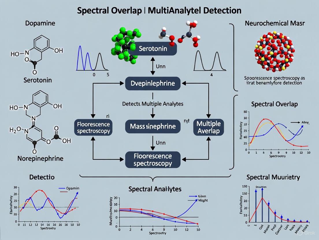
Overcoming Spectral Overlap: Advanced Strategies for Multianalyte Neurochemical Detection
This article provides a comprehensive resource for researchers and drug development professionals tackling the critical challenge of spectral overlap in multianalyte neurochemical detection.
Overcoming Spectral Overlap: Advanced Strategies for Multianalyte Neurochemical Detection
Abstract
This article provides a comprehensive resource for researchers and drug development professionals tackling the critical challenge of spectral overlap in multianalyte neurochemical detection. It explores the fundamental principles that cause signal crosstalk in techniques like fluorescence imaging and voltammetry and details cutting-edge methodological solutions, including computational approaches, AI-enhanced biosensors, and optimized chromatographic separations. The content further offers practical troubleshooting guidance for method optimization and a framework for the rigorous validation and comparative analysis of emerging multi-analyte platforms. The goal is to equip scientists with the knowledge to achieve precise, simultaneous quantification of neurochemicals, thereby accelerating research in neurology and therapeutic development.
The Spectral Overlap Problem: Fundamentals and Impact on Neurochemical Analysis
Defining Spectral Overlap and Signal Crosstalk in Analytical Neuroscience
Core Definitions and Their Impact on Neurochemical Detection
What are Spectral Overlap and Signal Crosstalk? Spectral overlap occurs when the absorption or emission spectra of two or more fluorescent indicators or electroactive species coincide within a similar wavelength or potential range [1]. In analytical neuroscience, this phenomenon is critical when using multiple fluorescent probes or electroactive neurotransmitters simultaneously.
In multianalyte detection, this overlap leads to signal crosstalk, where the measured signal in one detection channel contains contributions from non-target species [2] [3]. This interference can cause misidentification of neurochemical signals, inaccurate concentration measurements, and ultimately, flawed biological interpretations.
The fundamental challenge arises because most organic fluorescent dyes exhibit asymmetric spectral profiles that typically extend over 100 nm beyond their peak emission [2]. Similarly, in electrochemical methods like FSCV, electroactive neurochemicals can oxidize at similar potentials, creating analogous crosstalk issues [4].
Advanced Methodologies for Crosstalk Mitigation
Computational Approaches: Machine Learning
Machine learning (ML) models, particularly decision tree algorithms like XGBoost, can resolve complex, nested correlations in spectral data that conventional statistical methods cannot decipher [5].
Table 1: Machine Learning Workflow for Spectral Crosstalk Resolution
| Step | Implementation | Benefit in Neurochemical Detection |
|---|---|---|
| Data Acquisition | Hyperspectral imaging capturing 470-900 nm range with 3 nm resolution [5] | Provides large, high-quality spectral datasets for training |
| Feature Extraction | Full spectral signature from each image pixel [5] | Captures complex emission patterns traditional methods miss |
| Model Training | XGBoost algorithm on spectral data [5] | Learns to disentangle overlapping fluorescence signatures |
| Validation | Mean Absolute Error calculation for pH and O₂ predictions [5] | Ensures model reliability for quantitative measurements |
Experimental Protocol: ML Implementation for Multi-Analyte Sensing
- Sensor Fabrication: Create dual-analyte optodes by knife-coating successive polymer layers onto PET support foil [5]
- Data Collection: Use hyperspectral camera system with 460 nm excitation LED [5]
- Calibration: Acquire training data across full concentration ranges of target analytes [5]
- Model Optimization: Train XGBoost algorithm to predict analyte concentrations despite spectral overlap [5]
Experimental Approaches: Sequential and Concurrent Imaging
For wide-field fluorescence endoscopic imaging, researchers have developed multiple approaches to mitigate crosstalk:
Frame-Sequential Imaging
- Protocol: Capture images for each fluorophore separately using sequential excitation wavelengths [2]
- Advantage: Eliminates crosstalk by temporal separation
- Limitation: Potential image rendering lag in clinical workflows [2]
Concurrent Imaging with Cross-talk Ratio Subtraction (CRS)
- Protocol: Simultaneous excitation with mathematical subtraction of known crosstalk components [2]
- Implementation:
- Characterize crosstalk ratios between detection channels
- Apply CRS algorithm: Corrected Signal = Raw Signal - (Crosstalk Ratio × Reference Signal)
- Advantage: Maintains real-time imaging capability [2]
Table 2: Comparison of Spectral Overlap Mitigation Techniques
| Method | Temporal Resolution | Spatial Resolution | Implementation Complexity | Best Use Case |
|---|---|---|---|---|
| Machine Learning | High | High (pixel-level) | High | Complex heterogeneous environments [5] |
| Frame-Sequential Imaging | Reduced due to sequential capture | Preserved | Moderate | When timing artifacts are acceptable [2] |
| CRS Algorithm | Preserves real-time capability | Preserved | Low-Moderate | Live imaging requiring immediate feedback [2] |
| Microdialysis | Low (minutes) | Low (mm scale) | Low | When fast dynamics are not critical [4] |
The Scientist's Toolkit: Essential Reagents and Materials
Table 3: Key Research Reagents for Multi-Analyte Neurochemical Detection
| Reagent/Material | Function | Example Application |
|---|---|---|
| Pt-TPTBP (Platinum(II)-meso-tetraphenyl-tetrabenzoporphyrin) | O₂-sensitive indicator dye [5] | Dissolved oxygen sensing in neural tissue |
| Lipophilic HPTS derivatives | pH-sensitive fluorescent indicators [5] | Extracellular pH monitoring in brain microenvironments |
| Polystyrene (PS) & Polyurethane Hydrogel (D4) | Polymer matrices for sensor immobilization [5] | Creating stable, biocompatible sensor films |
| Fluorol 555 & Pyrromethene 597 | Model fluorescent dyes with spectral overlap [2] | Testing and validation of crosstalk correction methods |
| Artificial Cerebrospinal Fluid (aCSF) | Physiological perfusion fluid [4] | Microdialysis and maintaining tissue viability |
| Monocrystalline Diamond Powder | Signal enhancer in optodes [5] | Improving signal-to-noise in fluorescence detection |
Troubleshooting Guide: FAQs for Experimental Challenges
Q1: Our multi-fluorophore imaging shows persistent crosstalk despite using recommended filter sets. What optimization strategies can we implement?
A: Beyond filter optimization, consider these approaches:
- Spectral Unmixing: Use a hyperspectral imaging system to capture full emission spectra (470-900 nm) followed by computational separation of overlapping signals [5]
- Lifetime-Based Separation: Employ fluorescence lifetime measurements, as lifetimes are less affected by spectral overlap than intensity measurements [3]
- Concurrent Imaging with CRS: Implement Cross-talk Ratio Subtraction algorithm that mathematically removes known crosstalk components while preserving real-time imaging capability [2]
Q2: How can we validate that our crosstalk correction methods are working accurately in biological preparations?
A: Implement a multi-stage validation protocol:
- Phantom Validation: Create dye-in-polymer targets with known concentrations and spatial distributions to test your correction method [2]
- Single-Probe Controls: Perform control experiments with individual probes to establish baseline signals without crosstalk [2]
- Recovery Testing: Spike known concentrations of analytes into your biological preparation and verify your system accurately detects the expected changes
- Cross-Validation: When possible, validate with an orthogonal detection method (e.g., compare microdialysis with fluorescence imaging) [4]
Q3: We need to monitor multiple neurochemicals simultaneously but cannot achieve sufficient temporal resolution with microdialysis. What alternatives provide better time resolution?
A: Consider these alternatives based on your target analytes:
- Fast-Scan Cyclic Voltammetry (FSCV): Provides sub-second temporal resolution for electroactive neurotransmitters like dopamine [4]
- Genetically Encoded Fluorescent Sensors: Offer cell-type specific targeting with temporal resolution sufficient for tracking neural activity dynamics [4]
- Segmented-Flow Microfluidics with Online Analysis: Modern microdialysis adaptations that improve temporal resolution to seconds by collecting nanoliter-sized droplets [4]
Q4: What practical steps can we take to minimize spectral overlap during experimental design?
A: Implement proactive experimental design strategies:
- Fluorophore Selection: Choose dye combinations with minimal emission spectrum overlap, even if this requires compromising on brightness [2]
- Sequential Imaging Protocols: When dynamics allow, image fluorophores sequentially rather than simultaneously [2]
- Spectral Characterization: Precisely measure the emission spectra of your specific dye batches under experimental conditions, as spectra can vary based on environmental factors [1]
- Reference Measurements: Include single-label controls in each experiment to quantify crosstalk magnitudes for post-hoc correction [2]
Troubleshooting Guides and FAQs for Multianalyte Neurochemical Detection
This guide addresses common experimental challenges in multianalyte neurochemical detection research, with a focus on resolving spectral overlap and improving data fidelity.
Frequently Asked Questions (FAQs)
Q1: What is the primary challenge in simultaneously detecting dopamine, serotonin, glutamate, and GABA? The core challenge is spectral overlap, where the electrochemical or analytical signatures of these neurotransmitters interfere with one another, reducing the specificity and quantitative accuracy of measurements. While techniques like voltammetry excel for single-analyte detection (e.g., dopamine), distinguishing multiple analytes in a complex mixture requires advanced sensor design or data processing to deconvolve their overlapping signals [6].
Q2: How can I improve the specificity of my sensor for serotonin over dopamine? Employ advanced electrode materials and data processing. Chemically modified electrodes with specific coatings (e.g., Nafion) can impart selectivity based on charge or size. Furthermore, techniques like multiple cyclic voltammetry coupled with machine learning algorithms can train systems to recognize the unique "fingerprint" of each analyte's redox profile, effectively resolving their overlapping signals [6].
Q3: My results show high background noise in glutamate measurements. What could be the cause? In analytical methods like Magnetic Resonance Spectroscopy (MRS), a significant cause is the overlapping resonances of glutamate with other metabolites, particularly glutamine and GABA. This is often reported as a combined "Glx" signal. To address this, ensure you are using spectral editing sequences (e.g., MEGA-PRESS) that are specifically designed to isolate the GABA signal and can improve the resolution of glutamate [7].
Q4: Are there computational methods to model the interplay of these neurotransmitter systems? Yes, computational neuroscience increasingly uses quantitative models to understand these interactions. You can employ biophysically detailed models to simulate the excitatory-inhibitory balance between glutamate and GABA, or use reinforcement learning models to understand the role of dopamine and serotonin in reward and behavior. These models help generate testable hypotheses about neurotransmitter dynamics in health and disease [8].
Troubleshooting Common Experimental Issues
| Problem | Possible Cause | Solution |
|---|---|---|
| Poor Signal-to-Noise Ratio in Electrochemical Detection | Non-specific adsorption of proteins or other molecules on the electrode surface. | Use a microdialysis membrane or a size-selective polymer coating (e.g., cellulose acetate) to filter out large interferents [6]. |
| Inability to Resolve Glutamate from GABA via MRS | Standard pulse sequences cannot separate overlapping spectral peaks. | Implement specialized spectral editing sequences (e.g., MEGA-PRESS) for GABA and ensure voxel placement in large, homogeneous tissue volumes (8-20 cc) [7]. |
| Low Quantitative Accuracy in Sample-Multiplexed Proteomics | Stochastic precursor selection and co-isolation during mass spectrometry. | Adopt Intelligent Data Acquisition (IDA) strategies, such as Real-Time Library Searching (RTLS), to improve instrument efficiency and quantitative accuracy by triggering scans based on real-time spectral matching [9]. |
| Variable Recovery in Microdialysis | Inconsistent flow rate or membrane fouling. | Calibrate the perfusion pump regularly and use validated probes with appropriate molecular weight cut-offs. Analyze samples promptly or stabilize them to prevent analyte degradation [6]. |
Experimental Protocols for Neurotransmitter System Analysis
Protocol for Recording Neuronal Network Activity with Multi-Electrode Arrays (MEAs)
This protocol is adapted for studying the effects of neurotransmitter perturbations on network-level activity ex vivo [10].
1. Solutions and Reagents Preparation:
- Cutting Solution: Prepare an ice-cold, carbogenated (95% O₂/5% CO₂) solution suitable for acute brain slice preparation (e.g., sucrose-based artificial cerebrospinal fluid - ACSF). Final osmolarity should be 320-330 mOsm/L.
- Incubation/Recording ACSF: Prepare a standard ACSF (e.g., 126 mM NaCl, 3 mM KCl, 1.25 mM NaH₂PO₄, 2 mM MgSO₄, 26 mM NaHCO₃, 2 mM CaCl₂, and 10 mM glucose), continuously oxygenated with carbogen.
- Picrotoxin Stock Solution (500 mM): Dissolve 1.51 g of picrotoxin (PTX) in 5 mL of DMSO. Aliquot and store at -20°C, protected from light. CRITICAL: PTX is a GABAA receptor antagonist used to induce network bursting activity. The final concentration in the recording chamber is typically 100 µM [10].
2. Hippocampal Slice Preparation:
- Anesthetize a mouse (P16-P25) according to institutional guidelines and decapitate.
- Rapidly remove the brain and immerse it in ice-cold cutting solution.
- Using a vibratome, prepare 300-400 µm thick transverse hippocampal slices.
- Immediately transfer slices to an incubation chamber containing standard ACSF at 32-33°C for at least 1 hour for recovery.
3. MEA Recording and Pharmacological Induction of Bursting:
- Place a single hippocampal slice on the MEA probe, ensuring good contact between the tissue and the electrodes.
- Perfuse the slice with the incubation/recording ACSF containing 100 µM PTX to block GABAergic inhibition.
- Record extracellular field potentials from all electrodes for at least 30 minutes to capture stable network bursting activity.
- Analysis: Use a burst detection algorithm (e.g., in MATLAB) to identify network bursts. Key parameters to analyze include burst frequency, duration, and spike rate within bursts [10].
Protocol for Mapping Neurotransmitter Circuit Damage Using Structural MRI
This protocol outlines a computational method to estimate how focal brain lesions, like stroke, disrupt major neurotransmitter systems in a pre- or postsynaptic manner [11].
1. Data Acquisition:
- Acquire a high-resolution T1-weighted anatomical MRI and a T2-weighted lesion mask (e.g., from a FLAIR or DWI sequence) from the patient.
2. Lesion Mapping and Neurotransmitter Atlas Overlay:
- Normalize the patient's brain images to a standard stereotaxic space (e.g., MNI).
- Map the lesion mask onto a normative neurotransmitter atlas. This atlas contains voxel-wise maps of receptor and transporter density for acetylcholine, dopamine, noradrenaline, and serotonin systems, derived from PET scans of healthy individuals [11].
3. Calculating Pre- and Postsynaptic Damage Ratios:
- Presynaptic Ratio: Quantifies relative damage to the neuron producing the neurotransmitter. It is calculated as the proportion of the lesion overlapping with the transporter location density map and its white matter projections.
- Postsynaptic Ratio: Quantifies relative damage to the neuron receiving the signal. It is calculated as the proportion of the lesion overlapping with the receptor location density map and its white matter projections [11].
- A ratio >1 indicates a predominant disruption of that specific synaptic component.
Signaling Pathways and Experimental Workflows
Simplified Signaling Pathways of Key Neurotransmitters
The following diagram illustrates the core synthesis, receptor action, and termination mechanisms for dopamine, serotonin, glutamate, and GABA.
Workflow for Multianalyte Detection and Spectral Deconvolution
This diagram outlines a generalized experimental and computational workflow for overcoming spectral overlap in multianalyte detection.
The Scientist's Toolkit: Research Reagent Solutions
The following table details key reagents, materials, and computational tools used in experiments related to these neurotransmitter systems.
| Item Name | Function / Application | Specific Example / Note |
|---|---|---|
| Picrotoxin (PTX) | GABAA receptor antagonist. Used to block inhibitory GABAergic transmission, often to induce or study epileptiform network bursting activity in brain slices [10]. | Prepare a 500 mM stock in DMSO; light-sensitive. Final working concentration is typically 50-100 µM. |
| Flumazenil / Iomazenil | Radiolabeled ligands that bind to GABAA receptors containing α1, α2, α3, or α5 subunits. Used for PET or SPECT imaging to quantify GABAA receptor availability in vivo [7]. | 11C-flumazenil for PET; 123I-iomazenil for SPECT. |
| Raclopride / Fallypride | Radiolabeled ligands for dopamine D2/D3 receptors. Used in PET imaging to assess dopaminergic function in the context of addiction, Parkinson's disease, and psychosis [7]. | 11C-raclopride is restricted to striatum; 18F-fallypride can image extra-striatal regions. |
| Spectral Library (for Proteomics) | A curated collection of known peptide spectra used for matching and identifying compounds in mass spectrometry. Enables Real-Time Library Searching (RTLS) to improve quantification in multiplexed samples [9]. | Can be empirically derived or predicted in silico using tools like Prosit. |
| Functionnectome | A computational method that projects gray matter values (e.g., receptor densities) onto white matter voxels based on structural connection probability. Used to create maps of neurotransmitter circuit pathways [11]. | Allows estimation of neurotransmitter system damage from structural MRI lesions. |
| Multi-Electrode Array (MEA) | A grid of miniature electrodes for recording extracellular electrical activity from multiple sites simultaneously in ex vivo brain slices or cell cultures. Ideal for studying network-level dynamics [10]. | Used to record pharmacologically-induced network bursts in hippocampal slices. |
| MEGA-PRESS | An MRS spectral editing pulse sequence specifically designed to detect low-concentration metabolites with overlapping signals, most commonly used to isolate the GABA signal [7]. | Crucial for separating the GABA resonance from the much larger creatine and glutamate signals. |
Limitations of Single-Analyte Approaches in Understanding Complex Brain Circuitry
In the study of brain circuitry, a single-analyte approach focuses on measuring one specific neurochemical or biological marker at a time. While historically useful, this method is fundamentally limited when investigating complex neural systems, where the interactions between many different components—such as various neurotransmitter systems, cell types, and circuit motifs—govern function. The brain's operational logic arises from highly interconnected networks, and understanding it requires the simultaneous monitoring of multiple elements.
A primary technical challenge in moving beyond single-analyte methods is spectral overlap, which occurs in detection methodologies when the signals from different analytes are not sufficiently distinct and thus interfere with one another. For instance, in electrochemical detection, neurotransmitters like dopamine and serotonin have closely spaced oxidation potentials, leading to overlapping signals [12]. Similarly, in optical methods, the emission spectrum of one fluorophore can overlap with the excitation spectrum of another, causing crosstalk in the measured signals [13]. This phenomenon complicates the accurate isolation and quantification of individual components in a mixture, a problem that must be overcome to achieve reliable multianalyte detection.
Key Technical Challenges & Troubleshooting FAQs
FAQ 1: What is the primary limitation of single-analyte approaches in circuit neuroscience? The main limitation is the inability to capture interdependent signaling. Neural circuits rely on the coordinated activity and interaction of multiple neurotransmitters, neuromodulators, and cell types. Studying one element in isolation creates a fragmented and often misleading picture, as it misses the contextual interactions that define circuit operation and output [6] [14]. For example, subpopulations of dopamine neurons in the ventral tegmental area (VTA) are intermingled but project to different regions and can mediate opposing behaviors like reward and aversion. A single-analyte approach targeting "dopamine" would fail to resolve this critical functional heterogeneity [14].
FAQ 2: How does spectral overlap specifically hinder multianalyte neurochemical detection? Spectral overlap leads to signal crosstalk, where the measurement for one analyte is contaminated by the signal from another. This directly compromises the accuracy and reliability of quantification.
- In Electrochemical Detection: Dopamine and serotonin have similar oxidation potentials, which results in overlapping voltammetric peaks. This makes it extremely difficult to quantify each one accurately in a mixture without advanced data processing [12].
- In Optical Detection (e.g., Fluorescence Microscopy/Flow Cytometry): The emission light of one fluorophore (Donor) can spill over into the detector channel intended for a different fluorophore (Acceptor) [15]. This spillover, if not corrected, causes false-positive signals and misidentification of cell populations or molecular constituents [16] [15] [13].
FAQ 3: What are the standard methods to correct for spectral overlap? The standard method is mathematical compensation (also referred to as correction or deconvolution). This process calculates the amount of spillover from one channel into another and then subtracts that contribution from the contaminated signal [15].
- Procedure: The process requires control samples, each stained with only one of the fluorophores used in the multicolor experiment. The software measures the spillover coefficient from the single-color control and applies a correction matrix to the entire multicolor dataset [15].
- Critical Rules for Success:
- Brightness: Control samples must be at least as bright as the test samples.
- Purity: The positive and negative control populations must have matching background fluorescence.
- Matching Reagents: The fluorochrome used for compensation controls must be identical to the one used in the experiment [15].
FAQ 4: Beyond compensation, what advanced strategies can overcome these limitations? Emerging strategies integrate advanced materials science with machine learning (ML).
- Sensor Engineering: Modifying electrode surfaces with materials like laser-induced graphene (LIG) and coating them with selective polymers (e.g., Nafion) can improve sensitivity and partially reject interferents [12].
- Machine Learning-Powered Analytics: When material engineering alone is insufficient, machine learning models can be trained to deconvolute complex, overlapping signals. By feeding the algorithm multimodal voltammetry data, it can learn to accurately quantify individual analytes from a merged signal, dramatically improving detection limits and specificity in complex media like undiluted urine [12].
Experimental Protocols for Multianalyte Analysis
Protocol 1: Machine Learning-Integrated Electrochemical Detection of Dopamine and Serotonin
This protocol is adapted from research demonstrating simultaneous detection of dopamine (DA) and serotonin (SER) in undiluted human urine using laser-induced graphene (LIG) electrodes [12].
1. Sensor Fabrication:
- LIG Formation: Use a CO2 laser scriber to convert a polyimide substrate into porous graphene. Optimize laser power and pass number to maximize the electroactive surface area.
- Electrode Design: Define working, counter, and reference electrodes. Optimize the working electrode diameter for signal stability and minimal sample volume.
- Surface Functionalization: Coat the LIG working electrode with a Nafion solution (e.g., 0.5% in alcohol) by drop-casting and allow it to dry. This negatively charged coating repels interferents like ascorbic acid and uric acid.
2. Data Acquisition (Multimodal Voltammetry):
- Prepare standard solutions of DA and SER in a relevant buffer or biofluid (e.g., artificial urine or PBS).
- Record electrochemical measurements using multiple techniques on the same sensor:
- Cyclic Voltammetry (CV)
- Square Wave Voltammetry (SWV)
- Electrochemical Impedance Spectroscopy (EIS)
- Perform experiments with individual analytes and mixtures to build a comprehensive training dataset.
3. Machine Learning Training and Prediction:
- Feature Extraction: Extract peak currents, potentials, and other descriptive features from the voltammograms of all three techniques.
- Model Training: Train a regression model (e.g., Gaussian Process Regression) using the single-analyte data to learn the unique electrochemical "fingerprint" of each molecule.
- Quantification: Use the trained model to predict the concentrations of DA and SER in unknown mixture samples based on their combined voltammetric features.
Protocol 2: Mapping Functional Connectomes with FORCE Learning and Graph Theory
This protocol outlines a computational method to infer functional connectivity between thousands of individual neurons from calcium imaging data [17].
1. Data Collection:
- Use in vivo 2-photon calcium imaging to record the activity of thousands of individual neurons simultaneously in a live animal.
2. Network Modeling with FORCE Learning:
- Model Architecture: Represent the neural network as a chaotic recurrent neural network (RNN), where each node is a neuron and connections represent synaptic influences.
- Training: Train the RNN using a least-squares optimization rule (FORCE learning) to fine-tune the connection weights between neurons. The objective is for the RNN's output dynamics to closely match the experimentally recorded calcium traces.
- Output: The final output of this step is a directed graph of the network, where the edge weights represent the inferred strength and sign (excitatory/inhibitory) of influence from one neuron to another.
3. Higher-Order Network Mining:
- Hub Identification: Apply graph analytics to the inferred connectivity network to identify hub neurons—cells with an unusually high number or strength of connections.
- Motif Analysis: Use higher-order graph algorithms to search for over-represented subgraph patterns (motifs), such as specific three-node or four-node connection patterns. These can reveal common circuit building blocks.
- Superhub Discovery: The combination of these methods can identify "superhubs," neurons that are critical for network synchronization and may be prime targets for controlling pathological states like seizures [17].
Data Presentation: Quantitative Comparisons
Table 1: Performance Comparison of Single vs. Multianalyte Approaches for Neurotransmitter Detection
| Parameter | Single-Analyte Electrochemical Sensing | Multianalyte Sensing with Material Engineering | Multianalyte Sensing with ML Integration |
|---|---|---|---|
| Selectivity Challenge | Low; vulnerable to interferents | Moderate; improved via coatings | High; ML deconvolutes overlapping signals |
| Limit of Detection (LoD) in Urine | ~0.3 μM for DA [12] | Not specified | 5 nM for both DA and SER [12] |
| Key Advantage | Simplicity | Physical signal enhancement | Data-driven resolution of overlap |
| Primary Limitation | Provides a fragmented view | May not fully resolve co-localized analytes | Requires large, high-quality training datasets |
Table 2: Impact of Hub Neurons on Network Dynamics as Revealed by Multianalyte Circuit Analysis
| Neuron Classification | Definition | Role in Network Function | Implication for Disease |
|---|---|---|---|
| Regular Neuron | Standard connectivity | Participant in local circuit activity | --- |
| Hub Neuron | High number/strength of connections | Drives network synchronization [17] | Implicated in seizure generation [17] |
| Superhub Neuron | A hub embedded in higher-order motifs; exerts maximal influence | Predicted to be a critical regulator of dynamics [17] | A maximally selective target for seizure control [17] |
Essential Visualizations
Diagram 1: Spectral Overlap and Compensation in Detection
Diagram 2: Multimodal ML Workflow for Neurochemical Detection
The Scientist's Toolkit: Research Reagent Solutions
Table 3: Key Reagents and Materials for Advanced Multianalyte Circuit Research
| Item Name | Function/Description | Application Context |
|---|---|---|
| Laser-Induced Graphene (LIG) | A porous, highly conductive carbon material fabricated by laser-printing on polyimide; provides a large electroactive surface area. | Electrochemical sensor platform for sensitive neurotransmitter detection [12]. |
| Nafion Coating | A perfluorosulfonated ionomer; forms a negatively charged film that repels common anionic interferents (e.g., ascorbic acid, uric acid). | Selectivity enhancement for catecholamine detection on electrode surfaces [12]. |
| RNA Barcodes (MAPseq/BARseq) | Unique RNA sequences used to tag individual neurons, allowing their projections to be traced via sequencing. | High-throughput mapping of long-range neuronal projections with single-neuron resolution [14]. |
| Chaotic Recurrent Neural Network (RNN) | A type of computational model that is highly sensitive to initial conditions and can generate complex dynamics. | Inferring functional connectivity between neurons from time-series calcium imaging data [17]. |
| Spectral Overlap Compensation Matrix | A mathematical correction applied to raw data to subtract the contribution of signal spillover between detection channels. | Essential for accurate analysis in flow cytometry and multicolor fluorescence imaging [15]. |
FAQ: Addressing Common Technical Challenges
Why do organic fluorophores have broad, overlapping emission spectra? The broad emission spectra of organic fluorophores originate from their polyatomic molecular structures. Unlike monoatomic fluorophores that have discrete wavelengths, polyatomic organic dyes exhibit broad excitation and emission bands due to the numerous vibrational and rotational energy levels associated with their complex chemical structures [18]. This inherent physical property means that the emission spectra of common organic dyes like FITC and TAMRA can extend over 100 nm, creating significant overlap when using multiple fluorophores simultaneously [2].
What causes overlapping peaks in voltammetric detection of neurochemicals? Overlapping voltammetric peaks occur when multiple electroactive species have similar redox potentials. In multianalyte detection, compounds with similar chemical structures often oxidize or reduce at similar applied potentials, causing their current responses to merge into unresolved peaks. This is particularly challenging in neurochemical monitoring where neurotransmitters like dopamine, serotonin, and their metabolites may co-exist and have overlapping electrochemical signatures [19] [20].
How does spectral overlap impact multianalyte neurochemical detection? Spectral overlap significantly compromises data interpretation in multianalyte experiments. In fluorescence imaging, emission cross-talk between channels can lead to misidentification of labeled targets and false positive signals [2]. In voltammetry, overlapping peaks prevent accurate quantification of individual analytes, potentially leading to incorrect conclusions about neurochemical dynamics [19]. Both scenarios reduce the reliability of experimental data in drug development research.
Troubleshooting Guides
Problem: Fluorophore Emission Cross-Talk in Wide-Field Imaging
Symptoms: High background signal, bleed-through between detection channels, inability to distinguish specifically labeled targets in multiplexed experiments.
Solution: Implement sequential imaging or algorithmic separation techniques [2].
Experimental Protocol: Frame-Sequential Imaging
- Configure your imaging system to excite and detect one fluorophore at a time
- Set appropriate excitation wavelengths and emission filters for each fluorophore separately
- Acquire images for each channel in sequence rather than concurrently
- Merge the separate channel images during post-processing
- For the SFE platform tested, this approach successfully eliminated fluorophore cross-talk
Alternative Protocol: Concurrent Imaging with Cross-talk Ratio Subtraction Algorithm
- Image all fluorophores simultaneously using widefield detection
- Apply the CRS algorithm during image processing:
- Calculate the signal contribution from each fluorophore in each detection channel
- Use predetermined cross-talk ratios to mathematically separate the signals
- Reconstruct individual fluorophore distribution maps
- This approach maintains imaging speed while computationally removing cross-talk
Prevention Strategies:
- Select fluorophores with large Stokes shifts and minimal spectral overlap [18]
- Use optical filters with narrow bandpass ranges optimized for your specific fluorophore combination
- Consider quantum dots with their narrower emission bands if toxicity concerns are addressed [2]
Problem: Resolving Overlapping Voltammetric Peaks in Neurochemical Detection
Symptoms: Broad, poorly resolved peaks in cyclic voltammetry; inability to distinguish individual analytes in mixture samples; non-linear calibration curves for individual compounds in multianalyte solutions.
Solution: Apply advanced voltammetric techniques and mathematical deconvolution methods [19].
Experimental Protocol: Differential Pulse Voltammetry for Peak Separation
- Implement differential pulse voltammetry parameters:
- Pulse amplitude: 25-100 mV
- Pulse width: 10-100 ms
- Step height: 2-10 mV
- Step time: 0.1-1 s
- The differential current measurement enhances resolution of overlapping peaks
- Follow with mathematical modeling to deconvolute overlapping signals
Experimental Protocol: Mathematical Deconvolution of Overlapping Signals
- Record voltammetric data for individual analytes to establish characteristic profiles
- Collect experimental data for the unknown mixture
- Apply genetic algorithm or Marquardt-Levenberg method for curve fitting [21]
- Iteratively refine parameters until the combined individual profiles match the experimental mixture data
- Use the optimized fit to quantify individual analyte contributions
Prevention Strategies:
- Optimize electrochemical cell conditions (pH, solvent, supporting electrolyte) to maximize separation of redox potentials [19]
- Utilize modified electrodes with selective catalytic properties for target analytes [20]
- Implement scanning electrochemical techniques that provide additional dimensionality to data
Quantitative Data Reference Tables
Table 1: Spectral Characteristics of Common Organic Fluorophores and Overlap Potential
| Fluorophore | Excitation Max (nm) | Emission Max (nm) | Stokes Shift (nm) | Emission FWHM* (nm) | Overlap Risk |
|---|---|---|---|---|---|
| FITC | 495 | 519 | 24 | ~100 [2] | High |
| TRITC | 557 | 576 | 19 | ~100 [2] | High |
| Cy5.5 | 675 | 694 | 19 | ~100 [2] | High |
| Quantum Dots | Varies by size | Varies by size | 20-40 | ~25 [2] | Low |
*FWHM = Full Width at Half Maximum
Table 2: Comparison of Spectral Overlap Mitigation Approaches for Fluorescence Imaging
| Method | Spectral Resolution | Temporal Resolution | Implementation Complexity | Best Application Context |
|---|---|---|---|---|
| Image Stitching | Moderate | High | Low | Fixed samples, post-processing analysis |
| Frame-Sequential Imaging | High | Reduced | Moderate | Dynamic processes requiring high specificity |
| Concurrent with CRS* | High | High | High | Real-time in vivo imaging [2] |
| Optical Filter Optimization | Moderate | High | Low | Initial experimental design |
*CRS = Cross-talk Ratio Subtraction Algorithm
Table 3: Electrochemical Techniques for Resolving Overlapping Voltammetric Peaks
| Technique | Timescale | Detection Limit | Resolution Capability | Neurochemical Applications |
|---|---|---|---|---|
| Fast-Scan Cyclic Voltammetry (FSCV) | Subsecond | nM range [4] | Moderate | Dopamine, serotonin transients |
| Differential Pulse Voltammetry (DPV) | Seconds | µM-nM range | High | Catecholamines, metabolites |
| Square-Wave Voltammetry (SWV) | Seconds | µM-nM range | High | Multiplexed neurotransmitter detection |
| Mathematical Deconvolution | Post-processing | Dependent on base method | Very High | Complex mixtures [21] |
Research Reagent Solutions
Table 4: Essential Materials for Spectral Overlap Mitigation in Neurochemical Research
| Reagent/Material | Function | Application Context | Key Considerations |
|---|---|---|---|
| Organic Dyes (FITC, TRITC) | Fluorescent labeling | Immunofluorescence, receptor labeling | Broad emissions require careful filter selection [18] |
| Quantum Dots | Fluorescent labeling with narrow emission | Multiplexed imaging | Potential toxicity concerns for in vivo use [2] |
| Genetically-encoded Indicators | Direct neuromodulator sensing | Real-time neurochemical monitoring in specific cell types | High molecular specificity [4] |
| Fast-Scan Cyclic Voltammetry Electrodes | Electrochemical detection of electroactive neurochemicals | Monitoring dopamine, serotonin dynamics | Subsecond temporal resolution [20] |
| Cross-talk Ratio Subtraction Algorithm | Mathematical separation of overlapping signals | Computational resolution of spectral overlap | Maintains temporal resolution in dynamic imaging [2] |
Experimental Workflow Visualization
Experimental decision pathway for spectral and voltammetric overlap issues
Advanced Methodologies
Genetic Algorithm for Spectral Deconvolution
For complex overlapping signals where traditional fitting methods fail, genetic algorithms provide a powerful alternative for signal separation [21]. This evolutionary approach is particularly valuable when the number and identity of contributing analytes is unknown.
Implementation Protocol:
- Initialization: Create a population of potential solutions with random parameters
- Evaluation: Calculate how well each solution fits the experimental data
- Selection: Preferentially select the best-fitting solutions for reproduction
- Crossover: Combine parameters from parent solutions to create offspring
- Mutation: Introduce random changes to maintain diversity
- Iteration: Repeat for hundreds or thousands of generations until convergence
This method has demonstrated higher resolution capabilities compared to standard Marquardt-Levenberg fitting for challenging separations, particularly in energy-dispersive X-ray spectrometry and complex material analysis [21].
Cross-talk Ratio Subtraction Algorithm for Fluorescence Imaging
The CRS algorithm enables real-time separation of overlapping fluorescence signals without sacrificing temporal resolution [2]. This approach is particularly valuable for in vivo imaging where dynamic processes must be monitored.
Mathematical Framework:
- Determine the cross-talk ratio (R) for each fluorophore in each detection channel using control samples
- For a two-fluorophore system with signals detected in two channels:
- Channel A Signal = (S1A × C1) + (S2A × C2)
- Channel B Signal = (S1B × C1) + (S2B × C2) Where S1A, S1B, S2A, S2B are the characteristic signals, and C1, C2 are the actual concentrations
- Solve the system of equations to extract C1 and C2, the separated fluorophore contributions
This method has been successfully implemented in scanning fiber endoscopy for early cancer detection, demonstrating significant reduction of fluorophore emission cross-talk in wide-field multispectral fluorescence imaging [2].
FAQs on Data Integrity in Neurochemical Detection
Q1: What are the primary sources of data inaccuracy in multianalyte neurochemical detection?
Data inaccuracies primarily arise from the complex nature of biological samples and technical limitations of the analytical methods. Key challenges include:
- Spectral Overlap and Crosstalk: In techniques like LC-MS/MS, structurally similar neurochemicals (e.g., different monoamine neurotransmitters or their metabolites) can have overlapping mass transitions or chromatographic retention times, leading to misidentification and inaccurate quantification [22].
- Inadequate Separation of Polar Compounds: Highly polar neurochemicals, such as amino acid neurotransmitters (e.g., glutamate, GABA), are often poorly retained on conventional reversed-phase chromatography columns. This can result in co-elution, insufficient resolution from interferents, and increased matrix effects, compromising data integrity [22].
- Suboptimal Sample Preparation: Conventional sample preparation methods like liquid-liquid extraction (LLE) can be biased towards non-polar compounds, while solid-phase extraction (SPE) may require compound-specific optimization. Inefficient protein precipitation or analyte losses during multiple processing steps can lead to biased and non-reproducible results [22].
Q2: How can spectral overlap lead to misdiagnosis in a research or clinical context?
Spectral overlap directly compromises the specificity of an assay. If the detection method cannot reliably distinguish between two similar molecules, the reported concentration for a target biomarker will be artificially inflated. This inaccurate quantification can have significant downstream consequences [22]:
- In Preclinical Research: It can lead to incorrect conclusions about neurochemical pathways, disease mechanisms, or drug efficacy. For instance, failing to separate dopamine from a similar metabolite could misrepresent its role in a model of Parkinson's disease [22].
- In Clinical Diagnostics: While current guidelines focus on specialized care, the principles of accurate quantification are foundational. Reliable biomarker tests require high sensitivity and specificity (e.g., ≥90% for both, as per new guidelines for Alzheimer's blood-based biomarkers) to ensure correct patient stratification [23]. Inaccurate data could lead to a false positive or negative diagnosis, impacting a patient's treatment pathway.
Q3: What methodologies can improve data integrity when quantifying multiple analytes?
Implementing a rigorous, optimized workflow is crucial for ensuring data integrity. Key methodological improvements include:
- Advanced Chromatography: Using specialized stationary phases, such as fluorophenyl columns, can enhance the retention and separation of a wide range of neurochemicals with diverse polarities within a single analytical run, reducing the need for multiple columns and simplifying the process [22].
- Systematic Method Validation: Any developed method must be thoroughly validated against established bioanalytical guidelines. This includes assessing linearity, detection limits, precision, accuracy, recovery, matrix effects, and carry-over to ensure the reliability of the generated data [22].
- Robust Data Governance: Beyond the bench, maintaining data integrity requires a strong framework of policies and procedures. This includes clear data management protocols, audit trails, access controls, and comprehensive training for personnel to foster a culture of data integrity and accountability [24].
Experimental Protocol: Simultaneous Determination of Multiple Neurochemicals
This protocol is adapted from a validated method for the simultaneous determination of 55 neurochemicals in brain tissue using LC-MS/MS [22].
1. Sample Preparation (Optimized Protein Precipitation)
- Homogenization: Homogenize brain tissue samples in a chilled methanol:water (8:2, v/v) solution. Using a standardized, automated homogenizer can significantly improve reproducibility and recovery rates [22] [25].
- Precipitation: Vortex the homogenates vigorously for 2 minutes.
- Centrifugation: Centrifuge at 14,000 × g for 15 minutes at 4°C.
- Collection: Transfer the resulting supernatant to a new vial.
- Evaporation: Evaporate the supernatant to complete dryness under a gentle stream of nitrogen gas.
- Reconstitution: Reconstitute the dried residue in 100 µL of mobile phase A (e.g., 0.1% formic acid in water). Vortex thoroughly and centrifuge before LC-MS/MS analysis.
2. Liquid Chromatography (LC) Conditions
- Column: Fluorophenyl column (e.g., 2.1 x 100 mm, 1.8 µm).
- Mobile Phase A: 0.1% Formic acid in water.
- Mobile Phase B: 0.1% Formic acid in acetonitrile.
- Gradient:
- 0-1 min: 0% B
- 1-10 min: 0% B to 55% B (linear gradient)
- 10-11 min: 55% B to 100% B
- 11-13 min: 100% B
- 13-13.1 min: 100% B to 0% B
- 13.1-16 min: 0% B (re-equilibration)
- Flow Rate: 0.3 mL/min.
- Column Oven Temperature: 40°C.
- Injection Volume: 5 µL.
3. Mass Spectrometry (MS) Conditions
- Ionization: Electrospray Ionization (ESI), positive mode.
- Data Acquisition: Multiple Reaction Monitoring (MRM).
- Optimization: Systematically optimize MRM conditions (precursor ion, product ion, collision energy, etc.) for each target neurochemical to maximize sensitivity and selectivity.
- Source Parameters: Optimize parameters like desolvation temperature, capillary voltage, and gas flows for optimal ion generation and transmission.
Performance Metrics for Biomarker Assays
The table below summarizes key performance thresholds for biomarker assays to ensure data integrity, based on clinical guidelines and analytical best practices. These values provide a benchmark for evaluating methodological rigor.
Table 1: Key Performance Metrics for Biomarker Assays
| Metric | Recommended Threshold | Rationale |
|---|---|---|
| Analytical Sensitivity | Varies by analyte; LOD/LOQ should be well below physiological range [22] | Ensures detection of low-abundance biomarkers critical for early disease detection. |
| Diagnostic Sensitivity | ≥90% [23] | Minimizes false negatives, correctly identifying individuals with the disease. |
| Diagnostic Specificity | ≥90% (for confirmatory testing); ≥75% (for triage) [23] | Minimizes false positives, correctly identifying individuals without the disease. |
| Precision | <15% RSD for precision and accuracy [22] | Ensures reproducible and reliable results across multiple runs. |
The Scientist's Toolkit: Research Reagent Solutions
Table 2: Essential Materials for Multianalyte Neurochemical Analysis
| Item | Function |
|---|---|
| Fluorophenyl Chromatography Column | Provides enhanced retention and separation of neurochemicals with a wide range of polarities within a single analytical run, mitigating challenges of spectral overlap [22]. |
| LC-MS/MS Grade Solvents | High-purity solvents (water, acetonitrile, methanol) minimize background noise and ion suppression, ensuring optimal MS sensitivity and data quality [22]. |
| Stable Isotope-Labeled Internal Standards | Corrects for variability in sample preparation and matrix effects, significantly improving the accuracy and precision of quantification [22]. |
| Automated Homogenization System | Standardizes sample processing, increases throughput, and improves the reproducibility and recovery of analytes from complex tissue matrices [25]. |
Troubleshooting Workflow for Data Integrity
The following diagram outlines a systematic workflow for troubleshooting data integrity issues, from problem identification to resolution.
Methodological Solutions: From Computational Separation to Multi-Modal Sensing
Cross-talk Ratio Subtraction (CRS) and Deconvolution
Frequently Asked Questions (FAQs) & Troubleshooting Guides
FAQ 1: What is the primary source of crosstalk in multianalyte neurochemical detection, and how can it be identified?
Crosstalk primarily arises from the overlapping emission spectra of fluorescent probes or the overlapping electrochemical signatures of different neurochemicals. In fluorescence imaging, this occurs when the emission spectrum of one fluorophore is detected in the channel assigned to another [26]. In electrochemical techniques, different electroactive analytes can have similar redox potentials, causing their signals to overlap [6]. Identification involves collecting control samples for each individual analyte and measuring the signal bleed-through into other detection channels. A crosstalk matrix can be constructed to quantify the percentage of each analyte's signal that appears in other channels [26].
FAQ 2: My deconvolution results are poor. What are the common pitfalls?
Poor deconvolution typically stems from an inaccurate system model or low signal-to-noise ratio.
- Inaccurate Point Spread Function (PSF): The PSF used for deconvolution must be measured experimentally under the same conditions as your data acquisition. An incorrect or theoretical PSF will lead to artifacts. Solution: Measure the PSF directly using sub-resolution fluorescent beads immobilized in a similar medium to your sample [26].
- Excessive Noise: Deconvolution can amplify high-frequency noise. Solution: Ensure your original data has a high signal-to-noise ratio. For computational deconvolution of spectral data, apply appropriate smoothing or regularization parameters to prevent noise amplification during the inverse filtering process [6].
- Insufficient Spectral Unmixing: If the reference spectra for your fluorophores are incorrect or too similar, linear unmixing will fail. Solution: Acquire reference spectra from single-label control samples under identical imaging conditions. Use these pure spectra for the unmixing calculation [26].
FAQ 3: When should I use CRS versus more complex deconvolution methods?
The choice depends on the complexity of the crosstalk and the required accuracy.
- Use CRS when the crosstalk between channels is relatively low (e.g., <15%) and can be approximated as a linear, constant proportion. It is computationally simple, fast, and suitable for real-time applications [6].
- Use Spectral Unmixing or more advanced model-based deconvolution when crosstalk is severe, the system is non-linear, or when you have a large number of overlapping analytes. These methods are more accurate but computationally intensive and often performed offline [26] [4].
FAQ 4: How can I validate the effectiveness of my crosstalk correction method?
Validation requires testing with known samples.
- In vitro Validation: Prepare control samples containing known concentrations and mixtures of the target analytes. After applying your correction algorithm, compare the calculated concentrations to the known ground truth. A successful method will recover the expected values with high fidelity [4].
- Biological Validation: Use biological models where the outcome is predictable. For instance, if correcting for crosstalk in dopamine and serotonin detection, a pharmacological challenge specific to one system (e.g., a selective serotonin reuptake inhibitor) should predominantly affect the corrected serotonin signal with minimal impact on the dopamine signal [6].
Experimental Protocols for Key Scenarios
Protocol 1: Establishing a Crosstalk Matrix for Fluorescent Probes
This protocol is essential for setting up both CRS and deconvolution methods in imaging experiments [26].
Methodology:
- Sample Preparation: Prepare separate control samples for each fluorescent protein or dye used in your multicolor experiment (e.g., Turquoise2, YFP, tdTomato). Use transfected cells or labeled tissue sections.
- Image Acquisition: Image each control sample using the exact same acquisition settings (laser power, detector gain, filter sets) you plan to use for your experimental samples.
- Signal Measurement: For each control sample, measure the mean signal intensity in its primary detection channel and in all secondary channels.
- Matrix Calculation: Construct a crosstalk matrix
Mwhere each elementM(i,j)represents the signal in channelifrom the fluorophorej. Normalize the values so that each column (fluorophore) sums to 1.
Example Crosstalk Matrix Table:
| Fluorophore | Channel 1 (Blue) | Channel 2 (Green) | Channel 3 (Red) |
|---|---|---|---|
| Turquoise2 | 1.00 | 0.05 | 0.01 |
| YFP | 0.02 | 1.00 | 0.08 |
| tdTomato | 0.01 | 0.12 | 1.00 |
Protocol 2: Implementing Cross-talk Ratio Subtraction (CRS) for Electrochemical Data
This protocol provides a step-by-step guide for correcting crosstalk in real-time electrochemical detection, such as fast-scan cyclic voltammetry (FSCV) [6] [4].
Methodology:
- Characterize Crosstalk Ratios: From controlled injections of pure analytes, determine the constant ratio (α) at which Analyte B appears in the primary sensor for Analyte A. For example, α = SignalBinChannelA / TrueSignalBinChannel_B.
- Apply CRS during Acquisition: For an unknown sample, the corrected concentration of Analyte A, [A]ₜᵣᵤₑ, can be calculated from the raw signals as follows:
[A]ₜᵣᵤₑ = [A]ᵣₐ𝓌 - α * [B]ᵣₐ𝓌Where[A]ᵣₐ𝓌and[B]ᵣₐ𝓌are the raw, uncorrected measurements from their respective primary sensors. - Iterate for Multiple Analytes: In a three-analyte system, the calculation must be iterated. First, correct [A] using raw [B] and [C], then use the corrected [A] to recalculate [B], and so on.
Protocol 3: Linear Unmixing for Multispectral Image Data
This protocol is used for post-acquisition separation of signals in techniques like ChroMS microscopy [26].
Methodology:
- Obtain Reference Spectra: As in Protocol 1, acquire the emission spectrum for each pure fluorophore in your sample. This is your reference matrix, R.
- Acquire Experimental Data: Capture the mixed-spectrum image of your experimental sample. At each pixel, the detected signal S is a linear combination of the reference spectra.
- Compute Unmixing Coefficients: For each pixel, solve the linear equation S = R * C for the coefficient vector C, which represents the relative contribution of each fluorophore. This is typically done via a least-squares minimization algorithm.
- Generate Unmixed Images: Use the calculated coefficients C to generate a new image stack where each channel contains only the signal from its corresponding fluorophore.
The Scientist's Toolkit: Research Reagent Solutions
The following table details key materials and reagents essential for experiments in multianalyte neurochemical detection [26] [4].
Table: Essential Research Reagents for Multianalyte Neurochemical Detection
| Item | Function & Application |
|---|---|
| Genetically Encoded Fluorescent Indicators (e.g., GCaMP, RCaMP) | Engineered proteins that change fluorescence upon binding specific ions (Ca²⁺) or neurotransmitters. Enable cell-type-specific monitoring of neural activity with high spatiotemporal resolution [4]. |
| Combinatorial Fluorescent Labeling Systems (e.g., Brainbow, MAGIC Markers) | Transgenic or viral strategies to label individual neurons and their progeny with distinct, heritable color combinations. Facilitates tracing of neural circuits and analysis of cellular interactions in densely labeled tissue [26]. |
| Chroma Keyers / Background Removal Software (e.g., Descript, Adobe Premiere Pro) | Software tools using chroma keying or AI to remove video backgrounds. In a research context, analogous algorithms are used for spectral unmixing—separating a complex signal into its constituent parts based on their distinct "spectral signatures" [27]. |
| Artificial Cerebrospinal Fluid (aCSF) | A balanced salt solution mimicking the ionic composition of natural CSF. Used as a perfusate in microdialysis to collect extracellular analytes without perturbing the local chemical environment [4]. |
| Semi-Permeable Microdialysis Membrane | A hollow fiber membrane with a specific molecular weight cutoff (e.g., 20-60 kDa) implanted in the brain. Allows continuous sampling of low molecular weight neurochemicals from the extracellular space for subsequent analysis [4]. |
Workflow and Signaling Pathway Diagrams
Experimental Crosstalk Correction Workflow
Neurochemical Signaling & Crosstalk Pathways
Artificial Intelligence and Machine Learning for Signal Decoding and Deconvolution
Frequently Asked Questions (FAQs)
Q1: What is the fundamental difference between signal deconvolution and spectral unmixing in the context of neurochemical data? Deconvolution aims to reverse the blurring effect of a measurement system to recover the original, underlying signal, such as estimating neural activity from a measured BOLD signal in fMRI [28]. Spectral unmixing, however, separates a composite signal from multiple sources into its constituent components or "endmembers," such as isolating the fluorescence signatures of different neurotransmitters that have overlapping spectra [29] [30]. Both are critical for resolving mixtures in multianalyte detection.
Q2: Why does my deep learning model for spectral unmixing perform poorly on new data, and how can I improve it? Poor performance often stems from inadequate training data quality or quantity. Machine learning models, especially deep learning, are highly dependent on large volumes of high-quality, well-labeled data for training. If the model is trained on poor data, it will produce poor results—a "garbage in, garbage out" scenario [31]. To improve robustness, ensure your training data is highly curated and accurate. Furthermore, consider using techniques like sparsity-promoting regularization (e.g., L1-norm) during model training, which helps improve the interpretability and generalizability of the estimates by preventing overfitting to noise [28] [32].
Q3: My deconvolution algorithm produces results with high variability. What type of regularization should I use? High variability in deconvolution results often indicates an ill-posed inverse problem. Applying appropriate regularization is key.
- Use sparsity-promoting regularizers like the L1-norm (e.g., LASSO) if you expect the underlying neural activity signal to be composed of discrete, transient events [28]. This is common in task-based fMRI or when detecting spontaneous neural transients.
- Use L2-norm regularization (ridge regression) if you have less prior knowledge and seek a more stable, but potentially less interpretable, solution [28].
- For complex data, structured mixed-norm regularizers (e.g., L2,1-norm) can be applied to improve robustness across neighboring voxels or to account for variability in the shape of the expected hemodynamic response [28].
Q4: Can AI fully automate the analysis of chromatographic or neurochemical data? No, full automation is not currently advisable. AI will not replace human analysts but will augment their capabilities, reducing the effort needed to interpret every data output [31]. It is dangerous to trust AI models implicitly. Labs should always verify the results and not use AI for critical acquisition steps without fail-safes. A "person in the loop" is recommended to confirm the system is operating as intended and that decisions adhere to protocol [31].
Troubleshooting Guides
Issue 1: Poor Signal-to-Noise Ratio (SNR) in Spectral Unmixing
A low SNR can severely limit the ability to distinguish between different analytes.
- Problem: Unmixing algorithms fail to reliably identify endmembers, leading to high false positive rates and inaccurate abundance estimates [30].
- Solution:
- Data Pre-processing: Apply denoising techniques tailored to your data type. For multi-echo fMRI, methods like Multi-Echo Independent Component Analysis (ME-ICA) can effectively separate BOLD signal from noise [28].
- Leverage Multi-echo Data: If your acquisition system supports it, collect data at multiple echo times. Algorithms like Multi-echo Sparse Paradigm Free Mapping (ME-SPFM) directly use this information to yield more robust, quantifiable estimates of the apparent transverse relaxation rate (
ΔR2*), which is less sensitive to certain noise types [28]. - Algorithm Selection: Choose unsupervised machine learning methods designed for robustness. For example, the Learning Unsupervised Means of Spectra (LUMoS) method uses clustering to learn spectral signatures directly from mixed images, which can be more resilient to noise than methods requiring pre-defined libraries [29].
Issue 2: Failure to Resolve Overlapping Spectral Peaks
This is a core challenge in multianalyte neurochemical detection where fluorophores or analytes have similar spectral profiles.
- Problem: Traditional linear unmixing or derivative-based algorithms cannot distinguish between heavily overlapping peaks, resulting in co-localization errors and false positives [31] [29].
- Solution:
- Shift to Machine Learning-based Peak Deconvolution: Replace traditional mathematical algorithms with machine learning models. ML models are specifically trained for certain data sets and are better able to address overlapping and otherwise complex peaks, leading to fewer false positives [31].
- Implement Advanced Unmixing Algorithms: Use algorithms that go beyond simple linear models.
- Apply Spectral Deconvolution as Pre-processing: Before unmixing, use a spectral deconvolution step to artificially reduce the effective bandwidth of each spectral band. This can enhance spectral resolution and help resolve overlapping features, making subsequent unmixing more effective [33].
Issue 3: Suboptimal Parameter Tuning in AI/ML Decoding Models
The performance of neural decoding systems is highly sensitive to their many parameters.
- Problem: Manual parameter tuning is time-consuming, does not comprehensively explore the complex design space, and often leads to suboptimal trade-offs between decoding accuracy and computational efficiency [32].
- Solution:
- Adopt a Systematic Optimization Framework: Use an automated parameter optimization framework like NEDECO (NEural DEcoding COnfiguration) [32].
- Choose a Search Strategy: NEDECO can integrate population-based search strategies like:
- Define Objectives: The framework allows you to strategically define the optimization goal, such as maximizing accuracy for offline analysis or maximizing accuracy under strict execution time constraints for real-time decoding [32].
Experimental Protocols for Key Techniques
Protocol 1: Multi-Echo Sparse Paradigm Free Mapping (ME-SPFM) for fMRI Deconvolution
This protocol details the process for blind deconvolution of BOLD fMRI signals to estimate neural activity without prior knowledge of event timings [28].
Objective: To obtain voxel-wise, time-varying estimates of changes in the apparent transverse relaxation (ΔR2*) related to single BOLD events.
Materials and Reagents:
- Multi-echo fMRI dataset.
- Computing environment with ME-SPFM algorithm implementation.
Step-by-Step Methodology:
- Data Acquisition: Collect multi-echo fMRI data using a gradient-echo sequence with multiple echo times (TEs).
- Signal Modeling: For each voxel, model the MR signal at each echo time
TEkand timetusing the mono-exponential decay approximation:s(t,TEk) = S0(t) * e^(-R2*(t)*TEk) + n(t), whereS0is the net magnetization,R2*is the apparent transverse relaxation rate, andnis noise [28]. - Linear Approximation: Reformulate the model to express the signal in terms of relative changes in
S0andR2*. Using a first-order Taylor approximation, the model can be linearized to relate the measured signal to the quantityΔR2*[28]. - Sparse Deconvolution: Solve the resulting linear inverse problem using sparsity-promoting regularized least-squares estimation (e.g., employing an L1-norm penalty). This step assumes the underlying neural activity is composed of a sparse set of discrete events [28].
- Validation: Compare the estimated
ΔR2*maps at known stimulus times with activation maps generated by standard model-based analysis (e.g., general linear model) to assess spatial and temporal concordance [28].
Protocol 2: Unsupervised Spectral Unmixing with LUMoS for Fluorescence Microscopy
This protocol describes how to separate channels in fluorescence microscopy images without pre-knowledge of emission spectra or restrictions on the number of fluorophores [29].
Objective: To blindly separate multi-channel fluorescence images and remove autofluorescence.
Materials and Reagents:
- Fluorescence microscopy images (e.g., two-photon) with mixed signals from multiple fluorophores.
- ImageJ software with the integrated LUMoS tool.
Step-by-Step Methodology:
- Data Input: Load the mixed fluorescence image data into the LUMoS tool.
- Unsupervised Clustering: The algorithm applies an unsupervised machine learning clustering method (e.g., k-means) to the spectral data of all pixels to automatically learn the distinct spectral signatures of the individual fluorophores present in the sample [29].
- Blind Separation: Using the learned spectral signatures, LUMoS decomposes the mixed image into separate channels, each representing the spatial distribution of one fluorophore.
- Background Removal: The identified cluster corresponding to background or autofluorescence can be selectively removed.
- Output: Obtain clean, separated channel images and a colocalization analysis if required.
Research Reagent Solutions & Essential Materials
The following table lists key computational tools and algorithms used in advanced signal decoding and deconvolution.
| Item Name | Function/Application |
|---|---|
| Sparsity-Promoting Regularization (e.g., LASSO) | Regularizes deconvolution problems to produce interpretable, transient neural activity estimates by favoring solutions with few non-zero values [28]. |
| Multi-Echo Sparse Paradigm Free Mapping (ME-SPFM) | A deconvolution algorithm for multi-echo fMRI that estimates ΔR2* time series without prior timing information, improving accuracy over single-echo methods [28]. |
| Learning Unsupervised Means of Spectra (LUMoS) | An unsupervised learning tool for blind spectral unmixing in fluorescence microscopy, enabling separation of more fluorophores than detection channels [29]. |
| Nonlinear Unmixing Algorithms | Decomposes mixed pixels in spectral data where the mixture cannot be described by a linear model, crucial for complex neurochemical environments [30]. |
| NEural DEcoding COnfiguration (NEDECO) | A software package that uses PSO or GA to automatically optimize parameters in neural decoding systems, improving accuracy and time-efficiency trade-offs [32]. |
| Variational Autoencoders (VAEs) | A type of generative AI model used in chromatography to predict retention times and interpret mass spectral data, producing variations on training data [34]. |
Workflow and System Diagrams
AI for Spectral Unmixing Workflow
Neural Signal Deconvolution Process
Technical Support Center: Troubleshooting and FAQs
This technical support center is designed to assist researchers in overcoming common challenges in the LC-MS/MS analysis of polar neurochemicals, a critical step in advancing multianalyte detection research and resolving complex spectral overlaps.
Troubleshooting Guide: Common LC-MS/MS Issues for Polar Neurochemicals
The following table summarizes frequent issues, their potential causes, and solutions specific to the analysis of polar neurochemicals.
Table 1: Troubleshooting Guide for LC-MS/MS Analysis of Polar Neurochemicals
| Problem | Possible Cause | Solution |
|---|---|---|
| Inadequate Retention of Polar Analytes [35] | Reversed-phase (e.g., C18) column struggles to retain highly hydrophilic compounds. | Switch to a column designed for polar compounds, such as a HILIC (Hydrophilic Interaction Liquid Chromatography) column, a mixed-mode column, or a specialized C18 T3 column that reduces dewetting [35]. |
| Poor Peak Shape (Tailing) [36] | Active sites (e.g., residual silanols) on the column interacting with analytes. Prolonged analyte retention [36]. | Use a column with high-purity silica and advanced bonding technology. Modify the mobile phase with buffers or competing bases. Consider a different stationary phase chemistry [35] [36]. |
| Low Sensitivity / High Noise [37] [36] | Ion suppression from the sample matrix. Contaminated ionization source or detector flow cell. Air bubbles in the system [37] [36]. | Improve sample cleanup. Optimize chromatographic separation to separate analytes from interfering compounds. Clean or replace the source and flow cell. Degas mobile phases and purge the system [36]. |
| Retention Time Drift [36] | Poor temperature control. Incorrect mobile phase composition. Inadequate column equilibration, especially critical in HILIC methods [35] [36]. | Use a thermostat column oven. Prepare fresh mobile phase consistently. Allow sufficient column equilibration time with the starting mobile phase conditions [35] [36]. |
| High Backpressure [36] | Blockage in the system, often at the column inlet frit. Mobile phase precipitation. | Backflush the column if possible. Replace the guard column or analytical column. Flush the system with a strong solvent and ensure mobile phase components are miscible [36]. |
| Baseline Noise & Drift [36] | Mobile phase contamination or inconsistency. Detector lamp failure. Leaks in the system. | Prepare fresh, high-quality mobile phases. Replace the UV lamp. Check and tighten all fittings; replace pump seals if worn [36]. |
Frequently Asked Questions (FAQs)
Q1: My traditional C18 column does not retain key polar neurotransmitters like glutamate. What are my best options for column chemistry?
For highly polar neurochemicals, Reversed-Phase (RP) C18 columns are often inadequate [35]. The recommended alternatives are:
- HILIC Columns: Utilize a polar stationary phase (e.g., bare silica, zwitterionic, or amide) with an acetonitrile-rich mobile phase. This technique significantly improves the retention and peak shape of polar compounds like amino acids and sugars, and offers excellent MS compatibility [35] [38].
- Mixed-Mode Columns: These combine two separation mechanisms, such as reversed-phase and ion-exchange, in a single column. This is highly effective for analytes that are both polar and charged, allowing retention to be fine-tuned via pH and ionic strength [35].
- Specialized Reversed-Phase Columns: Columns like the Waters T3 are engineered with lower ligand density and larger pore size to enhance the retention of polar compounds and allow for 100% aqueous mobile phases, reducing the risk of "dewetting" [35].
Q2: I'm observing significant ion suppression in my complex biological samples. How can I mitigate this?
Ion suppression occurs when matrix components co-elute with and interfere with the ionization of your target analyte [37]. To minimize this:
- Improve Chromatographic Separation: The primary solution is to achieve better separation so that your analyte elutes away from the suppressing matrix components. This may involve optimizing the gradient elution program or using a column with different selectivity [37].
- Enhance Sample Cleanup: Employ more rigorous sample preparation techniques such as solid-phase extraction (SPE) or protein precipitation to remove proteins and phospholipids from the sample prior to injection [39].
- Optimize the Ionization Source: Regularly clean and maintain the ESI source to prevent contamination that can exacerbate ionization issues [37].
Q3: Why is column equilibration so critical in HILIC methods, and how long should it take?
HILIC separation relies on the formation of a stable, water-rich layer on the surface of the polar stationary phase [35]. This layer is essential for reproducible partitioning of analytes. If the column is not fully equilibrated, the thickness and consistency of this water layer will vary, leading to significant retention time drift and irreproducible results. Equilibration in HILIC often requires more time than in reversed-phase chromatography; it is not uncommon to require 10-20 column volumes or more. Monitor the baseline and retention times to determine the sufficient equilibration time for your specific system [35].
Q4: What is the biggest advantage of using 2D J-resolved NMR spectroscopy for neurochemical analysis like glutamate and glutamine?
The primary advantage is its superior ability to resolve overlapping J-coupled multiplet resonances [40]. Traditional 1D proton MRS struggles to distinguish the complex, overlapping signals of glutamate (Glu) and glutamine (Gln). The 2D J-resolved acquisition, combined with advanced spectral fitting models, disentangles this spectral information into a second dimension, providing dramatically improved discrimination and more accurate quantification of these critically important neurotransmitters [40].
Essential Experimental Protocols
Protocol 1: HILIC-MS/MS Method for Polar Neurochemicals
This protocol provides a foundation for analyzing polar neurochemicals such as amino acids and neurotransmitters.
- Column Selection: Use a dedicated HILIC column, such as a zwitterionic (e.g., Atlantis Premier BEH Z-HILIC) or amide stationary phase [35].
- Mobile Phase Preparation:
- Mobile Phase A: 95% Acetonitrile, 5% Aqueous Buffer (e.g., 10-50 mM ammonium acetate or ammonium formate).
- Mobile Phase B: 50% Acetonitrile, 50% Aqueous Buffer.
- Note: Adjust the buffer pH to optimize selectivity and peak shape for your target analytes. Always use high-purity, LC-MS grade solvents.
- Gradient Elution:
- Start at 100% Mobile Phase A.
- Ramp to a higher percentage of Mobile Phase B over 5-15 minutes to elute compounds in order of increasing hydrophilicity.
- Re-equilibrate the column with 100% A for at least 10-15 column volumes before the next injection [35].
- Sample Preparation:
- Precipitate proteins from biofluids (e.g., plasma) with cold acetonitrile (a 2:1 or 3:1 ratio of ACN:sample).
- Centrifuge, and dilute the supernatant with acetonitrile to achieve a solvent composition similar to the initial mobile phase (e.g., >75% ACN) to maintain peak shape [35].
- MS Detection: Employ electrospray ionization (ESI) in positive or negative mode, optimized for the target neurochemicals. Use Multiple Reaction Monitoring (MRM) for high sensitivity and specificity [39].
The workflow for this protocol is summarized in the following diagram:
Protocol 2: A 2D J-Resolved NMR Protocol for Resolving Glutamate and Glutamine
This protocol is used to address spectral overlap in multianalyte detection for neuroscience research [40].
- Data Acquisition:
- Use a 2D J-resolved pulse sequence on an NMR spectrometer.
- Acquire a series of spectra with incrementing echo times (TE). A typical range is from 30 ms to 180 ms in equal steps (e.g., 10 ms) [40].
- Repetition Time (TR): 2000 ms.
- Total acquisition time per sample is typically several minutes.
- Data Processing:
- Apply a low-pass filter to remove the residual water signal.
- Perform Fourier transformation in both the direct and indirect dimensions.
- The resulting 2D spectrum displays chemical shift (δ, ppm) on one axis and J-coupling (Hz) on the other, effectively spreading out the overlapping multiplets of Glu and Gln [40].
- Spectral Fitting and Quantification:
- Use a time-domain parametric model that incorporates prior knowledge of the metabolite spectra (e.g., using software like LCModel or similar custom algorithms).
- The model fits the entire 2D dataset, returning concentration estimates that are unweighted by transverse relaxation, providing improved accuracy over 1D methods [40].
Research Reagent Solutions
The following table lists key materials and their functions for setting up LC-MS/MS assays for neurochemicals.
Table 2: Essential Research Reagents and Materials for Neurochemical LC-MS/MS
| Item | Function / Application |
|---|---|
| HILIC Column (e.g., Zwitterionic, Amide) | The core component for retaining and separating highly polar neurochemicals that are unretained on standard C18 columns [35]. |
| Mixed-Mode Chromatography Column | Provides a combination of reversed-phase and ion-exchange mechanisms for flexible method development and analysis of charged polar compounds [35]. |
| Ammonium Acetate/Formate (LC-MS Grade) | Volatile buffers used in the mobile phase to control pH and ionic strength without causing ion suppression in the MS detector [35]. |
| Acetonitrile (LC-MS Grade) | The primary organic solvent for HILIC mobile phases and sample preparation. Its high organic content promotes retention of polar compounds in HILIC mode [35]. |
| Solid-Phase Extraction (SPE) Cartridges | For sample clean-up and pre-concentration of analytes from complex biological matrices like plasma or brain tissue homogenates, helping to reduce matrix effects [39]. |
| Deuterated Internal Standards | Isotope-labeled versions of the target analytes added to samples to correct for losses during sample preparation and variability during ionization in the MS [39]. |
The decision-making process for selecting the right chromatographic solution is outlined below:
In multianalyte neurochemical detection research, accurately identifying and quantifying multiple targets is often hampered by the substantial spectral overlap of fluorescent reporters. This spectral overlap leads to cross-talk and bleed-through, compromising the accuracy of signal classification and quantification. This technical support center document provides troubleshooting guides and detailed methodologies for overcoming these challenges through frame-sequential imaging and multi-wavelength detection techniques. The content is structured to assist researchers in implementing robust experimental protocols that leverage temporal and spectral separation for precise multianalyte detection.
Frequently Asked Questions (FAQs)
1. What is the primary cause of spectral bleed-through in multicolor fluorescence imaging? Spectral bleed-through occurs due to the broad excitation and emission spectra of organic fluorophores. When multiple fluorophores are used, a single excitation wavelength can activate more than one fluorophore, and their emitted signals can be detected in the same emission band. This phenomenon is a major challenge in localizing specific biological structures or molecules within cells and tissues [41].
2. How does multi-wavelength detection improve the discrimination of fluorophores with overlapping spectra? Multi-wavelength detection leverages the fact that fluorophores possess unique excitation spectral profiles in addition to their emission signatures. By recording emission spectra from the same field of view using multiple combinations of excitation wavelengths, you obtain multi-view data. This provides complementary information, allowing machine learning models to differentiate between fluorophores with highly overlapping emission spectra much more effectively than single-view methods that rely on emission data alone [41].
3. My fluorescent signals are bleaching quickly during a sequential acquisition. How can I mitigate this? To minimize fluorophore bleaching during sequential imaging, acquire your images in a descending order of excitation laser light wavelengths (e.g., from longest to shortest wavelength). This strategy has been shown to effectively reduce bleaching artifacts. Furthermore, when using two-photon temporal focusing, the out-of-focus fluorophores are not excited, which also reduces their bleaching and improves the signal-to-background ratio in thick samples [41] [42].
4. What are the key advantages of using a multi-view machine learning approach for spectral unmixing? A multi-view machine learning framework significantly improves the accuracy of fluorophore identification and abundance estimation by incorporating both excitation and emission spectral data. It allows for the flexible incorporation of noise information and abundance constraints, enabling the discrimination of a large number of fluorophores (e.g., up to 100) with highly overlapping spectra in a single image. This approach outperforms traditional single-view learning methods that use only emission spectra [41].
5. When should I choose fluorescent detection over chromogenic detection for my IHC experiment? Fluorescent detection is superior when you need to label more than two targets or identify co-localized targets in the same cellular structures. Chromogenic techniques, while offering higher sensitivity with signal amplification and more durable staining, have a limited selection of reagents. Fluorescent markers can be independently analyzed and overlaid to provide a complete picture of protein interactions without the confusing overlap that chromogenic stains can produce [43].
Troubleshooting Guides
Issue 1: Poor Unmixing Accuracy Due to Spectral Overlap
Problem: Inability to accurately distinguish signals from multiple fluorophores with heavily overlapping emission spectra, leading to misidentification and inaccurate quantification.
Solutions:
- Implement a Multi-View Acquisition Protocol: Acquire emission spectra using multiple, distinct excitation wavelengths for the same field of view. This generates complementary data views that capture the unique excitation profile of each fluorophore [41].
- Utilize a Multi-View Linear Mixture Model (MV-LMM): For analysis, use an MV-LMM that shares the same abundance matrix across all views (excitations) but uses view-specific endmember matrices. This model accounts for changes in spectral responses under different excitation conditions [41].
- Apply Advanced Unmixing Algorithms: Employ deep-learning-based spectral unmixing methods, such as multi-head-attention networks or self-supervised wavelet neural networks (SWAN), which have been shown to significantly outperform traditional methods like constrained least squares in both classification accuracy and abundance estimation error [44] [45].
Issue 2: Low Signal-to-Background Ratio in 3D Samples
Problem: In thick, dense samples, out-of-focus fluorescence creates a high background, reducing image contrast and compromising the quality of 3D reconstructions and super-resolution imaging.
Solutions:
- Combine SOFI with Temporal Focusing (TF): Integrate Super-resolution Optical Fluctuation Imaging (SOFI) with two-photon temporal focusing. TF provides optical sectioning by exciting only a thin disc within the sample, drastically reducing out-of-focus background and bleaching. SOFI then uses the blinking of emitters within that section to achieve super-resolution [42].
- Leverage Cumulant Analysis: Use higher-order cumulant analysis in SOFI (e.g., 2nd or 3rd order) to further improve resolution and eliminate non-fluctuating background signals [42].
Issue 3: Dynamic Range Limitations in Real-Time Reagent Identification
Problem: Conventional spectroscopic systems have a limited dynamic range, preventing accurate identification of reagents through thick shielding that causes high signal attenuation.
Solutions:
- Adopt a Machine Learning-Based Identification System: Use a system like multi-wavelength terahertz parametric generation coupled with a Convolutional Neural Network (CNN). Instead of quantifying absorption spectra, the CNN directly recognizes features from the raw "detection Stokes beam" images. This approach is tolerant to image saturation and overlapping beams, enabling reagent identification over a wide dynamic range (e.g., up to 60 dB attenuation) in real-time [46].
Research Reagent Solutions
The following table lists key reagents and materials used in advanced spectral imaging and unmixing experiments.
Table 1: Essential Research Reagents and Materials for Spectral Unmixing
| Reagent/Material | Function/Application | Example Use Case |
|---|---|---|
| BODIPY-based Probes [47] | Fluorogenic chemosensors for specific analytes (e.g., HOCl). | Bioimaging of hypochlorous acid in live macrophage cells [47]. |
| Quantum Dots [42] | Photostable, blinking fluorescent nanoparticles. | Used as labels for 3D super-resolution imaging with SOFI and Temporal Focusing [42]. |
| Enzyme-based Biosensors [48] | Microfluidic electrochemical sensors for neurochemicals. | Continuous monitoring of brain glucose, lactate, and glutamate in online microdialysis [48]. |
| Ion-Sensitive Field-Effect Transistor (ISFET) [48] | Potentiometric sensor for ions. | Measuring potassium levels in brain dialysate in a multimodal monitoring setup [48]. |
| Polyethylene Powder [46] | Matrix for pelletizing solid samples. | Mixing with reagents like maltose and lactose to create pellets for THz spectroscopic identification [46]. |
Experimental Protocols
Protocol 1: Multi-View Spectral Imaging for Fluorophore Unmixing
This protocol is designed to maximize the accuracy of unmixing multiple fluorophores with overlapping spectra by leveraging both excitation and emission information [41].
Key Materials:
- Confocal microscope with a spectral detector and multiple laser lines (e.g., 445, 488, 514, 561, 594, 639 nm).
- Reference samples labeled with single fluorophores.
- Cells or tissues of interest labeled with a mixture of fluorophores.
Procedure:
- Microscope Setup: Configure your confocal microscope to sequentially acquire images using individual excitation laser wavelengths in descending order (from longest to shortest, e.g., 639 nm to 445 nm) to minimize bleaching.
- Acquire Reference Images: For each fluorophore used in your experiment, image a reference sample containing only that fluorophore. Acquire the full emission spectrum for each excitation wavelength. This provides the pure spectral signature (endmember) of each fluorophore under each condition.
- Acquire Mixed Sample Image: Image your experimental sample (containing the mixture of fluorophores) using the same sequential excitation protocol and settings from Step 2.
- Spectral Unmixing with MV-LMM:
- Input the reference endmembers and the mixed sample image into a Multi-View Linear Mixture Model.
- The model, which uses a shared abundance matrix and view-specific endmember matrices, will compute the abundance (fractional contribution) of each fluorophore at every pixel.
Protocol 2: 3D Super-Resolution Imaging with SOFI and Temporal Focusing
This protocol details the procedure for achieving 3D super-resolution in thick samples by combining SOFI with temporal focusing for optical sectioning [42].
Key Materials:
- Femtosecond fiber laser (e.g., 1030 nm center wavelength).
- Diffraction grating (e.g., 600 lines/mm).
- Inverted microscope with a high-NA oil immersion objective (e.g., NA 1.4, 100x).
- NIR-sensitive camera (e.g., sCMOS).
- Sample stained with blinking fluorophores (e.g., quantum dots).
Procedure:
- Optical Setup: Integrate the temporal focusing module into the excitation path of a wide-field microscope. The setup should include a diffraction grating to disperse the femtosecond laser pulse, with a 4f telescope (tube lens and objective) imaging the grating onto the sample plane.
- Adjust Temporal Focus: Use a translation stage to adjust the mirrors reflecting the diffracted light, setting the axial position of the temporal focus to your desired imaging plane.
- Data Acquisition: Illuminate the sample with the temporally focused beam and record a long sequence of image frames (a movie) of the fluctuating fluorescence using the camera. Several thousand frames are typically required.
- SOFI Analysis: Compute the 2nd-order (or higher) temporal cross-cumulants of the acquired image stack. This calculation effectively shrinks the point spread function, yielding a super-resolved image with improved resolution in all three dimensions and suppressed background.
Workflow and System Diagrams
Multi-View Spectral Unmixing Workflow
Diagram 1: Multi-view spectral unmixing workflow for distinguishing overlapping fluorophores.
SOFI with Temporal Focusing Setup
Diagram 2: SOFI with temporal focusing system for 3D super-resolution in thick samples.
Technical Troubleshooting Guide: FAQs for Experimental Issues
This section addresses common challenges researchers face when working with nanomaterial-modified biosensors and multielectrode arrays (MEAs) for multianalyte neurochemical detection.
Q1: In our nanomaterial-based electrochemical dopamine (DA) sensor, we are observing poor selectivity against ascorbic acid (AA) and uric acid (UA). What are the primary strategies to resolve this?
- A: Poor selectivity often arises from the overlapping oxidation potentials of DA, AA, and UA on standard electrodes. Nanomaterials can be engineered to enhance selectivity through several mechanisms:
- Surface Charge Tuning: Modify the nanomaterial surface to carry a negative charge. At physiological pH, AA and UA (pKa ~4.2 and ~5.1) exist as anions and are electrostatically repelled, while DA (pKa ~8.9) remains cationic and is attracted to the surface [49].
- π-π Stacking Interactions: Utilize carbon-based nanomaterials like graphene or carbon nanotubes (CNTs). Their sp²-hybridized carbon structures allow for strong π-π stacking interactions with the catechol ring of dopamine, preferentially adsorbing DA over interferents [49].
- Electrocatalytic Enhancement: Incorporate noble metal nanoparticles (e.g., platinum) or metal oxides. These can catalyze the oxidation of DA at a lower potential, separating its electrochemical signal from that of AA and UA [49].
Q2: Our MEA recordings from neuronal cultures show a consistently high noise floor, obscuring single-unit activity. What are the key steps to mitigate electrical noise?
- A: High noise in MEA recordings can originate from multiple sources. A systematic approach to troubleshooting is essential:
- System and Environmental Noise: Ensure the MEA system is placed in a Faraday cage to block external electromagnetic interference. All equipment, including the MEA stage and manipulators, should be properly grounded.
- Biofouling and Electrode Impedance: Confirm that electrodes are clean before use. The accumulation of proteins or cell debris on electrode surfaces increases impedance and noise. Follow manufacturer protocols for cleaning (e.g., with enzymatic solutions like papain) [50] [51].
- Cell Culture Medium: The ionic composition of the cell culture medium is a significant source of thermal noise. For high-resolution HD-MEA recordings, consider temporarily replacing the standard medium with a low-chloride solution or plain buffer during recording sessions to significantly reduce background noise [51].
- Validation with a Blank Well: Record from a well without cells. If the noise persists, the issue is likely related to the MEA plate, the interface board, or the electronic setup itself, and technical support should be contacted [50].
Q3: A core challenge in our multianalyte detection is spectral overlap between fluorophores. Beyond careful filter selection, what advanced techniques can help resolve this?
- A: Spectral overlap, or bleed-through, is a fundamental limitation in fluorescence-based multiplexing. Advanced computational and acquisition methods can effectively address this:
- Spectral Unmixing: This is a computational technique that leverages the full emission spectrum of each fluorophore at every pixel. Even with overlapping spectra, each fluorophore has a unique spectral signature. By acquiring reference spectra from single-label controls, a linear unmixing algorithm can mathematically resolve the individual contribution of each fluorophore to the mixed signal in a multicolor sample [41] [52].
- Multi-View Machine Learning: A cutting-edge extension involves acquiring images of the same sample under multiple excitation wavelengths. This generates a multi-view dataset that provides richer information about the fluorophores' excitation and emission properties. Machine learning models can be trained on this data to differentiate between fluorophores with highly overlapping spectra with much greater accuracy than single-view unmixing [41].
Performance Metrics and Material Properties
This section provides quantitative data on sensor performance and the properties of key materials to aid in experimental design and material selection.
Table 1: Performance Metrics of Nanomaterial-Based Dopamine Biosensors
| Nanomaterial Platform | Detection Technique | Linear Range | Limit of Detection (LOD) | Key Interferents Addressed | Reference |
|---|---|---|---|---|---|
| N-doped Mesoporous Carbon Nanosheets | Amperometry | Up to 0.5 mM | 10 nM | Ascorbic Acid, Uric Acid | [49] |
| Reduced Graphene Oxide (rGO) / Layered Double Hydroxide | Differential Pulse Voltammetry | Not Specified | 0.1 nM | Ascorbic Acid, Uric Acid | [49] |
| rGO with Dendritic Pt Nanoparticles | Cyclic Voltammetry / DPV | Not Specified | Not Specified | Ascorbic Acid, Uric Acid | [49] |
Table 2: Key Properties and Functions of Nanomaterials in Biosensing
| Nanomaterial | Key Properties | Primary Function in Biosensing |
|---|---|---|
| Carbon Nanotubes (CNTs) | High electrical conductivity, large surface-to-volume ratio, strong adsorptive properties via π-π stacking [53] [49]. | Enhances electron transfer, increases electrode active area, preferentially concentrates analyte molecules. |
| Graphene & Derivatives | Exceptional electrical conductivity, high surface area, facile surface modification [49]. | Serves as a highly efficient scaffold for biomolecule immobilization and signal transduction. |
| Gold Nanoparticles | Excellent biocompatibility, high electrical conductivity, tunable plasmonic properties, stable in cell culture conditions [49]. | Improves signal amplification, facilitates electron tunneling, can be used for both detection and cell culture. |
| Quantum Dots (QDs) | Size-tunable fluorescence, high photostability, broad excitation and narrow emission spectra [53]. | Acts as a fluorescent label for optical biosensing and imaging. |
Essential Experimental Protocols
Protocol: Fabrication of a Carbon Nanotube-Modified Microelectrode for Dopamine Detection
This protocol outlines the steps to create a CNT-based electrochemical sensor for neurochemical detection [53] [49].
- Electrode Pretreatment: Clean the base electrode (e.g., glassy carbon or gold electrode) by polishing with alumina slurry (0.05 µm) on a microcloth pad. Rinse thoroughly with deionized water and then ethanol. Perform electrochemical cleaning via cyclic voltammetry (CV) in a suitable electrolyte (e.g., 0.5 M H₂SO₄) until a stable CV profile is obtained.
- CNT Dispersion Preparation: Dispense 1 mg of multi-walled or single-walled CNTs into 1 mL of a suitable solvent (e.g., dimethylformamide, DMF) or aqueous solution containing a surfactant (e.g., 1% sodium dodecyl sulfate, SDS). Sonicate the mixture for 30-60 minutes using a probe sonicator to achieve a homogeneous, black dispersion.
- Electrode Modification: Deposit a precise volume (e.g., 5-10 µL) of the well-dispersed CNT suspension onto the pre-treated electrode surface. Allow the solvent to evaporate slowly at room temperature or under a mild heat lamp, forming a uniform CNT film.
- Post-Treatment (Optional): To improve stability and electrochemical performance, the CNT-modified electrode may be subjected to potential cycling in a neutral pH buffer solution (e.g., 0.1 M phosphate buffer saline, PBS) over a suitable potential window until a stable background current is achieved.
- Calibration: Characterize the electrode's performance using standard solutions of dopamine in PBS via CV or Differential Pulse Voltammetry (DPV). Record the peak current response against dopamine concentration to establish a calibration curve and determine the sensor's sensitivity and LOD.
Protocol: Measuring Neuronal Network Activity on a Multielectrode Array (MEA)
This protocol describes a standard procedure for acquiring electrophysiological data from cultured neurons on an MEA system [50] [54].
- MEA Preparation and Cell Plating: Sterilize the MEA plate according to the manufacturer's instructions (e.g., UV exposure, ethanol rinse). Coat the electrode array surface with a biocompatible substrate like poly-D-lysine or laminin to promote cell adhesion. Seed a suspension of primary neurons or stem cell-derived neurons at an optimal density (e.g., 500-1000 cells/mm²) onto the coated MEA.
- Culture Maintenance: Maintain the cultures in a suitable neuronal medium in a humidified incubator at 37°C and 5% CO₂. Change half of the culture medium 2-3 times per week, taking care not to disturb the network on the electrodes. Allow the neuronal network to mature for at least 2-3 weeks until robust, synchronized activity is observed.
- Experimental Setup: On the day of recording, remove the MEA plate from the incubator and place it into the pre-warmed (37°C) MEA station headstage. Ensure the environmental control (temperature, CO₂ if available) is stable. Allow the system to equilibrate for at least 15 minutes to minimize thermal drift.
- Data Acquisition: Open the acquisition software. Select the electrodes for recording. Set appropriate acquisition parameters: a sampling rate of at least 20 kHz, and apply a hardware band-pass filter (e.g., 300-3000 Hz for detecting action potentials). Begin recording spontaneous activity for a defined period (e.g., 10 minutes).
- Stimulation (Optional): To probe network connectivity, use the MEA's integrated stimulator to deliver a biphasic current pulse (e.g., 100 µA, 200 µs per phase) through a selected electrode. Record the post-stimulus activity across the entire array to map functional connections.
- Data Analysis: Use the system's software or external tools (e.g., Neuroexplorer, Offline Sorter) to analyze the recorded data. Key metrics include: Mean Firing Rate (average spikes per second per electrode), Burstdetection (periods of high-frequency activity), and Network Burst Analysis (synchronized activity across multiple electrodes).
Workflow and Signaling Pathway Visualizations
Multianalyte Detection Workflow
Dopamine Detection Signaling
The Scientist's Toolkit: Research Reagent Solutions
Table 3: Essential Materials for Neurochemical Sensor Development
| Item | Function / Application | Key Characteristics |
|---|---|---|
| Carbon Nanotubes (CNTs) | Electrode modification to enhance sensitivity and selectivity for electroactive neurochemicals like dopamine [53] [49]. | High electrical conductivity, large surface area, strong adsorption via π-π stacking. |
| Gold Nanoparticles | Electrode modification for improved signal amplification and biocompatibility in cell-based assays [49]. | Excellent conductivity, electrocatalytic properties, stable and biocompatible. |
| Reduced Graphene Oxide (rGO) | Conductive scaffold in composite electrodes for high-sensitivity neurotransmitter detection [49]. | High surface area, excellent electrical properties, functionalizable surface. |
| Poly-D-Lysine | Coating substrate for MEA plates and culture vessels to promote adhesion of neuronal cells [50] [51]. | Biocompatible polymer that enhances cell attachment to glass and silicon surfaces. |
| Artificial Cerebrospinal Fluid (aCSF) | Ionic solution for perfusing cells during MEA recordings and microdialysis experiments [4]. | Mimics the electrolyte composition of the brain's extracellular fluid, minimizing background noise. |
| Fluorophore-conjugated Antibodies | Labeling specific cell types or biomarkers for multimodal (electrical + optical) experiments on HD-MEAs [41] [52]. | Enable visualization and correlation of cell location with electrophysiological activity. |
Troubleshooting and Optimization: A Practical Guide for Robust Assay Development
In multianalyte neurochemical detection research, particularly in studies focusing on neurotransmitters like glutamate and glutamine, sample preparation presents a substantial analytical challenge. These compounds exhibit significant spectral overlap in techniques like MR spectroscopy, where their multiplet resonance patterns overlap with signals from N-acetylaspartate (NAA) and other metabolites [40]. Effective sample preparation through protein precipitation (PP) or solid-phase extraction (SPE) is therefore not merely a preliminary step but a critical determinant in the success of subsequent analysis. This technical support center addresses the specific challenges researchers face when selecting and optimizing these techniques for complex matrices, with particular emphasis on applications in neurochemical and pharmaceutical research.
Core Principles: PP vs. SPE
Protein Precipitation (PP)
Protein precipitation is a process that separates and concentrates proteins from a solution by altering the protein's solubility through the addition of precipitation reagents [55]. The fundamental mechanisms include:
- Solvation Layer Disruption: The protective layer of solvent molecules surrounding a protein is disrupted when a salt or miscible solvent is added, displacing water from the protein surface and forcing precipitation [55].
- Hydrophobic Interactions: Adding salts, organic solvents, or acids increases water's hydrophobicity toward proteins, disrupting the bonds between water molecules and proteins and leading to precipitation [55].
Solid-Phase Extraction (SPE)
SPE is a sample preparation technique that separates analytes from a liquid matrix using a solid sorbent, offering higher recoveries, better separation from interferences, and greater reproducibility compared to liquid-liquid extraction [56]. The process relies on chromatographic principles where analytes are retained on a sorbent based on their physicochemical properties and then eluted with an appropriate solvent.
Troubleshooting Guide: Solving Common Experimental Problems
Protein Precipitation Troubleshooting
| Problem Symptom | Potential Cause | Recommended Solution |
|---|---|---|
| No visible pellet formed | Incorrect solvent ratio or volume; inefficient precipitation method | For urine samples, a commercial cleanup kit provided more reproducible pellets than traditional TCA/acetone methods [57]. |
| Low protein recovery | Protein solubility not sufficiently reduced; pellet loss during washing | PP with 3 volumes of ACN or EtOH showed highest overall recoveries (>50% for parent peptides and catabolites) [58]. |
| High sample variability | Inconsistent pellet formation or resolubilization | Use low-retention tubes and pipette tips to reduce protein binding to plastic [57]. |
Solid-Phase Extraction Troubleshooting
| Problem Symptom | Potential Cause | Recommended Solution |
|---|---|---|
| Low analyte recovery | Sorbent polarity mismatch; insufficient eluent strength or volume; column drying out | Choose sorbent with appropriate retention mechanism; increase eluent strength/volume; ensure column doesn't dry before sample addition [59] [56]. |
| Poor reproducibility | Variable flow rates; cartridge bed dried out; overloaded cartridge | Control loading flow (~1-2 mL/min); ensure proper conditioning; reduce sample amount or use higher capacity cartridge [56] [60]. |
| Inadequate cleanup | Wrong purification strategy; poorly chosen wash solvents | Retain analyte and remove matrix with selective washing; optimize wash composition, pH, ionic strength [56] [60]. |
| Slow/rapid flow rate | Particulate clogging; high sample viscosity; improper vacuum | Filter/centrifuge samples; dilute viscous samples; adjust vacuum/pressure [59] [56]. |
Frequently Asked Questions (FAQs)
Q1: Which technique generally provides better recovery for peptide catabolites in complex matrices like plasma?
Protein precipitation with three volumes of acetonitrile (ACN) or ethanol (EtOH) demonstrated the highest overall recoveries, exceeding 50% for four parent peptides and all their catabolites in human plasma. Mixed-mode anion exchange (MAX) SPE was the only sorbent that successfully extracted all peptides with recoveries above 20%, but PP generally showed superior recovery rates [58].
Q2: How do I choose between PP and SPE for my specific application?
The choice depends on your analytical goals. PP offers simplicity and high recovery for diverse compounds, making it suitable for initial sample treatment when dealing with catabolites of varying physicochemical properties [58]. SPE provides superior sample cleanup, reduced matrix effects, and better concentration capabilities, making it preferable when analyzing trace-level analytes or when significant interfering substances are present [58] [56] [60].
Q3: What are the key considerations for optimizing SPE methods for neurochemical analytes?
For neurochemical analytes, consider:
- Sorbent selectivity: Ion-exchange mechanisms often provide better selectivity for charged compounds [56].
- Solvent pH: Adjust to ensure analytes are in their non-retained form during elution [59] [56].
- Wash optimization: Use the strongest possible wash that doesn't elute your target analytes [60].
- Mixed-mode sorbents: These are particularly effective for analytes with both nonpolar and ionizable functional groups [60].
Q4: Why is my protein precipitation yielding inconsistent results even with the same protocol?
Inconsistent precipitation can result from several factors:
- Variable sample viscosity affecting solvent mixing [56].
- Incomplete pellet formation or loss during washing steps [57].
- Protein binding to plasticware, which can be mitigated by using low-retention tips and tubes [57].
- Inadequate control of precipitation temperature and time [57].
Q5: How does sample preparation impact the detection of overlapping neurochemical signals?
Proper sample preparation through PP or SPE reduces matrix interference and concentrates analytes, thereby improving the signal-to-noise ratio in subsequent analysis. This is particularly crucial for resolving overlapping signals, such as those from glutamate and glutamine, where matrix effects and low concentrations complicate spectral interpretation [40].
Experimental Protocols
Optimized Protein Precipitation Protocol for Urine Samples (Based on Method 3)
This protocol adapted from urine protein precipitation studies provides a reliable approach for complex matrices [57]:
- Sample Preparation: Thaw urine samples and vortex until no precipitate is visible. Centrifuge at 3,000 g for 10 minutes at 4°C to remove debris.
- Volume Reduction: Transfer 1 mL aliquot to a vacuum centrifuge and dry completely.
- Resuspension: Resuspend the dried sample in 100 μL water by repeated pipetting and place on ice.
- Precipitation: Use a commercial 2D Clean-up Kit following manufacturer's directions, with modification: incubate in wash buffer overnight at -20°C.
- Pellet Collection: The following day, vortex for 30 seconds and centrifuge at 12,000 g for 5 minutes at 4°C.
- Final Preparation: Remove supernatant, air dry pellet, and reconstitute in 100 mM ammonium bicarbonate with 5 minutes of sonication at room temperature.
Solid-Phase Extraction Protocol for Peptide Catabolites
This protocol is optimized for recovering diverse peptide catabolites from plasma samples [58]:
- Sorbent Selection: Use mixed-mode anion exchange (MAX) sorbent for broad recovery of peptide catabolites.
- Conditioning: Condition column with methanol followed by a buffer at pH that charges both sorbent and analyte.
- Sample Loading: Adjust sample pH to increase analyte affinity for sorbent. Control loading flow rate to prevent breakthrough.
- Washing: Wash with appropriate solvent to remove interferences without eluting analytes.
- Elution: Use stronger elution solvent (increased organic percentage or adjusted pH) with sufficient volume (typically 2-3 column volumes) to ensure complete analyte recovery.
LC-HRMS Analysis Parameters for Catabolite Identification
For analysis of peptide catabolites following extraction [58]:
- Instrumentation: Liquid Chromatography-High Resolution Mass Spectrometry (LC-HRMS)
- Chromatographic Separation: Reversed-phase column with gradient elution
- Mobile Phase: Combination of aqueous buffer (e.g., 0.1% formic acid) and organic modifier (acetonitrile or methanol)
- Detection: High-resolution mass spectrometry with electrospray ionization
- Data Analysis: Use in silico calculation of relative hydrophobicity (HR) and isoelectric point (pI) to predict catabolite behavior
Decision Framework: Selecting Sample Preparation Methods
The following workflow outlines a systematic approach for selecting between protein precipitation and solid-phase extraction:
The Scientist's Toolkit: Essential Research Reagents and Materials
| Item | Function | Application Notes |
|---|---|---|
| Mixed-mode Anion Exchange (MAX) SPE | Extracts diverse peptides with varying hydrophobicity and pI | Only sorbent recovering all tested peptides >20%; ideal for catabolites [58]. |
| Acetonitrile (ACN) / Ethanol (EtOH) | PP solvents for disrupting protein solvation | 3 volumes to sample yielded >50% recovery for parent peptides and catabolites [58]. |
| Low-retention pipette tips and tubes | Minimizes protein/peptide adsorption to plastic | Reduces variability in protein quantitation [57]. |
| Ammonium Sulfate | Salting-out agent for selective precipitation | High solubility, low toxicity; useful for enzyme fractionation [55]. |
| 2D Clean-up Kit | Commercial protein precipitation kit | Provided most reproducible results for urinary proteins (CV <10%) [57]. |
| BCA Assay Kit | Colorimetric protein quantitation | Measures total protein after precipitation; requires appropriate dilution [57]. |
| Reversed-phase SPE sorbents (C8, C18) | Retains nonpolar analytes from aqueous matrices | Capacity ~5% of sorbent mass; less selective than ion-exchange [56]. |
| Polymeric SPE sorbents (HLB) | Retains broad range of analytes | Higher capacity (~15% of sorbent mass) than silica-based sorbents [56]. |
Optimizing sample preparation is fundamental to success in multianalyte neurochemical detection research. The choice between protein precipitation and solid-phase extraction involves careful consideration of your specific analytical challenges, particularly when dealing with spectral overlap issues common in neurotransmitter analysis. Protein precipitation offers simplicity and excellent recovery for diverse compounds, while SPE provides superior cleanup and concentration capabilities. By applying the troubleshooting guidelines, experimental protocols, and decision framework presented in this technical support center, researchers can significantly improve the reliability and accuracy of their analytical results in complex matrices.
In multianalyte neurochemical detection research, achieving high specificity is paramount. Two pervasive challenges that compromise data integrity are electrode fouling and tissue autofluorescence. Electrode fouling occurs when proteins, lipids, and other biomolecules non-specifically adsorb to the sensor surface, leading to signal degradation and drift [61]. Autofluorescence, the background emission of light from endogenous tissue components such as lipofuscin, collagen, and red blood cells, can obscure specific immunofluorescence signals, resulting in poor signal-to-noise ratios and false positives [62] [63]. This technical support center article provides targeted troubleshooting guides and FAQs to help researchers overcome these critical obstacles, ensuring reliable and accurate experimental outcomes.
Troubleshooting Guide: Electrode Fouling
FAQ: What is electrode fouling and how does it impact my neurochemical detection experiments? Electrode fouling is the non-specific adsorption of molecules (e.g., proteins, amino acids, peptides, lipids) from complex biological matrices onto an electrode's sensing surface. This creates an impermeable layer that degrades analytical performance by increasing background noise, reducing sensitivity, and diminishing reproducibility. For neurochemicals like dopamine, fouling can be particularly severe as its oxidation leads to the formation of an insulating polymer called polydopamine (pDA), which passivates the electrode surface [64] [61].
FAQ: What strategies can I employ to protect my electrochemical sensors from fouling? Numerous antifouling strategies exist, primarily involving the application of a protective coating to the electrode. These layers act as passive barriers or create repellent surfaces. The table below summarizes the properties and performance of various coatings evaluated in a recent study [61].
Table 1: Evaluation of Antifouling Coatings for Electrochemical Sensors
| Antifouling Coating | Type/Mode of Action | Impact on Catalyst | Protective Performance in Cell Culture Medium |
|---|---|---|---|
| Sol-Gel Silicate | Porous, inorganic matrix; physical barrier [61] | Preserved catalyst performance [61] | Signal halved after 3 hours; still detectable after 6 weeks [61] |
| Poly-L-lactic Acid (PLLA) | Biodegradable polymer; physical barrier [61] | Preserved catalyst performance [61] | Low initial change; complete signal deterioration after 72 hours [61] |
| Poly(L-lysine)-g-poly(ethylene glycol) (PLL-g-PEG) | Polymer brush; creates repellent surface [61] | Preserved catalyst performance [61] | Sustained performance during prolonged incubation [61] |
| Carbon Nanotubes (CNT) | Carbon nanomaterial; high surface area & fouling resistance [64] | Improved electrical behavior & fouling resistance [64] | Effective mitigation of dopamine fouling in neuroanalysis [64] |
| Conductive Polymer (PEDOT:PSS) | Organic polymer; biocompatible & fouling-resistant [64] | Improved electron transfer rate [64] | Effective mitigation of dopamine fouling in neuroanalysis [64] |
Experimental Protocol: Applying a Sol-Gel Silicate Antifouling Layer This protocol is adapted from a study that demonstrated long-term (6-week) sensor protection [61].
- Electrode Preparation: Begin with a polished carbon electrode (e.g., glassy carbon, screen-printed, or pencil lead electrode).
- Catalyst Modification (Optional): For studies involving a catalyst, adsorb your chosen mediator (e.g., syringaldazine) onto the electrode surface by immersion in a 0.5 mg/mL solution in ethanol for 60 seconds, then dry under ambient conditions.
- Silicate Layer Deposition: Prepare a silicate sol-gel solution according to your specific formulation. Apply a thin layer onto the electrode surface via dip-coating, spin-coating, or drop-casting.
- Curing: Allow the coating to cure and solidify, typically under ambient conditions or mild heating, to form a stable porous network.
- Electrochemical Validation: Test the modified electrode's performance using cyclic voltammetry (CV) or differential pulse voltammetry (DPV) in a buffer solution to confirm the retention of electrochemical properties before proceeding to complex media.
The following workflow outlines the logical decision process for selecting and implementing an antifouling strategy:
Troubleshooting Guide: Autofluorescence
FAQ: What are the primary sources of autofluorescence in neural tissues? Neural and other tissues contain endogenous biomolecules that emit broad-spectrum light upon excitation. Key sources include:
- Lipofuscin: An "age pigment" that accumulates in lysosomes, with broad excitation and emission spectra that overlap with many common fluorophores [62].
- Aldehyde Fixation: Formalin and other aldehyde-based fixatives introduce autofluorescence, which is a predominant source in fixed tissues [65].
- Structural Elements: Collagen and elastin in connective tissues [62] [65].
- Red Blood Cells and intracellular molecules like flavins and flavoproteins [62].
FAQ: What are the most effective methods to quench autofluorescence? Methods can be categorized as chemical treatment, digital/image analysis, and optical separation. The choice depends on your tissue type, assay, and equipment. The table below compares the efficacy of several chemical quenchers.
Table 2: Efficacy of Autofluorescence Quenching Reagents on Fixed Tissue Sections
| Quenching Reagent | Reported Efficacy (Reduction in AF Intensity) | Key Characteristics & Best For |
|---|---|---|
| TrueBlack Lipofuscin Autofluorescence Quencher | 89-93% [62] | Most effective on lipofuscin; validated in diverse neuroscience applications [62] [66]. |
| MaxBlock Autofluorescence Reducing Reagent Kit | 90-95% [62] | Highly effective across multiple sources; preserves tissue integrity [62]. |
| Vector TrueVIEW Autofluorescence Quenching Kit | 70% (at 405 nm & 488 nm ex.) [62] | Effective against aldehyde-induced AF, RBCs, collagen; easy one-step method [65]. |
| Sudan Black B (SBB) | 82-88% [62] | Requires optimization; can leave uneven staining; cost-effective "home-brew" option [62]. |
| Copper Sulfate (CuSO₄) & Ammonia/Ethanol (NH₃) | 52-70% [62] | Moderate efficacy; well-documented "home-brew" methods [62]. |
| Trypan Blue | 12% (at 405 nm ex.) [62] | Low efficacy; best used on collagen-rich tissues; absorbs ~580-620nm light [62] [63]. |
Experimental Protocol: Using the Vector TrueVIEW Autofluorescence Quenching Kit This protocol is designed for use after completing your immunofluorescence staining procedure [65].
- Prepare Working Solution: Mix equal volumes of TrueVIEW Reagent A, B, and C in a tube. The prepared working solution can be stored at room temperature or 2-8°C for approximately 48 hours.
- Apply Quencher: Following the final wash step of your IF protocol, cover the tissue section with the TrueVIEW working solution. Incubate for 5-30 minutes at room temperature, protected from light.
- Rinse: Rinse the slide thoroughly with phosphate-buffered saline (PBS). Do not use TBS, HEPES, or detergents as they are incompatible.
- Mount: Coverslip the slide using VECTASHIELD Vibrance Antifade Mounting Medium (supplied with the kit). The mounting medium is a critical component for maintaining the high signal-to-noise ratio.
- Image Acquisition: You may need to increase exposure times to achieve optimal image acquisition, as the specific fluorescence signal will be clearer against a darker background.
Advanced Digital Strategy: Phasor-FLIM for Autofluorescence Separation Fluorescence Lifetime Imaging Microscopy (FLIM) leverages the distinct lifetime "fingerprints" of fluorophores to separate specific signal from autofluorescence digitally. Unlike chemical quenching, it requires no additional tissue treatment [67].
- Principle: The fluorescence lifetime decay curves of autofluorescence and your target immunofluorescence fluorophore are transformed into a 2D phasor plot. Their unique lifetimes cause them to occupy distinct regions on this plot [67].
- Procedure: The phasor of a mixed signal from a pixel lies on a line connecting the reference phasors of pure autofluorescence (from an unstained tissue section) and pure immunofluorescence (from the antibody solution). The fractional contribution of the specific IF signal is calculated geometrically based on its distance from the reference points [67].
- Benefit: This method effectively isolates, quantifies, and removes the autofluorescence component, providing a clear, autofluorescence-free image. High-speed FLIM systems using GPU acceleration have made this technique feasible for routine imaging workflows [67].
The logical workflow for addressing autofluorescence incorporates both chemical and digital solutions:
The Scientist's Toolkit: Key Research Reagent Solutions
Table 3: Essential Materials for Fouling and Autofluorescence Mitigation
| Item | Function/Benefit | Example Use Case |
|---|---|---|
| Carbon Nanotube (CNT) Ink | Electrode material conferring high fouling resistance for neurochemical detection [64]. | Coating paper-based electrodes for sensitive dopamine detection [64]. |
| PEDOT:PSS Conductive Polymer | Organic polymer coating for electrodes; improves conductivity and demonstrates biocompatibility and DA sensitivity [64]. | Creating composite (e.g., CNT/PEDOT:PSS) paper-based electrodes to enhance electron transfer and fouling resistance [64]. |
| TrueBlack Lipofuscin Autofluorescence Quencher | Specifically quenches autofluorescence from lipofuscin pigments [62] [66]. | Improving signal-to-noise in immunofluorescence of aged neuronal tissues or adrenal cortex [62]. |
| Vector TrueVIEW Autofluorescence Quenching Kit | Quenches autofluorescence from aldehyde fixation, red blood cells, and collagen [65]. | One-step quenching for multiplex IF on FFPE tissue sections from spleen, kidney, etc. [65]. |
| VECTASHIELD Vibrance Antifade Mounting Medium | Antifade mounting medium that preserves fluorescence signals and is optimized for use with TrueVIEW [65]. | Mounting slides after quenching to preserve signal integrity during microscopy [65]. |
| Sol-Gel Silicate Precursors | Forms a stable, porous inorganic layer on electrodes for long-term antifouling protection [61]. | Coating electrochemical sensors for sustained operation in cell culture media for weeks [61]. |
Experimental Design (DOE) and Response Surface Methodology for Parameter Optimization
In multianalyte neurochemical detection research, scientists face the significant challenge of spectral overlap, where the analytical signals of different neurotransmitters interfere with one another, potentially compromising data accuracy. Response Surface Methodology (RSM) provides a powerful statistical framework for systematically optimizing complex experimental parameters to overcome these challenges. As a collection of mathematical and statistical techniques, RSM enables researchers to model relationships between multiple input variables and desired responses, making it particularly valuable for developing robust analytical methods where multiple factors interact in complex ways [68].
Within the specific context of neurochemical detection, recent research has highlighted the pressing need for methods that can simultaneously quantify multiple neurochemicals despite their diverse physicochemical properties and varying concentration ranges in biological matrices [22]. The systematic approach offered by RSM allows researchers to efficiently navigate this multivariate parameter space, identifying optimal conditions that maximize signal resolution while minimizing interference—a crucial consideration when dealing with overlapping spectral data from compounds like dopamine, serotonin, glutamate, and other neurologically relevant molecules [6] [22].
Key Concepts: Understanding RSM Fundamentals
What is Response Surface Methodology?
Response Surface Methodology (RSM) is a specialized branch of experimental design focused on optimizing processes and products when multiple variables influence the outcomes. This methodology combines statistical and mathematical techniques to design experiments, build empirical models, analyze effects of factors, and identify optimal combinations of input variables [69] [68].
The core objective of RSM is to:
- Model the relationship between multiple input variables (factors) and one or more output responses
- Determine optimal factor settings that achieve desired response values
- Characterize how changes in input variables affect the response of interest
- Efficiently explore the experimental space with minimal experimental runs [69]
The Quadratic Model in RSM
RSM typically employs empirical models, most commonly second-order polynomial equations, to approximate the functional relationship between factors and responses. For a system with three factors (x₁, x₂, x₃), the standard quadratic model takes the form [70]:
y = β₀ + β₁x₁ + β₂x₂ + β₃x₃ + β₁₁x₁² + β₂₂x₂² + β₃₃x₃² + β₁₂x₁x₂ + β₁₃x₁x₃ + β₂₃x₂x₃ + ε
Where:
- y represents the predicted response
- β₀ is the constant term
- β₁, β₂, β₃ are linear effect coefficients
- β₁₁, β₂₂, β₃₃ are quadratic effect coefficients
- β₁₂, β₁₃, β₂₃ are interaction effect coefficients
- ε represents the error term [70] [68]
This model can capture curvature in the response surface, which is essential for identifying optimum conditions in complex systems like multianalyte detection where factors may exhibit non-linear effects on analytical performance.
Experimental Protocols: Implementing RSM for Method Optimization
Step-by-Step RSM Implementation
Implementing RSM follows a systematic sequence of steps that guide researchers from problem definition to validated optimization:
Define the Problem and Response Variables: Clearly identify the critical response variable(s) to optimize. In neurochemical detection, this might include signal-to-noise ratio, peak resolution, or quantitative recovery [69].
Screen Potential Factor Variables: Identify key input factors that may influence the response(s) through prior knowledge or screening experiments. For LC-MS/MS method development, factors could include mobile phase composition, gradient conditions, or column temperature [69] [22].
Code and Scale Factor Levels: Convert the selected factors to coded values (typically -1, 0, +1) to place them on a common scale and reduce multicollinearity [69].
Select an Experimental Design: Choose an appropriate RSM design based on the number of factors, resources, and objectives. Central Composite Design (CCD) and Box-Behnken Design (BBD) are most common [69].
Conduct Experiments: Execute the experimental runs according to the design matrix, randomizing the order to minimize confounding with external factors [69].
Develop the Response Surface Model: Fit a multiple regression model to the experimental data using regression analysis techniques [69] [70].
Check Model Adequacy: Evaluate the fitted model using statistical tests like ANOVA, lack-of-fit tests, R² values, and residual analysis [69] [70].
Optimize and Validate the Model: Use optimization techniques to determine optimal factor settings and verify through confirmation experiments [69].
Experimental Designs for RSM
The choice of experimental design is critical for efficient and effective response surface modeling. The most common designs include:
Table 1: Comparison of Common RSM Experimental Designs
| Design Type | Key Characteristics | Number of Runs for 3 Factors | Advantages | Limitations |
|---|---|---|---|---|
| Central Composite Design (CCD) | Includes factorial points, center points, and axial points | 14-20 runs depending on center points | Can estimate pure error; rotatable options available | Requires 5 levels for each factor |
| Box-Behnken Design (BBD) | Combines 2-level factorial with incomplete block design | 13-15 runs | Only 3 levels required; efficient for 3-7 factors | Cannot estimate axial effects independently |
| Three-Level Factorial | Full factorial with 3 levels per factor | 27 runs | Comprehensive; can model complex responses | Number of runs increases exponentially with factors |
Data compiled from [69] [70] [68]
For neurochemical detection optimization, BBD is often advantageous when extreme factor levels may cause analytical issues, as it avoids the vertices of the design space where conditions might be most extreme [71].
Troubleshooting Guides: Addressing Common RSM Challenges
Frequently Asked Questions (FAQs)
Q: My RSM model shows a high R² value but poor predictive performance. What might be wrong?
A: This discrepancy often indicates overfitting or issues with model validation. Focus on the predictive R² (R²pred) rather than the adjusted R², as it provides a better indication of how well your model predicts new observations. Ensure you're using proper validation techniques such as cross-validation or confirmation runs, and check for influential data points that might be distorting your model [70].
Q: How should I handle qualitative factors in my primarily quantitative RSM study?
A: For mixed factor types, use analysis techniques designed for qualitative factors, such as response modeling or combined array designs. These approaches properly account for the discrete nature of qualitative factors without treating them as continuous variables, which can lead to inaccurate models [69].
Q: What should I do if my residuals show non-constant variance or non-normality?
A: Residual problems often indicate need for data transformation or model reformulation. Test for normality using normal probability plots and for constant variance using plots of residuals versus predicted values. If issues are detected, consider transforming your response variable (e.g., log transformation) or applying weighted regression techniques [70].
Q: How can I effectively optimize multiple responses simultaneously in neurochemical detection?
A: Use desirability functions or overlaid contour plots to balance multiple objectives. For example, you might need to maximize sensitivity for low-abundance neurochemicals while maintaining resolution between co-eluting compounds. The desirability function approach converts each response into an individual desirability function (0-1 scale) and combines them into a composite metric for overall optimization [69] [68].
Q: My center points show unusual behavior compared to the rest of my experimental data. How should I proceed?
A: Unusual center point behavior may indicate curvature not captured by your model or potential experimental artifacts. Carefully investigate possible causes, including measurement errors, factor interactions, or underlying process dynamics. You may need to expand your experimental region or consider additional model terms [71].
Troubleshooting Common RSM Implementation Problems
Table 2: Troubleshooting Guide for RSM Challenges
| Problem | Potential Causes | Solutions |
|---|---|---|
| Model shows insignificant terms | Factor ranges too narrow; excessive noise | Widen factor ranges; increase replication; check measurement precision |
| Poor model fit despite significant factors | Missing important factors; incorrect model form | Conduct factor screening; consider additional terms or transformations |
| Conflicting optimization for multiple responses | Genuine trade-offs between responses | Use constrained optimization; establish priority weighting; explore compromise conditions |
| Failure to locate optimum in experimental region | Optimum outside current experimental space | Use sequential approach with steepest ascent/descent to redirect experimental focus |
| High correlation between factors (multicollinearity) | Improper factor scaling; inherent factor relationships | Use factor coding; consider principal component analysis; apply ridge regression |
Based on information from [69] [70] [71]
Visualization: RSM Workflows and Relationships
RSM Implementation Workflow
RSM Model Development and Validation Process
The Scientist's Toolkit: Essential Research Reagents and Materials
Research Reagent Solutions for Neurochemical Detection Optimization
Table 3: Essential Research Reagents and Materials for Neurochemical Detection Method Development
| Reagent/Material | Function in Neurochemical Detection | Application Notes |
|---|---|---|
| Fluorophenyl LC Columns | Enhanced separation of neurochemicals with wide polarity range | Provides alternative to conventional C18 columns; better retention of polar compounds without need for multiple columns [22] |
| Hydrophilic Interaction Liquid Chromatography (HILIC) Columns | Retention of highly polar neurochemicals | Useful for compounds poorly retained in reversed-phase; can suffer from reproducibility issues [22] |
| LC-MS/MS Mobile Phase Modifiers | Influence selectivity and sensitivity | Ammonium formate/acetic acid commonly used; composition critically affects ionization and separation [22] |
| Protein Precipitation Reagents | Sample cleanup for complex matrices | Acetonitrile, methanol commonly used; simple but may require optimization for specific neurochemical classes [22] |
| Solid Phase Extraction (SPE) Cartridges | Selective sample cleanup and concentration | Requires compound-specific optimization; can improve sensitivity but may introduce analyte losses [22] |
| Stable Isotope-Labeled Internal Standards | Quantification accuracy and compensation for matrix effects | Essential for precise quantification; should be added early in sample preparation process [22] |
| Chemical Derivatization Reagents | Enhance detection of low-response analytes | Can improve sensitivity and selectivity but adds complexity; may introduce variability [22] |
Advanced Applications: RSM in Neurochemical Detection Context
In multianalyte neurochemical detection, RSM finds particular utility in optimizing complex, multi-factor analytical methods. For example, when developing simultaneous detection methods for 55 neurochemicals in brain tissue, researchers must balance multiple competing objectives: achieving baseline separation of structurally similar compounds, maximizing sensitivity for low-abundance analytes, minimizing analytical run time, and reducing matrix effects [22].
A recent study demonstrated the application of systematic optimization approaches to develop an LC-MS/MS method for simultaneous determination of 55 neurochemicals. The researchers focused optimization efforts on several critical parameters:
- Mobile phase composition and gradient conditions
- Sample preparation protocols
- Column temperature
- Multiple-reaction monitoring (MRM) conditions [22]
This systematic approach resulted in a validated method capable of quantifying 55 neurochemicals within a 13-minute analytical run time, demonstrating the power of methodical parameter optimization in complex analytical challenges [22].
When dealing with spectral overlap issues in multianalyte detection, RSM can help identify factor settings that maximize resolution between interfering compounds while maintaining adequate sensitivity across all target analytes. This is particularly valuable in neurological research where understanding the coordinated activity of multiple neurotransmitters is essential for unraveling complex brain functions and dysfunctions [6].
The integration of RSM with emerging analytical technologies represents a promising direction for neurochemical detection. As new approaches like sequential resonance energy transfer emerge for studying complex biological interactions [72], the systematic optimization frameworks provided by RSM will become increasingly valuable for developing robust, reproducible analytical methods capable of elucidating the sophisticated neurochemical interplay underlying brain function and neurological disorders.
FAQ: Core Concepts and Common Challenges
1. Why is balancing speed and resolution a particular challenge in high-throughput screening (HTS) for neurochemical detection?
Neurochemical analysis often involves detecting multiple structurally similar molecules (like glutamate and glutamine) that exist in complex biological matrices and exhibit significant spectral overlap in many analytical techniques [40]. In High-Throughput Experimentation (HTE), where the goal is to execute hundreds to thousands of experiments per day, the analytical platform must be exceptionally fast to avoid becoming a bottleneck [73]. However, the techniques that offer the highest speed (such as some mass spectrometry methods) can struggle to distinguish between these overlapping neurochemical signatures without sophisticated data processing, creating a direct tension between throughput and data quality [73] [40].
2. What are the primary analytical techniques used for multianalyte neurochemical detection in HTS?
The field employs a suite of techniques, each with its own strengths in the speed-resolution balance:
- Liquid Chromatography-Mass Spectrometry (LC-MS/MS): This is a cornerstone technique due to its superior sensitivity and selectivity. Recent innovations, such as the use of fluorophenyl columns, allow for the simultaneous determination of dozens of neurochemicals in a single 13-minute run, offering a strong compromise between speed and resolution [22].
- 2D J-Resolved Spectroscopy: This magnetic resonance spectroscopy technique acquires data at multiple echo times, creating a second spectral dimension that helps disentangle the overlapping multiplet resonances of neurotransmitters like glutamate and glutamine. While it can require longer acquisition times, it provides high-resolution information that is difficult to obtain otherwise [40].
- Electroanalytical Techniques: Methods like amperometry and voltammetry are valuable for monitoring rapid neurochemical dynamics. A key advancement is the move toward multianalyte detection at single electrodes, which promises to reveal how different chemical signals fluctuate relative to one another in real-time [6].
3. During method development, how can I quickly assess if my HTS assay is robust enough for screening?
Before full validation, you should perform a Plate Uniformity and Signal Variability Assessment. This involves running plates where the signals for maximum (Max), minimum (Min), and mid-point (Mid) responses are systematically interleaved across the plate layout. This statistical design helps characterize the assay's signal window, variability, and reproducibility across different days and reagent preparations, ensuring it can reliably distinguish active compounds from background noise [74].
Troubleshooting Guide: Spectral Overlap and Resolution
| Problem | Possible Causes | Solutions & Strategies |
|---|---|---|
| Poor Chromatographic Separation | - Inadequate retention of highly polar neurochemicals [22].- Sub-optimal stationary phase or mobile phase [73]. | - Use superficially porous particles (SPP) for faster, efficient separations without ultra-high pressure [73].- Employ specialized columns (e.g., fluorophenyl, HILIC) to better retain a wide range of neurochemicals [22]. |
| Spectral Overlap in Detection | - Overlapping multiplet resonances in NMR/MRS (e.g., Glu & Gln) [40].- Fluorescence spillover in flow cytometry [75].- Isobaric interferences in MS. | - Implement 2D acquisition methods (e.g., J-resolved spectroscopy) and advanced time-domain parametric fitting [40].- Apply spectral compensation using control beads or samples [76] [75].- Use tandem MS (MS/MS) with Multiple Reaction Monitoring (MRM) for superior selectivity [22]. |
| Low Analytical Throughput | - Long LC run times [73].- Sample preparation is a bottleneck [77].- Slow data analysis. | - Implement ultrahigh-pressure LC (UHPLC) with very short columns packed with sub-2µm particles [73].- Adopt high-throughput microextraction techniques in 96-well plate formats for parallel sample processing [77].- Integrate AI and machine learning algorithms to accelerate data analysis [78]. |
| High Signal Variability & Background | - Inconsistent sample preparation [22].- Reagent instability [74].- Matrix effects or nonspecific binding [75]. | - Simplify and standardize protocols (e.g., protein precipitation without derivatization) [22].- Perform reagent stability studies under assay conditions [74].- Use QC beads (flow cytometry) and optimize washing steps to reduce background [76] [75]. |
Experimental Protocol: Implementing a High-Throughput LC-MS/MS Method for Neurochemicals
The following detailed protocol, adapted from Kim et al. (2026), outlines a validated method for the simultaneous quantification of 55 neurochemicals, balancing a rapid 13-minute run time with high selectivity [22].
1. Sample Preparation (Simplified Protein Precipitation)
- Homogenization: Homogenize brain tissue in a 1:4 (w/v) ratio of ice-cold extraction solvent (e.g., 50% methanol/50% ammonium formate buffer).
- Precipitation: Vortex the homogenate vigorously for 1 minute and incubate on ice for 10 minutes.
- Clearing: Centrifuge the sample at 4°C and 14,000 RPM for 15 minutes.
- Analysis: Transfer the clear supernatant to an LC vial for analysis. This simplified protocol maximizes recovery and reproducibility while minimizing steps [22].
2. Liquid Chromatography (LC) Conditions
- Column: Fortis XP HILIC-Amide (100 x 2.1 mm, 3.0 µm) or equivalent fluorophenyl column.
- Mobile Phase A: 20 mM ammonium formate in water, pH 3.0.
- Mobile Phase B: Acetonitrile.
- Gradient:
- 0-1 min: 90% B
- 1-8 min: 90% B → 40% B
- 8-10 min: 40% B
- 10-10.1 min: 40% B → 90% B
- 10.1-13 min: 90% B (column re-equilibration)
- Flow Rate: 0.4 mL/min
- Column Oven: 40°C
- Injection Volume: 5 µL
3. Mass Spectrometry (MS) Conditions
- Instrument: Triple quadrupole mass spectrometer with electrospray ionization (ESI) source.
- Ionization Mode: Positive and/or negative mode, depending on the analytes.
- Data Acquisition: Multiple Reaction Monitoring (MRM). Optimize MRM transitions, collision energies, and fragmentor voltages for each neurochemical using analytical standards.
- Source Parameters:
- Drying Gas Temperature: 300°C
- Drying Gas Flow: 10 L/min
- Nebulizer Pressure: 40 psi
- Sheath Gas Temperature: 350°C
- Sheath Gas Flow: 11 L/min
- Capillary Voltage: 3500 V (positive mode)
Workflow Visualization: Strategic Path for HTS Method Development
The following diagram illustrates a logical workflow for selecting and optimizing HTS methods to balance speed and resolution.
The Scientist's Toolkit: Key Research Reagent Solutions
| Item | Function in HTS Neurochemical Analysis |
|---|---|
| Fluorophenyl Chromatography Column | A specialized stationary phase that provides enhanced selectivity for separating a wide range of neurochemicals with diverse polarities in a single LC-MS/MS run, eliminating the need for multiple columns [22]. |
| Superficially Porous Particles (SPP) | Also known as core-shell particles, these provide high separation efficiency similar to sub-2µm particles but with lower backpressure, enabling faster analyses without requiring ultra-high-pressure instrumentation [73]. |
| QC & Compensation Beads | Non-biological microspheres used in flow cytometry and other plate-based assays for instrument calibration, quality control, and correcting for fluorescence spillover (compensation) in multicolor panels, ensuring data accuracy and reproducibility [76]. |
| Affinity Selection Mass Spectrometry (ASMS) Platforms | Label-free screening platforms (e.g., SAMDI) used to discover small molecules that engage a specific target. They are amenable to a broad spectrum of targets, including proteins and RNA, in a high-throughput format [78]. |
| 96-Well Plate Microextraction Devices | Platforms that adapt techniques like Solid Phase Microextraction (SPME) and Liquid Phase Microextraction (LPME) for parallel processing of dozens of samples simultaneously, dramatically reducing sample preparation time and solvent consumption [77]. |
FAQ: Understanding and Identifying Matrix Effects
What is a matrix effect and how does it impact my multi-analyte results? A matrix effect is the impact on an analytical assay caused by all other sample components except the specific compound (analyte) to be analyzed [79]. In mass spectrometry, this typically manifests as suppression or enhancement of the ionization efficiency of the analyte due to the presence of other compounds in the sample [80] [79]. These effects can lead to inaccurate quantification, reduced precision, decreased sensitivity, and even false positives or negatives, ultimately compromising the reliability of your data [79] [81]. In multi-analyte methods, these effects can be compound-specific, meaning some analytes may be severely affected while others are not, creating a false impression of data quality if not properly investigated [82].
Why are multi-analyte methods particularly susceptible to matrix and analyte effects? The drive for high-throughput analysis often leads to simplified sample preparation (like protein precipitation) and short chromatographic run times [80]. This creates a double compromise: first, between sample cleanliness and preparation speed, potentially introducing matrix effects from co-eluting endogenous substances like phospholipids; and second, between peak separation and run-time, which can result in analyte effects caused by co-eluting analytes themselves [80]. Although mass spectrometers can identify analytes based on mass even when they co-elute, the co-eluting substances can significantly affect the ionization process [80].
How can I quickly check if my method is suffering from matrix effects? A common qualitative assessment is the post-column infusion method [81] [83]. In this setup, an infusion pump delivers a constant stream of analyte into the LC stream post-column, while a blank matrix extract is injected. The resulting chromatogram shows a steady signal where no matrix interferents elute, and dips (suppression) or peaks (enhancement) in the baseline indicate regions where co-eluting matrix components affect ionization [81]. This helps identify critical regions in the chromatogram to manage during method development.
FAQ: Troubleshooting and Solving Specificity Issues
I've confirmed matrix effects are present. What are my options to fix this? Several strategies can be employed to mitigate or eliminate matrix effects:
- Sample Cleanup: Use techniques like solid-phase extraction (SPE) or liquid-liquid extraction (LLE) to remove interfering matrix components before analysis [79] [82].
- Sample Dilution: Simply diluting the sample can reduce the concentration of interfering components, provided the method sensitivity allows for it [79] [81].
- Chromatographic Optimization: Adjusting the chromatographic conditions (e.g., mobile phase gradient, column type) to achieve better separation of the analytes from each other and from interferences is a highly effective approach [80] [79].
- Internal Standards: Using a stable isotopically labeled internal standard is considered one of the best approaches [84] [81]. The labeled analog co-elutes with the analyte, experiences the same matrix effects, and allows for precise correction. This is the basis of the Stable Isotope Dilution Assay (SIDA) [84].
- Change Ionization Source: Electrospray ionization is more prone to ion suppression than Atmospheric Pressure Chemical Ionization. Switching to APCI can sometimes reduce matrix effects [80] [81].
Why do I have low sensitivity for some analytes in my panel but not others? When some, but not all, analytes show a loss in response, look for trends [82]. Key causes include:
- Detector Settings: Verify that settings like wavelength (for UV) or MRM transition (for MS) are optimal for each specific analyte [82].
- Chemical Activity (pH-related): Check the pKa of the affected analytes. If they are acidic or basic, improper buffering can lead to poor peak shape and response [82].
- Matrix Effects (Short-term): Components in the sample matrix can selectively suppress the ionization or detection of certain analytes [82].
- Incompatible Sample Solvent: If the sample solvent is stronger than the starting mobile phase, it can cause peak broadening and smearing for early-eluting compounds, reducing their apparent response [82].
How do spectral overlap and spillover in detection affect multi-analyte specificity? In techniques like flow cytometry, spectral overlap occurs when the emission spectrum of one fluorophore is detected in the channel intended for another [85]. This "spillover" can lead to inaccurate quantification and false positives if not corrected through a mathematical process called compensation [85] [86]. While this is a specific concern for optical detection, the underlying principle is analogous to chromatographic co-elution in LC-MS/MS: the signal from one entity is mistakenly assigned to another, compromising specificity.
Experimental Protocols for Validation
Protocol 1: Quantifying Matrix Effect Using the Post-Extraction Spike Method
This method provides a quantitative measure of the matrix effect [81] [83].
Prepare Solutions:
- Set A (in solvent): Prepare at least five (n=5) replicates of your analyte at a fixed concentration in a pure solvent.
- Set B (in matrix): Take a blank matrix sample (e.g., plasma, tissue homogenate), process it through your entire extraction and clean-up procedure, and then spike the same concentration of analyte into the final extracted matrix. Also prepare at least five replicates.
Analysis: Inject all samples from Set A and Set B in the same analytical run under identical conditions.
Calculation: Calculate the Matrix Effect (ME) for each analyte using the formula:
- ME (%) = (B / A) × 100
- Where A is the average peak response in solvent (Set A) and B is the average peak response in the post-extracted matrix (Set B) [83].
Interpretation:
- ME ≈ 100%: No significant matrix effect.
- ME < 100%: Signal suppression.
- ME > 100%: Signal enhancement. As a rule of thumb, if the matrix effect is outside the 80-120% range, action should be taken to compensate for it [83].
Protocol 2: Determining Analytical Recovery (Extractability)
This protocol evaluates the efficiency of your sample preparation in extracting the analyte from the matrix [83].
Prepare Solutions:
- Set A (in solvent): As in Protocol 1, prepare replicates in solvent.
- Set C (pre-extraction spike): Spike the analyte into the blank matrix at the same concentration before the extraction and clean-up procedure. Process these samples through the entire method. Prepare at least five replicates.
Analysis: Inject all samples from Set A and Set C.
Calculation: Calculate the Recovery (R) for each analyte using the formula:
- R (%) = (C / A) × 100
- Where C is the average peak response from the samples spiked before extraction (Set C) [83].
This measures the efficiency of the extraction process, independent of the ionization effects measured in Protocol 1.
Data Presentation: Key Calculations & Parameters
Table 1: Interpretation of Matrix Effect and Recovery Results
| Measurement | Calculation | Acceptance Criterion | Indicates |
|---|---|---|---|
| Matrix Effect (ME) | ME = (B / A) × 100 [83] | Typically 80-120% [83] | Ion suppression/enhancement in the ion source. |
| Recovery (R) | R = (C / A) × 100 [83] | Varies by method; should be consistent and precise. | Efficiency of the extraction process. |
Table 2: Troubleshooting Guide for Specific Problems
| Problem | Possible Causes | Recommended Solutions |
|---|---|---|
| Signal suppression for all analytes | General ion suppression, loss of detector sensitivity. | Optimize sample cleanup, use APCI source, check instrument calibration [80] [87]. |
| Signal suppression for some analytes | Selective matrix effect, incompatible solvent, analyte-specific issues [82]. | Improve chromatographic separation, use stable isotope IS for affected analytes, check detector settings [80] [84] [82]. |
| Poor peak shape for ionizable compounds | Incorrect mobile phase pH, lack of buffering capacity, active sites on column [82]. | Use a buffer with pKa within ±1.0 unit of target pH, consider a different column chemistry [82]. |
| High background noise in complex matrices | Co-eluting matrix components. | Enhance chromatographic separation, implement more selective sample cleanup (e.g., SPE) [81]. |
The Scientist's Toolkit: Essential Research Reagents & Materials
Table 3: Key Reagents and Materials for Mitigating Matrix Effects
| Item | Function / Purpose |
|---|---|
| Stable Isotopically Labeled Internal Standards | Corrects for analyte loss during preparation and matrix effects during ionization; considered the gold standard [84] [81]. |
| Solid-Phase Extraction (SPE) Cartridges | Removes proteins, phospholipids, and other endogenous interferents from samples, reducing matrix effects [84] [79]. |
| LC-MS Grade Solvents & Reagents | Minimizes chemical background noise and contamination that can contribute to signal interference. |
| U/HPLC Columns (e.g., C18, HILIC) | Provides the chromatographic resolution needed to separate analytes from each other and from matrix interferences [80] [84]. |
| Guard Columns / Security Guard Cartridges | Protects the expensive analytical column from contamination and buildup of matrix components, extending column life [80] [82]. |
Workflow and Signaling Pathways
Matrix Effect Assessment Workflow
Troubleshooting Specificity Issues
Validation and Comparative Analysis of Multianalyte Platforms
FAQs: Core Concepts and Metric Definitions
Q1: What is the critical distinction between sensitivity and selectivity in multianalyte detection?
In multianalyte detection, sensitivity and selectivity are distinct but equally critical performance parameters [88]:
- Sensitivity refers to a method's ability to reliably detect low concentrations of a target analyte. In a classification context, this is analogous to the Recall metric, which measures the proportion of actual positives that are correctly identified [89].
- Selectivity (or Specificity) is the ability to distinguish and quantify a specific analyte in the presence of other interfering substances. High selectivity ensures that the signal measured for one analyte is not influenced by other analytes, which is a central challenge when dealing with spectral overlap [88] [6].
Q2: What are the minimum statistical criteria for a validated sensor or assay?
Based on a scoping review of validation studies for wearable sensors, a validated method should meet the following three criteria [90]:
- Precision: A calculated precision value of ≥ 85%, or the use of high-correlation proxies such as Lin’s Concordance Correlation Coefficient (CCC) or a Pearson correlation coefficient of ≥ 0.85.
- No Bias: Bias must be statistically assessed and shown to be absent, using methods such as Bland-Altman plots, deviations from the mean, or bias correction factors.
- Reproducibility: The study must define the sensor type, commercial name, sample size, and observed behaviors in a detailed ethogram or protocol table.
The review found that only 14% of studies met all these validity criteria, highlighting the importance of rigorous statistical reporting [90].
Q3: How can reproducibility be quantitatively reported in a study?
Reproducibility is demonstrated by providing sufficient methodological detail and statistical evidence. Key requirements include [90]:
- Clearly defining the sensor type and commercial name.
- Reporting the sample size used for validation.
- Providing a detailed description of the measured behaviors or analytes.
- Adhering to the same protocol across experimental runs to ensure consistent results. Variations in protocol are a common source of poor assay-to-assay reproducibility [91].
Troubleshooting Guides
Problem 1: High Background Signal
A high background signal can mask true positive results and reduce the effective sensitivity of your assay.
| Possible Source | Test or Action |
|---|---|
| Insufficient washing | Increase the number of wash steps. Incorporate a 30-second soak period between washes to improve the removal of unbound materials [91]. |
| Contaminated buffers | Prepare fresh buffers to eliminate contamination that could cause non-specific signaling [91]. |
| Plate sealers or reagent reservoirs reused | Use fresh, disposable plate sealers and reagent reservoirs for each assay step to prevent carryover of reactive components [91]. |
Problem 2: Poor Discrimination Between Analytes (Spectral Overlap)
Suboptimal separation of signals from different analytes, often due to overlapping spectral signatures, severely impacts selectivity.
| Possible Source | Test or Action |
|---|---|
| Single-view data acquisition | Move to a multi-view data strategy. Acquire emission spectra using multiple excitation wavelengths instead of a single combination. This provides complementary information for better discrimination [41]. |
| Limited fluorophore spectral profiles | Leverage a multi-view machine learning framework. This approach uses both excitation and emission spectral signatures to differentiate between fluorophores with highly overlapping spectra, potentially discriminating dozens of targets [41]. |
| Insufficient spectral unmixing | Employ a two-step process using reference samples. First, obtain multi-view reference images for each fluorophore to extract pure spectral signatures (endmembers). Second, use these signatures to unmix signals from complex samples [41]. |
Problem 3: Poor Assay-to-Assay Reproducibility
Inconsistent results between experimental runs undermine the reliability of your data.
| Possible Source | Test or Action |
|---|---|
| Variations in protocol | Adhere strictly to the same protocol from run to run. Eliminate any unvalidated modifications [91]. |
| Insufficient washing | Standardize the washing procedure. If using an automatic plate washer, ensure all ports are clean and unobstructed [91]. |
| Variations in incubation temperature | Perform incubations in a temperature-controlled environment and adhere to recommended temperatures to minimize variability [91]. |
| Improper calculation of standard curves | Check calculations and prepare new standard curves for each assay. The use of internal controls is also recommended [91]. |
Experimental Protocols
Protocol: A Multi-View Machine Learning Workflow for Spectral Unmixing
This protocol provides a methodology to overcome spectral overlap by leveraging multi-view data and machine learning, adapted from a framework for biological spectral unmixing [41].
1. Sample Preparation and Labeling
- Grow and fix your cell culture (e.g., Escherichia coli K12) as required by your experimental model [41].
- Label target structures with fluorescent reporters. The protocol demonstrated labeling with a general bacteria probe (EUB338) conjugated to a fluorescent dye [41].
2. Multi-View Spectral Image Acquisition
- Use a confocal microscope equipped with a spectral detector and multiple laser lines.
- Key Step: Acquire images of the same field of interest sequentially using different excitation wavelengths (e.g., 445, 488, 514, 561, 594, and 639-nm). Acquiring images in descending order of excitation wavelength can help minimize fluorophore bleaching [41].
- Maintain consistent laser power settings across all reference and mixture samples for a given view.
3. Reference Endmember Extraction
- Prepare reference samples, each containing a single fluorophore.
- Acquire multi-view images for each reference fluorophore.
- Use the Multi-View Linear Mixture Model (MV-LMM) to extract the spectral signature (endmember) for each fluorophore from each view (excitation wavelength). The model assumes a consistent spatial distribution of fluorophores (abundance matrix) across views but allows the spectral signature (endmember matrix) to vary with excitation conditions [41].
4. Unmixing of Unknown Samples
- Acquire multi-view images of your experimental sample containing a mixture of fluorophores.
- Using the pre-extracted endmembers from the reference data, apply the MV-LMM to estimate the abundance of each fluorophore in every pixel of the unknown sample image.
The following diagram illustrates the logical workflow and data flow for this protocol:
Protocol: Validation of Sensor Performance for Behavioral Monitoring
This protocol outlines the key steps for validating the performance of wearable sensors, synthesized from a scoping review on the topic [90]. The workflow can be adapted for other continuous monitoring devices.
1. Define the Gold Standard and Behaviors
- Establish a reliable "gold standard" method for observation (e.g., direct human observation, video recording).
- Clearly define the behaviors or analytes to be measured in an ethogram or protocol table.
2. Conduct Simultaneous Data Collection
- Collect data from the wearable sensor and the gold standard method simultaneously from the same subjects.
3. Calculate Precision and Assess Bias
- Precision: Calculate the percentage agreement between the sensor and the gold standard. A value of ≥ 85% is considered precise. Alternatively, use Lin’s Concordance Correlation Coefficient (CCC) or Pearson correlation, where a value of ≥ 0.85 indicates high precision [90].
- Bias: Use statistical methods like Bland-Altman plots to identify and quantify any systematic bias (deviation from the gold standard). A valid study should demonstrate no significant bias [90].
4. Ensure Reproducibility Reporting
- Report the sensor type, commercial name, and sample size.
- Ensure the experimental protocol is repeatable by providing sufficient detail.
The logical sequence for this validation protocol is shown below:
The Scientist's Toolkit: Key Research Reagent Solutions
| Item | Function in the Context of Spectral Unmixing |
|---|---|
| Confocal Microscope with Spectral Detector | Essential for acquiring high-resolution spectral images. It allows for the recording of full emission spectra at each pixel and provides multiple laser lines for multi-view excitation [41]. |
| Fluorescent Reporters (Fluorophores) | Organic dyes or proteins used to label specific biological targets. Their unique but overlapping excitation and emission spectra are the source of the multiplexing challenge and opportunity [41]. |
| Reference Samples | Samples containing a single, known fluorophore. These are critical for the first step of the multi-view learning workflow, where they are used to extract the pure spectral signature (endmember) for each fluorophore [41]. |
| Multi-View Linear Mixture Model (MV-LMM) | A computational model that forms the core of the advanced unmixing framework. It leverages data from multiple excitation wavelengths to significantly improve the accuracy of differentiating between fluorophores [41]. |
Platform Comparison Tables
The table below summarizes the core operational characteristics of the three analytical platforms for neurochemical detection.
Table 1: Core Platform Characteristics Comparison
| Feature | Electrochemical Platforms | Chromatographic Platforms | Fluorescence-Based Platforms |
|---|---|---|---|
| Primary Principle | Measures current from redox reactions of electroactive species [92] [6] | Separates analytes based on interaction with stationary and mobile phases [92] [93] | Detects emitted light from fluorescently labeled or native analytes upon excitation [92] [94] |
| Key Advantage | High temporal resolution (sub-second), portability for POC use [6] [95] | High specificity and ability to separate multiple analytes in complex mixtures [92] [4] | Extremely high sensitivity, can achieve single-molecule detection, spatial mapping [4] [94] |
| Key Disadvantage | Limited to electroactive species; surface fouling [6] | Low temporal resolution; bulky equipment; complex operation [92] [4] | Susceptible to photobleaching and autofluorescence; requires labeling for many analytes [95] |
| Sensitivity | Pico- to nanomolar range [95] | Pico- to nanomolar range (coupled with MS or FLD) [92] [4] | Can detect single molecules; picomolar range common [4] [94] |
| Temporal Resolution | Very High (Milliseconds to seconds) [6] [4] | Low (Minutes to hours) [92] [4] | Moderate to High (Seconds to minutes, depending on method) [4] |
| Multiplexing Capability | Moderate (with electrode arrays or advanced waveforms) [6] | High (inherent in separation process) [92] | Very High (with spectral unmixing, e.g., PICASSO) [94] |
The table below compares the platforms' susceptibility to and handling of spectral interference, a critical consideration in multianalyte detection.
Table 2: Handling of Spectral Overlap & Matrix Effects
| Aspect | Electrochemical Platforms | Chromatographic Platforms | Fluorescence-Based Platforms |
|---|---|---|---|
| Spectral Overlap Issue | Overlapping oxidation/reduction potentials [6] | Co-elution of analytes [93] | Emission spectrum overlap of fluorophores [94] |
| Primary Resolution Strategy | Using selective waveforms (e.g., FSCV) or chemical surface modification [6] | Optimizing mobile phase composition, gradient, and column chemistry [93] | Computational unmixing (e.g., Linear Unmixing, PICASSO) [94] |
| Impact of Complex Matrix | High (Protein fouling and competing reactions) [6] | Moderate (Can be mitigated with sample cleanup and separation) [92] [93] | High (Background autofluorescence and signal quenching) [95] |
Troubleshooting Guides & FAQs
Frequently Asked Questions
Q1: In my fluorescence imaging of brain tissue, I see high background and unclear signals from my target fluorophores. What could be the cause and how can I resolve this?
- Causes: This is often caused by autofluorescence from the tissue itself or from components in the medium. Another common cause is spectral bleed-through (crosstalk) when the emission of one fluorophore is detected in the channel of another [95] [94].
- Solutions:
- Use Spectral Unmixing: Employ techniques like PICASSO or linear unmixing to computationally separate the signals of overlapping fluorophores, which allows the use of more optimal filter sets that capture more signal but also more crosstalk [94].
- Optimize Sample Preparation: Use tissue clearing protocols to reduce light scattering and autofluorescence.
- Validate Specificity: Include controls without primary antibodies to assess the level of non-specific background signal.
Q2: My electrochemical sensor shows a declining signal response over time when used in a biological fluid. What is happening and how can I fix it?
- Causes: This is typically a result of surface fouling, where proteins or other macromolecules in the sample adsorb to the electrode surface, blocking the electron transfer process [6].
- Solutions:
- Apply a Anti-fouling Membrane: Modify the electrode surface with a protective layer, such as Nafion or a porous hydrogel, that repels proteins while allowing small analyte molecules to diffuse through.
- Use Pulsed Potentials: Employ electrochemical waveforms that include cleaning potentials (e.g., in Fast-Scan Cyclic Voltammetry) to oxidize and desorb foulants from the electrode surface between measurements [6].
- Nanomaterial Modification: Use nanomaterials like graphene or carbon nanotubes that can offer higher resistance to fouling and regenerate the surface more easily [92].
Q3: My chromatographic separation shows broad or tailing peaks, leading to poor resolution. What are the main culprits and how do I address them?
- Causes:
- Chemical Interactions: For basic compounds, secondary interactions with acidic silanol groups on the silica column can cause peak tailing [93].
- Column Degradation: A void has formed at the head of the column, or the frit is blocked by particles [93].
- Instrumental Issues: Excessive extra-column volume in tubing or detector flow cells can cause peak broadening [93].
- Solutions:
- Choose the Right Column: Use high-purity silica (Type B) or polar-embedded phase columns to minimize silanol interactions. For severe cases, switch to a polymeric column [93].
- Use Mobile Phase Additives: Add a competing base like triethylamine (TEA) to the mobile phase to saturate active sites on the column [93].
- Check Instrument Connections: Ensure all capillary connections are tight and use tubing with the smallest appropriate internal diameter to minimize extra-column volume [93].
Troubleshooting Flowcharts
The following diagram outlines a logical workflow for diagnosing and resolving the common issue of peak tailing in chromatographic systems.
Diagnosing Chromatographic Peak Tailing
The following diagram illustrates the core principle of the PICASSO method for resolving spectral overlap in fluorescence imaging.
Resolving Spectral Overlap with PICASSO
Experimental Protocols
Protocol: PICASSO for Ultra-multiplexed Fluorescence Imaging
This protocol enables the imaging of over 15 spatially overlapping proteins in a single round without measuring reference spectra, directly addressing spectral overlap [94].
Fluorophore and Antibody Complex Formation:
- Select N spectrally overlapping fluorophores that can be excited by the same laser source.
- For each target protein, pre-form a complex by assembling the primary antibody with a Fab fragment of a secondary antibody that is conjugated to one of the selected fluorophores.
- Pool all assembled antibody complexes into a single staining solution [94].
Staining:
- Apply the pooled antibody complex solution to the specimen (e.g., brain tissue section) and incubate as per standard immunofluorescence protocols [94].
Image Acquisition:
- Image the specimen using a standard fluorescence microscope. Acquire N images, each at a different emission range (channel) that corresponds to the emission peak of each of the N fluorophores used [94].
Image Unmixing with PICASSO:
- Input the N mixed images into the PICASSO software.
- The algorithm initializes by setting the unmixed image for each channel equal to the acquired mixed image.
- It then iteratively updates each unmixed image by subtracting a scaled version of the other images from it. The scaling factors are determined by minimizing the mutual information between the unmixed images.
- The final output is a set of N unmixed images, each representing the spatial distribution of a single target protein [94].
Protocol: Fast-Scan Cyclic Voltammetry (FSCV) for Neurochemical Detection
FSCV is an electrochemical technique prized for its high temporal resolution in detecting dynamic neurochemical changes, such as dopamine release [6] [4].
Electrode Preparation:
- Use a carbon-fiber microelectrode as the working electrode.
- Place Ag/AgCl reference and auxiliary electrodes in the solution or brain tissue.
Instrument Setup:
- Apply a triangular waveform to the working electrode. A typical waveform for dopamine scans from a holding potential of -0.4 V to a switching potential of +1.3 V and back, at a scan rate of 400 V/s.
- Repeat this waveform at a frequency of 10 Hz [6].
Data Collection:
- Measure the resulting current at the working electrode. The current is a function of the applied potential and the redox reactions of electroactive species at the electrode surface.
- Background currents are subtracted to isolate the faradaic current from analyte oxidation/reduction.
Signal Identification & Quantification:
- Identify neurotransmitters based on their characteristic cyclic voltammogram (current vs. potential plot), which serves as an electrochemical fingerprint.
- Quantify concentration by comparing the peak oxidation current to a calibration curve [6].
The Scientist's Toolkit
Table 3: Key Research Reagent Solutions for Featured Experiments
| Item Name | Function/Application | Example Use Case |
|---|---|---|
| Carbon-Fiber Microelectrode | The working electrode for in vivo electrochemical measurements due to its small size, biocompatibility, and favorable electrochemistry for neurotransmitters [6]. | Fast-Scan Cyclic Voltammetry (FSCV) for detecting real-time dopamine release [6] [4]. |
| Fab Fragments of Secondary Antibodies | Conjugated to fluorophores and used to pre-form complexes with primary antibodies. This allows multiple primary antibodies from the same host species to be used simultaneously [94]. | Enabling highly multiplexed imaging in a single round for techniques like PICASSO [94]. |
| Type B (High-Purity) Silica HPLC Column | A stationary phase for chromatographic separation with reduced acidic silanol groups, minimizing undesirable interactions with basic analytes [93]. | Improving peak shape and resolution for basic compounds like certain neurotransmitters or drugs in HPLC analysis [93]. |
| Nafion Perfluorinated Polymer | A cation-exchange polymer coated onto electrode surfaces. It repels large anionic molecules like proteins and ascorbate, reducing fouling and improving selectivity for cationic neurotransmitters [6]. | Coating carbon-fiber microelectrodes for selective detection of dopamine in the presence of ascorbic acid in vivo. |
The simultaneous quantification of a wide panel of neurochemicals in brain tissue represents a significant analytical challenge in neuroscience research and drug development. The complex interplay between different neurochemical systems in health and disease necessitates methods that can profile multiple biomarkers to understand disease mechanisms and monitor therapeutic responses. However, the diverse physicochemical properties of neurochemicals, their varying concentration ranges in biological matrices, and the presence of endogenous interferents create substantial hurdles for analytical chemists. A primary obstacle in multianalyte detection is spectral overlap, where similar compounds interfere with each other's detection signals, potentially compromising data accuracy. This case study examines the development and validation of a robust liquid chromatography-tandem mass spectrometry (LC-MS/MS) method for the simultaneous determination of 55 neurochemicals in mouse brain tissue, with particular emphasis on strategies to overcome spectral overlap and matrix effects.
Method Development and Optimization
Chromatographic Separation Strategy
The cornerstone of addressing spectral overlap begins with effective chromatographic separation. Rather than relying on conventional columns that often fail to adequately retain highly polar neurochemicals, the developed method employed a fluorophenyl column to achieve high selectivity across a wide range of molecular polarities [22]. This single-column approach eliminated the need for multiple columns while providing enhanced separation capabilities.
Mobile phase optimization was systematically performed, ultimately selecting water with 0.1% formic acid as the aqueous phase and methanol as the organic phase [96]. The ratio was optimized to 85:15 (aqueous:methanol) to achieve optimal retention and separation of the 55 neurochemicals within a 13-minute analytical run time [22] [96]. Column oven temperature was additionally optimized to improve chromatographic sensitivity and reproducibility [22].
Mass Spectrometric Detection
Detection was performed using multiple reaction monitoring (MRM) mode on a triple quadrupole mass spectrometer [22] [97]. For each neurochemical, MS/MS parameters were meticulously optimized, including:
- Ionization mode (positive/negative)
- Parent ion selection
- Daughter ion selection (two per analyte)
- Cone voltage
- Collision energy [96]
The daughter ion with the largest response signal was selected as the quantitative ion, while the second served as a qualifier ion to confirm compound identity [96]. This approach provided the specificity needed to distinguish between co-eluting compounds with similar mass transitions.
Sample Preparation Protocol
Sample preparation is critical when analyzing trace-level neurochemicals in complex matrices like brain tissue. The validated method utilized a simplified protein precipitation procedure without derivatization [22]. Brain tissue samples were manually ground with acetonitrile, followed by:
- Addition of isotopically labelled internal standards
- Sonication in a water bath at 4°C
- Incubation at 4°C to facilitate protein precipitation
- Vortexing and centrifugation [97]
This approach maximized recovery while minimizing analyte losses often associated with more complex extraction methods like liquid-liquid extraction or solid-phase extraction [22].
Table 1: Key Method Validation Parameters
| Validation Parameter | Performance Characteristics | Significance |
|---|---|---|
| Linearity | R² > 0.9941 [96] | Demonstrates reliable quantification across concentration ranges |
| Analytical Run Time | 13 minutes [22] | Enables high-throughput analysis |
| Precision | Intra-day: 0.12-11.53% [97] | Indicates method reproducibility |
| Accuracy | Inter-day: 0.28-9.11% [97] | Shows consistency over time |
| Recovery | 94.04-107.53% (RSD < 4.21%) [96] | Confirms efficient extraction |
| Number of Neurochemicals | 55 compounds [22] | Demonstrates comprehensive profiling capability |
Troubleshooting Spectral Overlap and Matrix Effects
Understanding Spectral Overlap
Spectral overlap occurs when the emission spectra or mass transitions of different compounds interfere with each other's detection signals [2]. In multianalyte detection, this can lead to:
- Misidentification of compounds
- Inaccurate quantification due to signal contribution from interfering compounds
- Reduced method sensitivity and specificity [2] [98]
In LC-MS/MS, spectral overlap can manifest as isobaric interference where compounds with similar mass-to-charge ratios co-elute and produce overlapping signals, or as matrix effects where co-eluting compounds suppress or enhance ionization efficiency [99].
Strategies for Mitigating Spectral Overlap
Chromatographic Resolution Enhancement The fundamental approach to addressing spectral overlap is achieving adequate chromatographic separation. A peak resolution (Rₛ) of at least ≥1.5 is recommended to sufficiently separate analyte peaks from potential interferents [100]. Optimization strategies include:
- Adjusting the percentage of organic solvent in the mobile phase
- Modifying pH of the eluent (within column limits)
- Testing different column chemistries
- Fine-tuning temperature [100]
For isomeric compounds with identical molecular masses and fragmentation patterns (such as morphine-3-glucuronide and morphine-6-glucuronide), chromatographic separation becomes the only viable method for discrimination, as mass spectrometry cannot distinguish between them [100].
Internal Standard Correction The use of stable isotope-labeled analogues as internal standards is crucial for correcting matrix effects. However, complete co-elution of the analyte and its internal standard is essential for effective correction [99]. When partial separation occurs, the internal standard cannot accurately reflect the matrix effects experienced by the analyte, leading to inaccurate quantification and data scatter [99].
Computational Peak Separation When chromatographic separation is incomplete, computational methods can help resolve overlapping peaks:
- Clustering algorithms separate convolved chromatogram fragments into groups based on peak shape similarity [98]
- Functional Principal Component Analysis detects sub-peaks with the greatest variability, providing multidimensional peak representation [98]
- Exponentially Modified Gaussian functions model peak shapes for deconvolution [98]
These computational approaches are particularly valuable for large datasets where complete chromatographic separation of all compounds is impractical.
Table 2: Troubleshooting Guide for Common LC-MS/MS Issues in Neurochemical Analysis
| Problem | Potential Causes | Solutions |
|---|---|---|
| Peak Overlap | Inadequate chromatographic separation, similar compound properties | Optimize mobile phase composition, gradient, or column chemistry; utilize computational deconvolution [98] [100] |
| Matrix Effects | Co-elution of interfering compounds from complex brain matrix | Improve sample cleanup; ensure complete co-elution of stable isotope-labeled internal standard with analyte [99] |
| Signal Suppression/Enhancement | Ion competition in MS interface | Dilute samples; enhance chromatographic separation; use appropriate internal standards [99] |
| Poor Peak Shape | Suboptimal mobile phase pH, column degradation | Adjust pH; use additives; replace column [100] [96] |
| Inconsistent Retention Times | Mobile phase or temperature fluctuations | Implement rigorous mobile phase preparation protocols; use column oven [22] |
Experimental Protocols for Key Validation Experiments
Method Selectivity and Specificity
Protocol:
- Prepare analyte standards individually to confirm unique retention times and mass transitions [100]
- Analyze blank brain matrix samples to check for endogenous interferents at the retention times of analytes
- Confirm peak resolution (Rₛ ≥ 1.5) between critical analyte pairs [100]
- For isomers with identical mass transitions, verify baseline chromatographic separation [100]
Acceptance Criteria: No significant interference (typically <20% of LLOQ response) at analyte retention times in blank samples [100].
Matrix Effect Evaluation
Protocol:
- Prepare post-extraction spiked samples at low and high concentrations
- Compare peak areas with neat standards at equivalent concentrations
- Calculate matrix factor (MF) = Peak area (post-extraction spiked) / Peak area (neat standard)
- Normalize MF using internal standard: IS-normalized MF = MF (analyte) / MF (IS) [99]
Acceptance Criteria: IS-normalized MF should be consistent across lots and concentrations (typically 85-115%) [99].
FAQs: Addressing Common Challenges
Q1: How can we distinguish between isomeric compounds with identical mass transitions? A: Isomeric compounds with identical molecular formulae and mass transitions, such as M3G and M6G, cannot be distinguished by mass spectrometry alone. Complete reliance must be placed on chromatographic separation using optimized conditions [100].
Q2: Why is complete co-elution of stable isotope-labeled internal standards with their analytes critical? A: Complete co-elution ensures that the internal standard experiences the same matrix effects as the analyte during ionization. Even slight retention time differences can result in the internal standard and analyte encountering different co-eluting matrix components, compromising correction accuracy [99].
Q3: What computational approaches can help resolve overlapping peaks? A: Two effective methods are (1) clustering-based separation that groups similar peaks across chromatograms, and (2) Functional Principal Component Analysis that identifies sub-peaks with the greatest variability. Both methods can separate co-eluted compounds in large datasets [98].
Q4: How does a fluorophenyl column improve neurochemical analysis compared to conventional C18 columns? A: Fluorophenyl columns provide enhanced retention and selectivity for polar compounds that often show poor retention on conventional reversed-phase columns. This eliminates the need for multiple columns while maintaining separation efficiency across diverse neurochemical classes [22].
Q5: What strategies can reduce matrix effects in complex brain tissue samples? A: Effective strategies include: (1) simplified protein precipitation without derivatization, (2) optimized chromatographic separation to resolve analytes from interferents, (3) use of stable isotope-labeled internal standards that co-elute completely with analytes, and (4) adequate dilution of extracts [22] [99].
The Scientist's Toolkit: Essential Research Reagents and Materials
Table 3: Essential Research Reagents and Materials for Neurochemical LC-MS/MS Analysis
| Item | Function | Application Notes |
|---|---|---|
| Fluorophenyl Column | Chromatographic separation | Provides enhanced retention of polar neurochemicals; alternative to C18 and HILIC columns [22] |
| Stable Isotope-Labeled Analytes | Internal standards | Correct for matrix effects; must co-elute completely with corresponding analytes [99] |
| Formic Acid | Mobile phase additive | Improves ionization efficiency in positive ion mode; concentration typically 0.1% [96] |
| Ammonium Formate | Mobile phase buffer | Volatile salt compatible with MS detection; used at ~3mM concentration [97] |
| Acetonitrile (LC-MS Grade) | Protein precipitation solvent | Effectively precipitates proteins while maintaining analyte stability [97] |
| Methanol (LC-MS Grade) | Mobile phase component | Organic modifier with elution strength appropriate for neurochemical separation [96] |
Workflow and Signaling Pathway Diagrams
Diagram 1: LC-MS/MS Method Development and Troubleshooting Workflow
Diagram 2: Spectral Overlap Resolution Strategies
The successful validation of an LC-MS/MS method for 55 neurochemicals in brain tissue requires a systematic approach to address the inherent challenges of multianalyte detection, particularly spectral overlap and matrix effects. The combination of a fluorophenyl stationary phase, optimized mobile phase conditions, stable isotope-labeled internal standards, and simplified sample preparation provides a robust foundation for comprehensive neurochemical profiling. When spectral overlap persists despite chromatographic and mass spectrometric optimization, computational deconvolution methods offer valuable alternatives for data recovery. This multifaceted approach enables researchers to generate reliable, reproducible data for advancing our understanding of neurological function and dysfunction, ultimately supporting drug development efforts for neurological and psychiatric disorders.
A central challenge in modern neuroscience is the accurate, real-time measurement of neurotransmitters in the living brain. Electrochemical techniques, particularly voltammetry, provide the temporal and spatial resolution necessary for monitoring rapid neurochemical fluctuations. However, a significant limitation persists: spectral overlap, where multiple electroactive species with similar oxidation potentials produce indistinguishable signals [101] [102]. This is especially problematic for in vivo dopamine sensing, where common interferents like ascorbic acid (AA) and uric acid (UA) oxidize at potentials close to dopamine, complicating data interpretation [12] [102]. This case study explores how the integration of artificial intelligence (AI) with advanced voltammetric methods is creating robust solutions to this persistent problem, enabling selective dopamine detection in complex biological environments.
Technical Support Center
Frequently Asked Questions (FAQs)
Q1: What is the primary cause of signal overlap in dopamine sensing experiments, and how can I confirm it's affecting my data? Signal overlap primarily occurs because dopamine, ascorbic acid, uric acid, and other monoamine neurotransmitters like serotonin have similar redox potentials [12] [102]. Your cyclic voltammograms will show broad, poorly resolved peaks or a single merged oxidation peak instead of distinct peaks for each analyte. To confirm, run control experiments by introducing common interferents like AA individually and observe if they produce a signal at your target dopamine detection potential.
Q2: My carbon-fiber electrode (CFE) performance has degraded after implantation. What could be the cause? Electrode fouling is a common issue caused by protein adsorption and biofouling from the brain's inflammatory response to implantation [103]. This inflammatory response upregulates reactive oxygen species (ROS), which can degrade electrode performance and foul the surface [103]. Using coatings like Nafion or advanced materials like single-atom catalysts with antioxidative properties can mitigate this effect [101] [103].
Q3: How can I differentiate between phasic (rapid) and tonic (basal) dopamine release in my experiments? The choice of voltammetric technique determines what you can measure. Fast-scan cyclic voltammetry (FSCV) is excellent for monitoring phasic, sub-second dopamine release but cannot measure steady-state tonic levels because it is a differential technique that subtracts the baseline current [104] [105]. To measure tonic levels, techniques like multiple cyclic square wave voltammetry (M-CSWV) are required, as they can quantify absolute basal concentrations with a temporal resolution of about 10 seconds [104] [105].
Q4: Can machine learning really improve the selectivity of my sensor, and what kind of data does it need? Yes. Machine learning models, particularly when trained on multimodal voltammetric data, can deconvolve overlapping signals from multiple analytes [12]. For effective model training, you need comprehensive training data from your sensor, including voltammograms collected across a range of known concentrations of dopamine and key interferents. The model learns the unique "fingerprint" of each analyte, enabling it to accurately quantify them even in a mixture [12].
Troubleshooting Guides
Problem: Unusual or Drifting Baseline in Voltammograms A non-flat or drifting baseline can stem from several issues [106]:
- Capacitive Charging Currents: The electrode-solution interface acts as a capacitor. High scan rates can lead to significant charging currents, causing a reproducible hysteresis in the baseline.
- Solution: Reduce the scan rate, increase the analyte concentration, or use a working electrode with a smaller surface area [106].
- Working Electrode Issues: Contamination or poor electrical contacts can cause a sloping baseline.
- Solution: Polish the working electrode with 0.05 μm alumina slurry and rinse thoroughly. For Pt electrodes, cycling in 1 M H₂SO₄ solution can help clean the surface [106].
- Reference Electrode Problems: A blocked frit or air bubbles can break electrical contact.
- Solution: Check for blockages and ensure no air bubbles are trapped. Test by temporarily replacing the reference electrode with a quasi-reference electrode (e.g., a bare silver wire) [106].
Problem: Inconsistent or No Response Upon In Vivo Implantation
- Cause 1: Electrode Fouling. The inflammatory response has biofouled the electrode surface [103].
- Solution: Utilize anti-fouling coatings. Nafion is a common ion-exchange membrane that repels negatively charged interferents like ascorbate [101] [102]. Emerging solutions include single-atom nanozymes (e.g., FeN4 catalysts) that actively scavenge ROS at the implantation site, reducing inflammation and fouling [103].
- Cause 2: Poor Electrical Connection.
- Solution: Perform a system check by disconnecting the electrochemical cell and connecting the electrode cables to a 10 kΩ resistor. Scan the potentiostat over a small range (e.g., ±0.5 V). The result should be a straight line obeying Ohm's law (V=IR), confirming the potentiostat and cables are functional [106].
Problem: Inability to Discriminate Dopamine from Serotonin or Norepinephrine
- Cause: Inherent Electrochemical Similarity. These monoamines have nearly identical oxidation potentials on bare carbon electrodes [101].
- Solution 1: Biosensor Modification. Coat the electrode with enzyme layers. A probe coated with monoamine oxidase B (MAO-B), which selectively metabolizes dopamine, has been shown to successfully discriminate dopamine from serotonin and norepinephrine [101].
- Solution 2: Leverage Advanced Data Analysis. Use machine learning-powered analysis of the entire voltammogram, not just peak currents or potentials. This approach can extract subtle features that are unique to each analyte, enabling multiplexed detection from a single sensor [12].
Core Methodologies and Data
Featured Voltammetry Techniques for Dopamine Sensing
The table below summarizes key voltammetric techniques used in modern dopamine sensing research.
Table 1: Comparison of Voltammetric Techniques for In Vivo Dopamine Detection
| Technique | Key Principle | Temporal Resolution | Dopamine Measurement Type | Reported Tonic DA Level (Striatum) | Key Advantage |
|---|---|---|---|---|---|
| Fast-Scan Cyclic Voltammetry (FSCV) [101] [105] | Applies a rapid triangular waveform; uses background subtraction. | Sub-second (~100 ms) | Phasic (transient) changes only | Not measurable (differential method) | Excellent temporal resolution for rapid release events. |
| Multiple Cyclic Square Wave Voltammetry (M-CSWV) [104] [105] | Applies tandem square wave waveforms; measures absolute current. | ~10 seconds | Tonic (basal) concentration | 120 ± 18 nM [104] / 274 ± 49 nM [105] | Directly quantifies basal dopamine levels. |
| AI-Enhanced Multimodal Voltammetry [12] | Combines multiple voltammetric scans; uses ML for signal deconvolution. | Varies with protocol | Both phasic and tonic (model-dependent) | Sub-micromolar in urine [12] | High selectivity for multiplexed detection in complex media. |
Experimental Protocol: MAO-B Biosensor for Selective Dopamine Detection
This protocol is adapted from a study demonstrating a novel biosensor to discriminate dopamine from other monoamines [101].
1. Carbon-Fiber Electrode (CFE) Preparation:
- Aspirate two carbon fibers (7-20 μm diameter) into a glass capillary.
- Heat and pull the capillary to a fine taper, sealing it with epoxy resin. Trim the carbon fiber to extend 100 μm from the tip.
2. Enzyme Immobilization:
- Coat the exposed carbon fiber by soaking it in a solution of 20% MAO-B (Monoamine Oxidase B) and 5% cellulose.
- Air dry for 30-60 minutes at room temperature.
- Expose the coated tip to glutaraldehyde vapor for 30 minutes to cross-link the enzyme layer.
3. Nafion Coating:
- Apply a final layer of Nafion over the cross-linked MAO-B/cellulose layer to create an ion-exchange membrane that improves selectivity against anionic interferents like DOPAC and ascorbic acid [101] [102].
4. In Vivo Validation:
- Implant the finished biosensor into the target brain region (e.g., striatum of an anesthetized rat).
- For selectivity confirmation, administer a selective serotonin reuptake inhibitor (SSRI) or serotonin-norepinephrine reuptake inhibitor (SNRI). A true dopamine-specific sensor will not respond to these challenges, confirming no cross-talk from serotonin or norepinephrine signals [101].
The Scientist's Toolkit: Essential Research Reagents & Materials
Table 2: Key Materials for In Vivo Dopamine Sensing Experiments
| Item | Function / Rationale | Example Use |
|---|---|---|
| Carbon-Fiber Microelectrode | The working electrode; provides a small, conductive, and biocompatible sensing surface [101] [105]. | Fundamental component for all in vivo voltammetry measurements. |
| Nafion | A perfluorosulfonated ionomer coating; repels negatively charged molecules (ascorbate, DOPAC) while allowing cationic dopamine to pass, enhancing selectivity [101] [102]. | Applied as a final coating on electrodes to reduce interferent signals [101] [12]. |
| Monoamine Oxidase B (MAO-B) | An enzyme that selectively metabolizes dopamine. Used in a biosensor design to create specificity [101]. | Coated on electrodes to selectively eliminate the dopamine signal, confirming identity [101]. |
| Alpha-Methyl-p-tyrosine (AMPT) | A tyrosine hydroxylase inhibitor that blocks the synthesis of new dopamine [105]. | Used pharmacologically to deplete dopamine pools and validate the dopaminergic nature of a measured signal [105]. |
| Laser-Induced Graphene (LIG) | A highly porous, high-surface-area carbon material that enhances electrochemical signal sensitivity [12]. | Used as the electrode material in next-generation, disposable sensors, often paired with ML [12]. |
| FeN4 Single-Atom Nanozyme | An advanced carbon-based nanomaterial that mimics antioxidative enzymes (catalase, SOD) [103]. | Coated on implants to scavenge ROS at the implantation site, reducing inflammation and electrode fouling for more reliable long-term sensing [103]. |
AI Integration and Data Visualization
The Workflow of AI-Enhanced Dopamine Sensing
The integration of machine learning transforms the traditional voltammetry workflow, moving from simple peak analysis to a holistic, multi-feature data modeling approach. The following diagram illustrates this enhanced process.
Signaling and Experimental Logic in a Dopamine Sensing Experiment
A well-designed experiment accounts for the neurochemical environment and uses pharmacological validation to ensure signal specificity. The diagram below outlines the key logical components and their relationships.
Troubleshooting Guides and FAQs
Frequently Asked Questions (FAQs)
Q1: What are the main limitations of earlier green chemistry assessment tools that AGREE and AGREEprep aim to address? Earlier metrics, such as the National Environmental Methods Index (NEMI) and the Green Analytical Procedure Index (GAPI), were primarily designed for qualitative analysis only. Others, like the Analytical Eco-scale, lack a visual pictogram for quick interpretation. A significant limitation of many older tools is their complex calculation process. AGREE and AGREEprep were developed to be more comprehensive, user-friendly, and to provide both quantitative scores and intuitive pictograms [107].
Q2: Can AGREEprep be used to assess the greenness of an entire analytical method? No, AGREEprep is specifically designed to evaluate the greenness of the sample preparation stage only. For an assessment of the entire analytical method, the AGREE metric should be used. The two tools are complementary, with AGREEprep offering a detailed focus on the sample preparation workflow, which is often a significant contributor to an method's environmental impact [107].
Q3: In a multianalyte context, how can green chemistry principles be applied to minimize analysis time and waste? A core principle of green chemistry is to maximize the information obtained per single analytical run. The GEMAM metric, for instance, includes a criterion for the "Number of analytes determined in a single run." Methods that can separate, detect, and quantify multiple analytes simultaneously are inherently greener because they conserve energy, reagents, and time compared to running individual assays for each analyte [107].
Q4: What are some common data analysis techniques for resolving spectral overlap in multianalyte detection? Spectral overlap, where signals from different analytes interfere, is a common challenge. Techniques to resolve this include:
- Multivariate Curve Resolution (MCR): A chemometric technique that decomposes complex data into individual component spectra.
- Peak Deconvolution: Uses mathematical algorithms to separate overlapping peaks in chromatographic or spectroscopic data.
- Machine Learning Algorithms: Neural networks and support vector machines can be trained to classify and quantify analytes in complex samples [108].
Troubleshooting Guide for Green Metric Implementation
| Problem | Possible Cause | Recommended Action |
|---|---|---|
| Consistently Low Overall Score | High reagent toxicity and/or large waste generation. | Review and substitute reagents with safer alternatives (e.g., ethanol instead of acetonitrile); miniaturize the method to reduce volumes [107]. |
| Low Score in "Energy Consumption" | Use of energy-intensive instrumentation or long analysis times. | Optimize method parameters to shorten run time; switch to equipment with lower energy demands where possible [107]. |
| Difficulty Interpreting Pictogram | Unfamiliarity with the color scoring system. | Refer to the metric's guide: red areas (scores 0-2) indicate major issues, yellow (scores 3-7) need improvement, and green sections (scores 8-10) represent acceptable greenness [107]. |
| Poor Score for Sample Preparation | Manual, off-line preparation methods that are destructive and use large sample sizes. | Automate sample preparation, employ micro-extraction techniques, and use in-line or on-site methods to improve this score [107]. |
Comparison of Green Chemistry Assessment Metrics
The following table summarizes key characteristics of modern green analytical chemistry metrics, including AGREE and AGREEprep.
| Metric Name | Scope of Assessment | Output Type | Calculation Complexity | Key Strengths |
|---|---|---|---|---|
| AGREE | Entire Analytical Method | Quantitative (0-1 scale) & Pictogram | Moderate | Comprehensive, based on the 12 GAC principles, free software available [107]. |
| AGREEprep | Sample Preparation Only | Quantitative (0-1 scale) & Pictogram | Moderate | Detailed evaluation of sample prep, a often problematic stage [107]. |
| GEMAM | Entire Analytical Method | Quantitative (0-10 scale) & Pictogram | Simple to Flexible | New, comprehensive, flexible weighting; covers sample, reagent, instrument, waste, operator [107]. |
| NEMI | Entire Analytical Method | Qualitative (Pictogram only) | Simple | Easy to use, but lacks detail and is only qualitative [107]. |
| Analytical Eco-Scale | Entire Analytical Method | Quantitative Score Only | Complex | Provides a numerical score, but no pictogram and calculations are complex [107]. |
| GAPI | Entire Analytical Method | Qualitative (Pictogram only) | Simple | More detailed than NEMI, but still only qualitative [107]. |
GEMAM Evaluation Criteria and Default Weights
The newer GEMAM metric uses a detailed scoring system across six sections. The default weights for each section are listed below, highlighting their relative importance in the overall greenness score [107].
| Section | Default Weight (%) | Key Evaluation Criteria |
|---|---|---|
| Reagent | 25% | Toxicity, amount used, need for derivatization. |
| Waste | 25% | Amount generated, toxicity, and treatment. |
| Instrument | 15% | Energy consumption per analysis, automation, miniaturization. |
| Method | 15% | Number of analytes per run, sample throughput, number of procedural steps. |
| Sample | 10% | Preparation site, sample destruction, sample size. |
| Operator | 10% | Hermetic sealing of the process, noise generation. |
Experimental Protocols
Protocol 1: Assessing an Analytical Method Using the AGREE Metric
This protocol outlines the steps to evaluate the greenness of a complete analytical method using the AGREE calculator.
1. Principle: The AGREE metric translates the 12 principles of Green Analytical Chemistry (GAC) into a comprehensive assessment, resulting in a circular pictogram with 12 segments and an overall score between 0 and 1.
2. Requirements:
- A detailed description of the analytical method to be assessed.
- Access to the free AGREE software available online.
3. Procedure: 1. Compile Method Data: Gather all relevant information about the method, including: sample preparation type (in-line, on-site, ex-situ), scale (micro, macro), sample size, reagent types and quantities, energy consumption of instruments, analysis time, and waste production. 2. Input Data into Software: Launch the AGREE calculator and input the compiled data into the corresponding fields for each of the 12 principles. 3. Generate and Interpret Results: The software will generate a pictogram. The overall score (closer to 1 is better) and the color of each segment (green to red) quickly identify which aspects of the method are environmentally friendly and which require improvement [107].
Protocol 2: Systematic Greenness Evaluation with GEMAM
GEMAM is a recently proposed metric that offers a flexible and detailed assessment.
1. Principle: GEMAM evaluates the greenness of an analytical assay based on 21 criteria distributed across six key sections. It allows for user-defined weighting of these sections based on their relative importance.
2. Requirements:
- A complete description of the analytical assay.
- Access to the freely available GEMAM software.
3. Procedure: 1. Define the Workflow: Break down the method into its constituent steps: sample collection, storage, preparation, instrumentation, and waste disposal. 2. Score Each Criterion: For each of the 21 criteria (e.g., sample preparation site, reagent toxicity, energy consumption), assign a score based on the guidelines in the GEMAM transformation summary. For example, an in-line sample preparation site scores 1.0, while an ex-situ site scores 0.25 [107]. 3. Input and Calculate: Enter the scores and any desired custom weights into the GEMAM software. 4. Review Output: The tool outputs a hexagonal pictogram. The central hexagon shows the total score (0-10), and the six surrounding hexagons show the scores for each section (Sample, Reagent, Instrument, Method, Waste, Operator), allowing for easy pinpointing of environmental hotspots [107].
Signaling Pathways and Workflows
Analytical Greenness Assessment Workflow
This diagram illustrates the logical workflow for selecting and applying a green chemistry metric to improve an analytical method.
Multianalyte Detection and Spectral Overlap
This diagram outlines the challenges and solutions in multianalyte detection, linking to the thesis context of handling spectral overlap.
The Scientist's Toolkit
Research Reagent Solutions for Multianalyte Neurochemical Detection
This table lists key materials and their functions in the development of advanced sensors for multianalyte detection, particularly in neurochemical research.
| Item | Function & Application |
|---|---|
| Electrocatalysts | Materials (e.g., metallophthalocyanines, metal nanoparticles) that modify electrodes to enhance sensitivity and selectivity for specific gasotransmitters (NO, CO, H₂S) in electrochemical sensors [109]. |
| Semi-permeable Membranes | Coatings (e.g., Nafion, cellulose acetate) applied to sensor surfaces to block interfering substances (like ascorbate or urate) in biological samples, thereby improving selectivity for target analytes [109]. |
| Microelectrode Arrays | Miniaturized platforms that enable the simultaneous detection of multiple analytes from a single sample, crucial for studying co-modulatory signaling between neurotransmitters and gasotransmitters [6] [109]. |
| Chemometric Software | Data analysis tools that employ algorithms (e.g., Principal Component Analysis, Machine Learning) to resolve overlapping signals from multiple analytes, a key technique for overcoming spectral overlap [108]. |
| Solventless Extraction Techniques | Sample preparation methods (e.g., solid-phase microextraction) that align with green chemistry principles by eliminating or reducing the use of toxic solvents, improving the greenness profile of the analysis [107]. |
Conclusion
Spectral overlap is a surmountable barrier that is being addressed through a powerful convergence of computational, instrumental, and AI-driven strategies. The key takeaway is that no single method is universally superior; rather, the choice depends on the specific application, required temporal resolution, and the panel of target neurochemicals. The future of multianalyte neurochemical detection lies in the deeper integration of artificial intelligence for real-time signal interpretation, the development of more stable and selective biosensor materials, and the creation of unified analytical platforms that combine the strengths of multiple techniques. These advancements promise to unlock a more holistic understanding of brain function and dysfunction, directly informing the development of precise diagnostics and targeted therapies for neurological and psychiatric disorders.
References
- 1. Spectral Overlap - (Inorganic Chemistry II) [https://fiveable.me/key-terms/inorganic-chemistry-ii/spectral-overlap]
- 2. Mitigating fluorescence spectral overlap in wide-field ... [https://pmc.ncbi.nlm.nih.gov/articles/PMC3767456/]
- 3. Representation Method for Spectrally Overlapping Signals in ... [https://pmc.ncbi.nlm.nih.gov/articles/PMC5134636/]
- 4. Advances and future directions in neurochemical detection [https://pmc.ncbi.nlm.nih.gov/articles/PMC9508992/]
- 5. Machine learning for optical chemical multi-analyte imaging [https://pmc.ncbi.nlm.nih.gov/articles/PMC10185573/]
- 6. Progress toward Multianalyte Neurochemical Detection [https://pubmed.ncbi.nlm.nih.gov/40852965/]
- 7. How Imaging Glutamate, GABA, and Dopamine Can Inform ... [https://pmc.ncbi.nlm.nih.gov/articles/PMC4712074/]
- 8. Computational Neuroscience's Influence on Autism Neuro- ... [https://www.mdpi.com/2227-9059/13/6/1420]
- 9. Real-time spectral library matching for sample multiplexed ... [https://pmc.ncbi.nlm.nih.gov/articles/PMC11554524/]
- 10. Protocol for recording and analyzing neuronal network ... [https://pmc.ncbi.nlm.nih.gov/articles/PMC12357157/]
- 11. Neurotransmitters' white matter mapping unveils the ... [https://www.nature.com/articles/s41467-025-57680-2]
- 12. Multiplexed Detection of Dopamine and Serotonin Using ... [https://www.sciencedirect.com/science/article/abs/pii/S0956566325008577]
- 13. What does spectral overlap mean? [https://www.aatbio.com/resources/faq-frequently-asked-questions/what-does-spectral-overlap-mean]
- 14. Leveraging single-cell spatial transcriptomics and ... [https://www.nature.com/articles/s41386-025-02178-0]
- 15. An Introduction to Spectral Overlap and Compensation ... [https://bitesizebio.com/13696/introduction-to-spectral-overlap-and-compensation-flow-cytometry-protocol/]
- 16. Spectral Overlap - an overview [https://www.sciencedirect.com/topics/engineering/spectral-overlap]
- 17. From single-neuron dynamics to higher-order circuit motifs in ... [https://pmc.ncbi.nlm.nih.gov/articles/PMC10655857/]
- 18. Fluorescent Probes | Thermo Fisher Scientific - ES [https://www.thermofisher.com/us/en/home/life-science/protein-biology/protein-biology-learning-center/protein-biology-resource-library/pierce-protein-methods/fluorescent-probes.html]
- 19. Recent Advances in Voltammetry - PMC [https://pmc.ncbi.nlm.nih.gov/articles/PMC4522172/]
- 20. Recent Advances in In Vivo Neurochemical Monitoring [https://www.mdpi.com/2072-666X/12/2/208]
- 21. [ Resolution of overlapped spectra in polarization X-ray fluorescence ...] [https://pubmed.ncbi.nlm.nih.gov/18536448/]
- 22. Simultaneous determination of multiple neurochemicals in ... [https://www.sciencedirect.com/science/article/abs/pii/S0039914025009117]
- 23. New Clinical Practice Guideline for Blood-Based Biomarkers [https://aaic.alz.org/releases-2025/clinical-practice-guideline-blood-based-biomarkers.asp]
- 24. Data integrity considerations in pharma and life sciences [https://www.europeanpharmaceuticalreview.com/article/219686/data-integrity-considerations-in-pharma-and-life-sciences/]
- 25. What Are Biomarkers? Definition, Applications and ... [https://blog.omni-inc.com/blog/what-are-biomarkers-and-whats-new-in-2025]
- 26. Multicolor multiscale brain imaging with chromatic ... [https://www.nature.com/articles/s41467-019-09552-9]
- 27. 9 Best Green Screen Software Tools in 2025 [https://www.descript.com/blog/article/best-green-screen-software]
- 28. A deconvolution algorithm for multi-echo functional MRI [https://pmc.ncbi.nlm.nih.gov/articles/PMC6819276/]
- 29. Robust blind spectral for fluorescence microscopy using... unmixing [https://pubmed.ncbi.nlm.nih.gov/31790435/]
- 30. Advanced Spectral Techniques Unmixing [https://www.numberanalytics.com/blog/advanced-spectral-unmixing-techniques-environmental-analysis]
- 31. Advancing Method Development and Data Interpretation [https://www.chromatographyonline.com/view/artificial-intelligence-in-chromatography-advancing-method-development-and-data-interpretation]
- 32. A parameter-optimization framework for neural decoding ... [https://www.frontiersin.org/journals/neuroinformatics/articles/10.3389/fninf.2023.938689/full]
- 33. Spectral Deconvolution and Non-Overlap Bands Sampling ... [https://www.semanticscholar.org/paper/Spectral-Deconvolution-and-Non-Overlap-Bands-for-Kumar-Radhika/974a49bc7cde27d6a24a670994cb70bac70d7d69]
- 34. Generative AI in Chromatographic Analysis [https://chemanager-online.com/en/news/generative-ai-in-chromatographic-analysis]
- 35. Improving, Retaining, and Separating Polar Compounds ... [https://www.waters.com/blog/improving-retaining-and-separating-polar-compounds-using-chromatographic-techniques/?srsltid=AfmBOopZH7Ie4yP87_oyxnUHQuxKp7bDho_zeSR6_p9IASDYwn2wbvkf]
- 36. HPLC Troubleshooting Guide [https://scioninstruments.com/us/blog/hplc-troubleshooting-guide-2/]
- 37. Mastering LC - MS Techniques for Enhanced Instrumental Analysis [https://www.numberanalytics.com/blog/mastering-lc-ms-techniques-instrumental-analysis]
- 38. Separation Technique for the Determination of Highly Polar ... [https://pmc.ncbi.nlm.nih.gov/articles/PMC3901216/]
- 39. Recent Advances in Mass Spectrometry for the Identification of ... [https://pmc.ncbi.nlm.nih.gov/articles/PMC3744906/]
- 40. Comparison of Spectral Fitting Methods for Overlapping J ... [https://pmc.ncbi.nlm.nih.gov/articles/PMC2932802/]
- 41. A framework of multi-view machine learning for biological ... [https://pmc.ncbi.nlm.nih.gov/articles/PMC11726699/]
- 42. 3D super-resolution optical fluctuation imaging with temporal ... [https://pmc.ncbi.nlm.nih.gov/articles/PMC11249675/]
- 43. Fluorescent vs. Chromogenic Detection in ... [https://www.biossusa.com/blogs/news/fluorescent-vs-chromogenic-detection-ihc?srsltid=AfmBOop6Sl8GfKefGIPmCRl3ho2rOA33LCvvkp0WeN6Tf4Zhp8lUCiCb]
- 44. Evaluation of deep-learning-based approaches for spectral ... [https://www.spiedigitallibrary.org/conference-proceedings-of-spie/13455/134550P/Evaluation-of-deep-learning-based-approaches-for-spectral-unmixing/10.1117/12.3053529.full]
- 45. Self-supervised Wavelet Neural Network for Hyperspectral ... [https://arxiv.org/html/2510.22607v1]
- 46. Wide dynamic range and real-time reagent identification ... [https://www.nature.com/articles/s41598-023-40013-y]
- 47. Recent progress in chromogenic and fluorogenic ... [https://pmc.ncbi.nlm.nih.gov/articles/PMC4789306/]
- 48. The Neurochemical Signature of Cardiac Arrest [https://pmc.ncbi.nlm.nih.gov/articles/PMC11969431/]
- 49. Recent advances in nanomaterial-modified electrical ... [https://nanoconvergencejournal.springeropen.com/articles/10.1186/s40580-020-00250-7]
- 50. Multielectrode Array [https://www.axionbiosystems.com/multielectrode-array]
- 51. Our Technology [https://www.mxwbio.com/our-technology]
- 52. Introduction to Spectral Overlap and Compensation in Flow ... [https://fluorofinder.com/newsletter-introduction-to-spectral-overlap-and-compensation-in-flow-cytometry/]
- 53. Nanomaterial-Powered Biosensors: A Cutting-Edge ... [https://pmc.ncbi.nlm.nih.gov/articles/PMC12471708/]
- 54. What Is an MEA (Multielectrode Array)? [https://www.harvardbioscience.com/blog/what-is-an-mea-multielectrode-array-or-microelectrode-array]
- 55. Protein precipitation: A comprehensive guide [https://www.abcam.com/en-us/knowledge-center/proteins-and-protein-analysis/protein-precipitation]
- 56. Common Problems in Solid-Phase Extraction: A Practical ... [https://www.welch-us.com/blogs/knowleage-base/common-problems-in-solid-phase-extraction-a-practical-troubleshooting-guide]
- 57. Evaluation of Urinary Protein Precipitation Protocols for the ... [https://pmc.ncbi.nlm.nih.gov/articles/PMC4211279/]
- 58. Comparison of different protein precipitation and solid ... [https://pubmed.ncbi.nlm.nih.gov/32633064/]
- 59. Chromatography Troubleshooting Guides-Solid Phase ... [https://www.thermofisher.com/us/en/home/industrial/chromatography/chromatography-learning-center/chromatography-consumables-resources/chromatography-troubleshooting-guides/chromatography-troubleshooting-guides-spe.html]
- 60. Three Common SPE Problems [https://www.chromatographyonline.com/view/three-common-spe-problems-0]
- 61. Methods of Protection of Electrochemical Sensors against ... [https://pmc.ncbi.nlm.nih.gov/articles/PMC10831843/]
- 62. Characterizing and Quenching Autofluorescence in Fixed ... [https://pmc.ncbi.nlm.nih.gov/articles/PMC9968082/]
- 63. Combating Autofluorescence in Multiplex IF Image Analysis [https://oraclebio.com/overcoming-autofluorescence-variability-innovative-strategies-for-accurate-data/]
- 64. Tuning the properties of paper-based electrodes for ... [https://www.sciencedirect.com/science/article/abs/pii/S0013468623016997]
- 65. Vector TrueVIEW Autofluorescence Quenching Kit [https://vectorlabs.com/products/trueview-autofluorescence-quenching-kit/?srsltid=AfmBOor5eUj8a20PyRtH8KIoCJvgpCRrL2HyJB_5GMI-viTO0ahYXixQ]
- 66. Literature Digest: TrueBlack® Lipofuscin Autofluorescence ... [https://biotium.com/resource/literature-digest-trueblack-lipofuscin-autofluorescence-quencher-in-diverse-neuroscience-applications/]
- 67. A robust method for autofluorescence-free ... [https://www.nature.com/articles/s41598-025-89142-6]
- 68. Response Surface Methodology - Overview & Applications [https://www.neuralconcept.com/post/response-surface-methodology-overview-applications]
- 69. Response Surface Methodology (RSM) in Design of ... [https://www.6sigma.us/six-sigma-in-focus/response-surface-methodology-rsm/]
- 70. A Study of the Response Surface Methodology Model ... [https://www.mdpi.com/2571-5577/8/4/99]
- 71. Help for analyzing data DOE Response Surface [https://community.jmp.com/t5/Discussions/Help-for-analyzing-data-DOE-Response-Surface/td-p/803614]
- 72. Multicolored sequential resonance energy transfer for ... [https://www.nature.com/articles/s41467-025-61690-5]
- 73. Recent advances in analytical techniques for high ... [https://pmc.ncbi.nlm.nih.gov/articles/PMC10989611/]
- 74. HTS Assay Validation - NCBI [https://www.ncbi.nlm.nih.gov/books/NBK83783/]
- 75. Flow Cytometry Troubleshooting Guide [https://www.sigmaaldrich.com/US/en/technical-documents/technical-article/protein-biology/flow-cytometry/flow-cytometry-troubleshooting-guide?srsltid=AfmBOoplRvf6ETtlTN6hRpH-L2Xl1jEytNqaNCc3YDXdnhlvNNWBXR9U]
- 76. The Basics of High-Throughput Screening (HTS), through ... [https://www.sartorius.com/en/products/high-throughput-cytometry/high-throughput-cytometry-resources/the-basics-of-high-throughput-screening-through-flow-cytometry]
- 77. High-throughput analytical methods employing ... [https://www.sciencedirect.com/science/article/pii/S2772582023000451]
- 78. Accelerating Discovery and Development with Advances in ... [https://www.pharmtech.com/view/accelerating-discovery-and-development-with-advances-in-high-throughput-screening]
- 79. Understanding The Matrix Effect in Bioprocessing [https://bataviabiosciences.com/matrix-effect/]
- 80. Aspects of Matrix and Analyte Effects in Clinical ... [https://pmc.ncbi.nlm.nih.gov/articles/PMC7216566/]
- 81. Validation of bioanalytical LC–MS/MS assays: Evaluation ... [https://www.sciencedirect.com/science/article/abs/pii/S1570023209000166]
- 82. Loss in Response for Some, but Not All Analytes [https://www.restek.com/chromablography/2troubleshooting-hplc--loss-in-response-for-some-but-not-all-analytes?srsltid=AfmBOopi5qBJSpkxzF38Muc5HkBdOHvjfPHrDXykd48bgqMdDoMXwLoh]
- 83. Determining Matrix Effects in Complex Food Samples [https://www.waters.com/blog/understanding-sample-complexity-determining-matrix-effects-in-complex-food-samples/?srsltid=AfmBOorgD8-imuxJBSWoK0aVp3H5rbmDb8QsNuyniNbO6XWI-Gm4omIp]
- 84. Important Considerations Regarding Matrix Effects When ... [https://www.chromatographyonline.com/view/important-considerations-regarding-matrix-effects-when-developing-reliable-analytical-residue-method]
- 85. Spectral Overlap | Cytometry [https://cytometry.mlsascp.com/spectral-overlap.html]
- 86. Spectral Spillover in Flow Cytometry [https://fluorofinder.com/spectral-spillover/]
- 87. Essentials of LC Troubleshooting, Part V: What Happened ... [https://www.chromatographyonline.com/view/essentials-of-lc-troubleshooting-part-v-what-happened-to-my-sensitivity-]
- 88. Emerging multianalyte biosensors for the simultaneous ... [https://www.sciencedirect.com/science/article/pii/S095656632300742X]
- 89. 12 Important Model Evaluation Metrics for Machine ... [https://www.analyticsvidhya.com/blog/2019/08/11-important-model-evaluation-error-metrics/]
- 90. Review: Establishing precision, bias, and reproducibility ... [https://www.sciencedirect.com/science/article/pii/S175173112500196X]
- 91. Troubleshooting Guide: ELISA [https://www.rndsystems.com/resources/troubleshooting-guide-elisa-development]
- 92. Chromatographic, fluorescence, and electrochemical ... [https://www.sciencedirect.com/science/article/abs/pii/S0026265X25030309]
- 93. HPLC Troubleshooting Guide [https://www.thermofisher.com/us/en/home/industrial/chromatography/chromatography-learning-center/high-performance-liquid-chromatography-hplc-support/hplc-troubleshooting.html]
- 94. PICASSO allows ultra-multiplexed fluorescence imaging of ... [https://www.nature.com/articles/s41467-022-30168-z]
- 95. A comparison of optical, electrochemical, magnetic, and ... [https://pmc.ncbi.nlm.nih.gov/articles/PMC6736529/]
- 96. Determination of Multiple Neurotransmitters through LC- ... [https://www.mdpi.com/1420-3049/28/10/4158]
- 97. A highly sensitive LC-MS/MS method for quantitative ... [https://pmc.ncbi.nlm.nih.gov/articles/PMC9928823/]
- 98. Separation of Chromatographic Co-Eluted Compounds by ... [https://pmc.ncbi.nlm.nih.gov/articles/PMC8065729/]
- 99. The Importance of Complete Overlapping of Analyte and ... [https://www.chromatographyonline.com/view/the-importance-of-complete-overlapping-of-analyte-and-internal-standard-peaks-in-eliminating-matrix-effects-with-liquid-chromatography-mass-spectrometry-lc-ms-]
- 100. 2.3. Selectivity examples – Validation of liquid ... [https://sisu.ut.ee/lcms_method_validation/23-selectivity-examples/]
- 101. A novel biosensor with high signal‐to‐noise ratio for real ‐ time ... [https://pmc.ncbi.nlm.nih.gov/articles/PMC5873456/]
- 102. New trends in the electrochemical sensing of dopamine - PMC [https://pmc.ncbi.nlm.nih.gov/articles/PMC3608872/]
- 103. Inflammation-free electrochemical in vivo sensing ... - Nature [https://www.nature.com/articles/s41467-024-52279-5]
- 104. Tracking tonic dopamine levels in using multiple cyclic square... vivo [https://experts.arizona.edu/en/publications/tracking-tonic-dopamine-levels-in-vivo-using-multiple-cyclic-squa]
- 105. Frontiers | Hypoxia-Associated Changes in Striatal Tonic Dopamine ... [https://www.frontiersin.org/journals/neuroscience/articles/10.3389/fnins.2020.00869/full]
- 106. Troubleshooting Cyclic Voltammetry and Voltammograms [https://www.ossila.com/pages/cyclic-voltammetry-troubleshooting]
- 107. Greenness evaluation metric for analytical methods and ... [https://pmc.ncbi.nlm.nih.gov/articles/PMC12318332/]
- 108. Mastering Spectral Overlap [https://www.numberanalytics.com/blog/mastering-spectral-overlap-instrumental-analysis]
- 109. Electrochemical Detection of Gasotransmitters - PubMed Central [https://pmc.ncbi.nlm.nih.gov/articles/PMC11196117/]