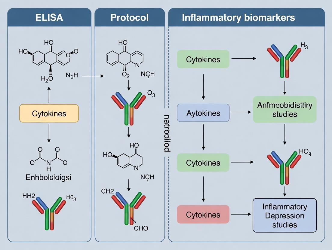
Mastering ELISA Protocols for Inflammatory Biomarkers in Depression Research: A Comprehensive Guide for Translational Scientists
This comprehensive guide provides researchers and drug development professionals with an expert-level overview of ELISA (Enzyme-Linked Immunosorbent Assay) protocols tailored specifically for quantifying inflammatory biomarkers in depression studies.
Mastering ELISA Protocols for Inflammatory Biomarkers in Depression Research: A Comprehensive Guide for Translational Scientists
Abstract
This comprehensive guide provides researchers and drug development professionals with an expert-level overview of ELISA (Enzyme-Linked Immunosorbent Assay) protocols tailored specifically for quantifying inflammatory biomarkers in depression studies. Covering the foundational role of cytokines like IL-6, TNF-α, and CRP in the immuno-inflammatory hypothesis of depression, it delivers detailed, step-by-step methodological workflows for plasma, serum, and CSF sample analysis. The article addresses common troubleshooting challenges in psychiatric biomarker research, offers optimization strategies for sensitivity and specificity, and critically compares ELISA with emerging multiplex platforms like Luminex and MSD. By synthesizing current best practices, this resource aims to enhance the reliability and translational value of inflammatory biomarker data in mood disorder research and clinical trials.
The Inflammatory Basis of Depression: Key Biomarkers and Rationale for ELISA Analysis
The immuno-inflammatory hypothesis posits that dysregulation of the immune system, particularly elevated pro-inflammatory cytokines, contributes to the pathophysiology of major depressive disorder (MDD). This chronic, low-grade inflammation can disrupt neurotransmitter metabolism, neuroendocrine function, and neural plasticity. This document provides application notes and protocols for investigating this hypothesis within a thesis focused on ELISA-based analysis of inflammatory biomarkers in depression research.
The following cytokines and acute-phase proteins are most consistently associated with MDD in clinical research.
Table 1: Key Inflammatory Biomarkers Elevated in Major Depressive Disorder
| Biomarker | Full Name | Primary Cellular Source | Typical Assay Range in Serum/Plasma (pg/mL) in MDD Studies | Reported Average Fold-Change vs. Controls (Meta-Analysis) |
|---|---|---|---|---|
| IL-6 | Interleukin-6 | Macrophages, T cells, Astrocytes | 1.0 - 10.0 | 1.5 - 2.5 |
| TNF-α | Tumor Necrosis Factor-alpha | Macrophages, Microglia | 0.5 - 5.0 | 1.3 - 2.2 |
| CRP | C-Reactive Protein | Hepatocytes (induced by IL-6) | 500 - 10,000 (ng/mL) | 1.5 - 2.0 |
| IL-1β | Interleukin-1 beta | Monocytes, Microglia | 0.1 - 2.0 | 1.2 - 1.8 |
| sIL-2R | Soluble IL-2 Receptor | Activated T cells | 200 - 1000 (U/mL) | ~1.4 |
Detailed Experimental Protocols
Protocol 3.1: Human Serum/Plasma Collection for Cytokine Analysis
Objective: To obtain high-quality, cytokine-stable samples from depressed patients and matched controls. Materials: Serum separator tubes (SST), EDTA or heparin plasma tubes, centrifuge, -80°C freezer. Procedure:
- Patient Cohort: Recruit MDD patients (diagnosed via DSM-5/ICD-10 criteria) and age-, sex-, BMI-matched healthy controls. Exclude individuals with autoimmune disorders, acute infection, or chronic inflammatory conditions.
- Fasting Draw: Perform venipuncture after an 8-hour fasting period, preferably between 8-10 AM to control for diurnal variation.
- Tube Handling:
- For serum: Draw blood into SST. Allow to clot upright at room temperature (RT) for 30 minutes. Centrifuge at 1000-2000 x g for 15 minutes at 4°C.
- For plasma: Draw blood into EDTA/K2EDTA tubes. Invert gently 8-10 times. Centrifuge at 500-2000 x g for 15 minutes at 4°C within 30 minutes of collection.
- Aliquot & Storage: Carefully pipette the supernatant (serum/plasma) into sterile, low-protein-binding microcentrifuge tubes. Create 100-200 µL aliquots to avoid freeze-thaw cycles. Flash-freeze in liquid nitrogen or on dry ice and store at -80°C.
Protocol 3.2: Quantitative Analysis of IL-6 and TNF-α via High-Sensitivity ELISA
Objective: To accurately quantify circulating levels of IL-6 and TNF-α. Materials: Commercial high-sensitivity ELISA kit (e.g., R&D Systems Quantikine HS, ThermoFisher Scientific), microplate reader (450 nm with 540/570 nm correction), adjustable pipettes, wash buffer, graph plotting software. Procedure:
- Kit & Sample Preparation: Equilibrate all kit components to RT (20-25°C) for 20 minutes. Gently mix all reagents. Dilute samples as per kit instructions (typical dilution 1:2 or 1:4 for serum/plasma in calibrator diluent).
- Assay Setup:
- Add 50 µL of Assay Diluent to each well of the antibody-coated microplate.
- Add 50 µL of standard, control, or sample to each well. Cover with adhesive strip. Incubate for 2 hours at RT on a horizontal orbital microplate shaker (~500 rpm).
- Washing: Aspirate and wash each well 4-6 times with 400 µL Wash Buffer using a squirt bottle or automated washer. Blot plate on clean absorbent paper.
- Detection Antibody: Add 100 µL of conjugated detection antibody to each well. Cover, incubate for 2 hours at RT on the shaker.
- Washing: Repeat wash step as in #3.
- Substrate Incubation: Add 100 µL of substrate solution (e.g., TMB) to each well. Incubate for 20-30 minutes at RT in the dark.
- Stop Reaction: Add 50 µL of Stop Solution (e.g., sulfuric acid). Gently tap plate to mix.
- Reading & Analysis: Read optical density (OD) at 450 nm within 30 minutes, using wavelength correction. Generate a 4- or 5-parameter logistic (4PL/5PL) standard curve. Interpolate sample concentrations.
Signaling Pathways & Experimental Workflows
Title: Cytokine-Mediated Pathways from Stress to Depression
Title: ELISA-Based Biomarker Study Workflow
The Scientist's Toolkit: Research Reagent Solutions
Table 2: Essential Materials for Cytokine Analysis in Depression Research
| Item / Reagent Solution | Function & Rationale |
|---|---|
| High-Sensitivity ELISA Kits (e.g., Quantikine HS) | Detect low physiological levels (pg/mL) of cytokines (IL-6, TNF-α) in serum/plasma with high specificity. |
| Multiplex Immunoassay Panels (Luminex/MSD) | Allow simultaneous quantification of 20+ analytes from a single small-volume sample, conserving precious clinical material. |
| C-Reactive Protein (hs-CRP) Assay | Measures low-grade inflammation; a key stable biomarker integrating IL-6 signaling. |
| Protease/Phosphatase Inhibitor Cocktails | Added during sample processing to prevent degradation of proteins/phosphoproteins in biospecimens. |
| Low-Protein-Binding Microtubes & Tips | Minimize analyte adsorption to plastic surfaces, improving recovery and accuracy. |
| Standardized Clinical Rating Scales (HAMD, MADRS) | Essential for quantifying depression severity to correlate with biomarker levels. |
| Luminex xMAP or MSD SECTOR Imager | Instrumentation for running and reading multiplex assays, offering broad dynamic range. |
| GraphPad Prism or R Statistical Software | For generating 4-5 parameter logistic standard curves and performing correlation/regression analyses. |
1. Introduction & Context Within a thesis investigating ELISA protocols for inflammatory biomarker research in depression, establishing a robust, reproducible core panel is paramount. This document details application notes and standardized protocols for quantifying key peripheral inflammatory mediators: Interleukin-6 (IL-6), Tumor Necrosis Factor-alpha (TNF-α), C-Reactive Protein (CRP), and Interleukin-1 beta (IL-1β). These analytes represent a consensus core panel, as meta-analyses consistently show their elevation in a significant subset of patients with Major Depressive Disorder (MDD). This resource provides researchers with the methodological rigor necessary to generate high-quality, comparable data across studies.
2. Key Inflammatory Biomarkers in Depression: Quantitative Summary Table 1: Core Panel Biomarkers - Summary of Evidence and Typical Assay Ranges
| Biomarker | Primary Cellular Source | Meta-Analysis Findings in MDD (vs. Controls) | Typical Serum/Plasma Range in Healthy Controls | Common ELISA Dynamic Range |
|---|---|---|---|---|
| CRP | Hepatocytes (induced by IL-6) | Significant elevation (Hedge's g ~0.15-0.46) | 0.1–3.0 mg/L (low-grade); >3 mg/L (elevated) | 0.01–50 mg/L (High-sensitivity) |
| IL-6 | Macrophages, T cells, Adipocytes | Significant elevation (SMD ~0.45-0.63) | 0.5–5.0 pg/mL | 0.5–200 pg/mL |
| TNF-α | Macrophages, Microglia | Significant elevation (SMD ~0.35-0.55) | 0.5–10.0 pg/mL | 0.5–500 pg/mL |
| IL-1β | Monocytes, Microglia | Moderate elevation (SMD ~0.20-0.40); less consistent | <1.0 pg/mL (often undetectable) | 0.1–100 pg/mL |
Table 2: Extended & Emerging Biomarkers of Interest
| Biomarker | Rationale for Inclusion in Depression Research | Typical Assay Range |
|---|---|---|
| IL-10 | Anti-inflammatory; imbalance with pro-inflammatory cytokines may be critical. | 0.5–200 pg/mL |
| sTNFR1/2 | Soluble TNF receptors; more stable markers of TNF system activity. | 100–5000 pg/mL |
| IL-17A | Links Th17 cell activity to neuroinflammation and anhedonia. | 0.5–500 pg/mL |
| MCP-1/CCL2 | Key chemokine for monocyte recruitment; implicated in blood-brain barrier disruption. | 5–1000 pg/mL |
3. Standardized Pre-Analytical & Sample Collection Protocol Critical for reproducibility across study sites. Title: Standard Operating Procedure (SOP) for Blood Collection & Processing for Cytokine Analysis. Objective: To minimize pre-analytical variability in cytokine and CRP measurement. Materials: Serum Separator Tubes (SST) and EDTA Plasma Tubes, tourniquet, needles, centrifuge (refrigerated, 4°C), 0.5 mL polypropylene cryovials, -80°C freezer. Workflow:
- Fasting: Collect blood after an overnight fast (8-12 hours), preferably between 7-9 AM to control for diurnal variation.
- Venipuncture: Perform with minimal stasis (<1 min tourniquet time).
- Tube Allocation:
- For Serum (CRP, cytokines): Collect in SST. Invert 5 times gently. Allow to clot upright at room temperature (RT) for 30 min. Do not exceed 60 min.
- For Plasma (cytokines): Collect in EDTA tube. Invert 8 times gently. Process immediately on wet ice.
- Centrifugation: Spin at 2000 x g for 15 min at 4°C.
- Aliquoting: Immediately aliquot supernatant (serum/plasma) into pre-cooled cryovials. Avoid pipetting from the buffy coat or pellet.
- Storage: Flash-freeze aliquots in liquid nitrogen or on dry ice, then store at -80°C. Avoid repeated freeze-thaw cycles (max 1-2 cycles).
4. Detailed ELISA Protocols (Sandwich Assay Principle) General Principle: Capture antibody → sample analyte → detection antibody → enzyme conjugate → substrate → colorimetric readout.
Table 3: Research Reagent Solutions Toolkit
| Item | Function & Critical Notes |
|---|---|
| High-Sensitivity ELISA Kits | Commercial kits (e.g., R&D Systems DuoSet, ThermoFisher ELISA, Millipore MILLIPLEX) are recommended for standardization. They provide matched antibody pairs, standards, and optimized buffers. |
| Matrix Solution (Assay Diluent) | Mimics the sample matrix (e.g., serum/plasma) to dilute standards and samples, reducing background and non-specific binding. |
| Wash Buffer (PBS/Tween-20) | Removes unbound proteins. Consistent and thorough washing is critical for low background. |
| Streptavidin-HRP Conjugate | Binds to biotinylated detection antibody. Provides enzymatic amplification. Must be fresh. |
| TMB Substrate | (3,3',5,5'-Tetramethylbenzidine). Colorimetric HRP substrate. Turns blue upon oxidation. Reaction stopped with acid. |
| Microplate Reader | Spectrophotometer capable of reading absorbance at 450 nm (and 540/570 nm for wavelength correction). |
| Polypropylene Labware | For sample/reagent dilution. Polystyrene can adsorb proteins. |
Protocol A: Quantification of IL-6, TNF-α, and IL-1β Title: Protocol for Sandwich ELISA of Serum/Plasma Cytokines.
- Coating: Dilute capture antibody in PBS. Add 100 µL/well to a 96-well plate. Seal, incubate overnight at RT.
- Wash & Block: Wash plate 3x with Wash Buffer. Add 300 µL/well of Block Buffer (1% BSA in PBS). Incubate 1-2 hours at RT.
- Standard & Sample Incubation: Prepare serial dilutions of the recombinant protein standard in Matrix Solution. Dilute samples (typical dilution 1:2 to 1:5). Add 100 µL of standard or sample per well. Incubate 2 hours at RT or overnight at 4°C.
- Detection Antibody Incubation: Wash 3x. Add 100 µL/well of biotinylated detection antibody. Incubate 2 hours at RT.
- Enzyme Conjugate Incubation: Wash 3x. Add 100 µL/well of Streptavidin-HRP. Incubate 20-30 min at RT (protected from light).
- Substrate & Stop: Wash 3x. Add 100 µL/well of TMB. Incubate for 5-20 min (develop in the dark). Add 50 µL/well of Stop Solution (e.g., 2N H₂SO₄).
- Readout: Measure absorbance at 450 nm immediately. Subtract readings at 540 nm or 570 nm for correction.
Protocol B: Quantification of High-Sensitivity CRP (hsCRP) Note: hsCRP requires kits with lower detection limits (<0.01 mg/L). Protocol similar to above but often uses different antibody pairs and may require higher sample dilution (e.g., 1:1000). Follow manufacturer instructions precisely.
5. Data Analysis & Quality Control
- Generate a 4- or 5-parameter logistic (4PL/5PL) standard curve for each plate.
- QC Samples: Include at least two levels of quality control samples (low and high) on each plate. Accept if values fall within 20% of expected concentration.
- Assay Performance: Report the Lower Limit of Detection (LLOD) and Lower Limit of Quantification (LLOQ) for each analyte. Samples below LLOQ should be handled statistically (e.g., imputed as LLOQ/√2).
6. Signaling Pathways & Experimental Workflow Visualizations
Title: Inflammatory Pathway from Stress to Depressive Symptoms
Title: ELISA Workflow for Biomarker Quantification
This application note provides a critical comparative analysis of plasma, serum, and CSF as biological matrices for the quantification of inflammatory biomarkers (e.g., IL-6, TNF-α, CRP) via ELISA. The selection of an appropriate sample type is a foundational step in our broader thesis investigating the role of neuroinflammation in Major Depressive Disorder (MDD), directly impacting the reliability, biological relevance, and translational potential of our findings.
Comparative Analysis: Plasma vs. Serum vs. CSF
Table 1: Key Characteristics and Comparative Pros & Cons
| Parameter | Plasma | Serum | Cerebrospinal Fluid (CSF) |
|---|---|---|---|
| Definition | Liquid fraction of blood with anticoagulant, contains fibrinogen. | Liquid fraction of blood after coagulation, fibrinogen removed. | Ultrafiltrate of plasma circulating in the brain ventricles and subarachnoid space. |
| Primary Pros | - Faster processing; avoids clotting time.- Reflects in vivo state with clotting factors.- Larger volume typically obtainable. | - No anticoagulant interference.- Standard for many clinical assays.- Higher concentration of some biomarkers. | - Direct window into CNS.- Low protein content reduces interference.- Essential for CNS-specific biomarkers. |
| Primary Cons | - Anticoagulant can interfere with some ELISAs.- Requires immediate processing.- Contains platelets that may release analytes. | - Longer processing time.- Clotting can sequester/release analytes (e.g., platelets release cytokines).- Potential for greater hemolysis. | - Invasive collection (lumbar puncture).- Very low volumes.- Dilutional effects from plasma; requires correction (e.g., albumin ratio). |
| Key Inflammatory Biomarkers | IL-6, TNF-α, CRP, IL-1β. | IL-6, TNF-α, CRP, IL-1β. | IL-6, TNF-α, QCK, MCP-1, GFAP. |
| Typical Sample Volume | 0.5 - 1 mL per analysis. | 0.5 - 1 mL per analysis. | 50 - 200 µL per analysis. |
| Stability at -80°C | Generally >2 years for cytokines (with protease inhibitors). | Generally >2 years for cytokines. | Variable; some labile biomarkers degrade faster. |
| Relevance to MDD Research | Assesses peripheral inflammatory status. | Assesses peripheral inflammatory status. | Gold standard for assessing neuroinflammation directly. |
Table 2: Quantitative Data Summary for Common Biomarkers
| Biomarker | Typical Plasma/Serum Concentration (Healthy) | Typical CSF Concentration (Healthy) | Reported Change in MDD Studies |
|---|---|---|---|
| IL-6 | 1-5 pg/mL | 0.5-2 pg/mL | Often elevated in serum/plasma; CSF findings inconsistent. |
| TNF-α | 5-15 pg/mL | 0.5-3 pg/mL | Frequently elevated in serum/plasma. |
| CRP | 0.5-3 µg/mL | < 0.01 µg/mL | Elevated hs-CRP is a robust peripheral finding. |
| Albumin | 35-50 mg/mL | 0.15-0.25 mg/mL | Used to calculate Albumin Ratio (QAlb) for blood-CSF barrier function. |
Detailed Experimental Protocols
Protocol 1: Collection and Processing of Paired Plasma and Serum for Cytokine ELISA
Objective: To obtain matched plasma and serum samples from MDD patients and healthy controls for comparative biomarker analysis.
Materials:
- Blood collection tubes: Serum separator tubes (SST) and K2EDTA or Lithium Heparin tubes.
- Centrifuge (refrigerated, capable of 2000g).
- Pipettes, aliquoting tubes (cryovials).
- Labels, permanent markers.
- Personal protective equipment (PPE).
Procedure:
- Collection: Draw blood via venipuncture. Fill the serum tube first, then the plasma (anticoagulant) tube to avoid anticoagulant carryover. Invert tubes gently as per manufacturer instructions.
- Serum Processing:
- Allow the serum tube to clot at room temperature (RT) for 30 minutes.
- Centrifuge at 2000g for 10 minutes at 4°C.
- Carefully pipette the clear supernatant (serum) into pre-labeled cryovials, avoiding the buffy coat and clot.
- Immediately flash-freeze in liquid nitrogen and store at -80°C.
- Plasma Processing:
- Centrifuge the anticoagulant tube at 2000g for 10 minutes at 4°C within 30 minutes of collection.
- Carefully pipette the plasma layer into cryovials, avoiding the buffy coat and platelets.
- Immediately flash-freeze and store at -80°C.
Protocol 2: CSF Collection, Processing, and Albumin Ratio Calculation
Objective: To collect and process CSF for the analysis of central inflammatory biomarkers and assess blood-CSF barrier integrity.
Materials:
- Sterile lumbar puncture kit.
- Polypropylene collection tubes (low protein binding).
- Refrigerated centrifuge.
- Pipettes, aliquoting tubes.
Procedure:
- Collection: Perform lumbar puncture (L3/L4 or L4/L5) under aseptic conditions. Collect CSF into serial polypropylene tubes.
- Processing: Centrifuge CSF at 2000g for 10 minutes at 4°C to remove cells and debris. Aliquot the supernatant into small-volume cryovials (e.g., 100 µL aliquots) to avoid freeze-thaw cycles.
- Storage: Immediately flash-freeze and store at -80°C.
- Albumin Ratio (QAlb) Calculation:
- Measure albumin concentration in paired CSF and serum samples via immunoassay.
- Calculate QAlb = [Albumin]CSF (mg/L) / [Albumin]Serum (g/L).
- Interpret: QAlb > 0.007 suggests blood-CSF barrier dysfunction, which must be considered when interpreting CSF biomarker levels.
Protocol 3: Multiplex ELISA for Inflammatory Panels
Objective: To quantify a panel of inflammatory cytokines (IL-6, TNF-α, IL-1β, IL-10) in plasma, serum, and CSF samples.
Materials:
- Validated multiplex ELISA kit (e.g., MSD, Luminex, or high-sensitivity singleplex).
- Plate washer.
- Microplate reader capable of chemiluminescence/fluorescence.
- Orbital plate shaker.
- Analytical software.
Procedure:
- Thawing: Thaw samples slowly on ice. Centrifuge briefly at 10,000g to pellet any aggregates.
- Assay Setup: Follow kit protocol precisely. For CSF, samples may be run neat and in a 1:2 dilution. For plasma/serum, use the recommended dilution (often 1:2 or 1:4).
- Incubation: Add standards, controls, and samples to the plate. Incubate with primary antibodies, then detection antibodies with washing steps as specified.
- Reading & Analysis: Add substrate and read immediately on the plate reader. Generate a standard curve (4- or 5-parameter logistic) and interpolate sample concentrations. Apply dilution factors.
Visualization: Pathways and Workflow
Diagram Title: Biomarker Source Selection Workflow for MDD Research
Diagram Title: Pathways Contributing to CSF Biomarker Levels
The Scientist's Toolkit: Research Reagent Solutions
Table 3: Essential Materials for Sample & ELISA Analysis
| Item | Function & Importance in MDD Biomarker Studies |
|---|---|
| K2EDTA/Li-Heparin Tubes | Anticoagulants for plasma collection; choice can affect analyte stability and assay interference. |
| Serum Separator Tubes (SST) | Facilitate clean serum separation; gel barrier must be compatible with target analytes. |
| Protease/Phosphatase Inhibitors | Cocktails added to samples (especially plasma) to prevent protein degradation post-collection. |
| High-Sensitivity ELISA Kits | Essential for detecting low-abundance cytokines in plasma, serum, and particularly CSF. |
| Albumin Assay Kit | For quantifying albumin in serum and CSF to calculate QAlb and assess barrier integrity. |
| Polypropylene Tubes | Low protein-binding material for storing CSF and making sample aliquots to prevent analyte loss. |
| Multiplex Analyzer (e.g., MSD) | Platform allowing simultaneous quantification of multiple biomarkers from low-volume samples like CSF. |
| Matched Antibody Pairs | For developing in-house ELISAs, offering flexibility but requiring rigorous validation. |
The reliability of ELISA-based quantification of inflammatory biomarkers (e.g., IL-6, TNF-α, CRP) in depression studies is fundamentally contingent on rigorous pre-analytical control. Variability introduced during patient preparation, sample collection, and storage can significantly confound results, leading to erroneous conclusions about the inflammation-depression nexus. This document provides detailed application notes and protocols to standardize these critical pre-analytical phases.
Patient Preparation Protocols
Key Variables and Control Measures
Patient-specific factors profoundly influence inflammatory biomarker levels. Standardization is essential.
Table 1: Patient Preparation Variables & Control Protocols
| Variable | Impact on Inflammatory Biomarkers | Recommended Control Protocol |
|---|---|---|
| Diurnal Rhythm | Cytokine levels (e.g., IL-6) peak in the afternoon. Variations up to 30-50% possible. | Schedule all blood draws between 7:00 AM and 9:00 AM. |
| Fasting Status | Recent food intake can increase CRP and IL-6. Postprandial increases up to 25% reported. | Mandatory 10-12 hour overnight fast. Water intake permitted. |
| Physical Activity | Strenuous exercise acutely elevates IL-6, TNF-α. | Rest seated for 20-30 minutes prior to phlebotomy. Avoid exercise 24h prior. |
| Medications | NSAIDs, statins, corticosteroids suppress cytokine levels. Antidepressants may modulate inflammation. | Document all medications. Consider washout periods if ethically & clinically feasible. |
| Stress | Acute stress can rapidly increase pro-inflammatory cytokines. | Conduct collection in a calm environment. Incorporate a 5-minute quiet rest. |
| Smoking & Caffeine | Both can transiently elevate biomarker levels. | Abstain from smoking ≥2 hours, caffeine ≥4 hours prior. |
Standard Operating Procedure (SOP): Patient Preparation
- Screening Visit: Instruct participants on preparation requirements using a standardized information sheet.
- Day Prior: Confirm compliance with fasting, exercise, and medication restrictions via checklist.
- Collection Day: Upon arrival, verify fasting duration and rest status. Escort participant to a quiet phlebotomy area.
- Pre-Phlebotomy Rest: Participant remains seated for a minimum of 20 minutes.
- Documentation: Record exact time of draw, last meal, medication intake, and any protocol deviations.
Sample Collection & Handling Protocols
Material Selection and Initial Processing
The choice of collection tube is critical for analyte stability.
Table 2: Sample Collection Tubes for Inflammatory Biomarker Analysis
| Tube Type | Additive | Primary Use | Processing Protocol for Plasma/Serum |
|---|---|---|---|
| Serum Clot Activator | Silica particles, gel separator | Serum collection (CRP, cytokines). | Allow to clot 30 min at RT. Centrifuge at 1000-2000 RCF for 10 min. Aliquot immediately. |
| EDTA (K2/K3) | EDTA (1.5-2.2 mg/mL) | Plasma collection (preferred for cytokine stability). | Mix gently by inversion x8. Centrifuge at 1000-2000 RCF for 10 min at 4°C within 30 min. |
| Citrate | Sodium citrate (3.2%) | Alternative plasma collection. | As per EDTA, but note dilution factor (1:9) in calculations. |
| PST/Li Heparin | Lithium heparin, gel separator | Plasma chemistry; less common for cytokines. | Centrifuge at 1000-2000 RCF for 10 min. Avoid use for IL-6/TNF-α unless validated. |
SOP: Blood Collection and Processing for Plasma (EDTA)
Materials: Tourniquet, alcohol swabs, 21G needle, EDTA vacutainers, labels, timer, centrifuge (pre-cooled to 4°C), cryovials, permanent marker.
- Venipuncture: Perform standard venipuncture. Fill EDTA tube to nominal volume.
- Immediate Mixing: Invert tube gently 8 times to ensure anticoagulant mixing.
- Timing: Place tube in an upright rack in crushed ice or a 4°C chill block. Processing clock starts at draw.
- Centrifugation: Within 30 minutes of draw, centrifuge at 1500 RCF for 10 minutes at 4°C.
- Aliquotting: Using a pipette, carefully transfer the plasma layer (upper 2/3rds, avoid buffy coat) into pre-labeled polypropylene cryovials.
- Immediate Storage: Place aliquots on dry ice or directly into a -80°C freezer. Do not leave at room temperature.
Diagram 1: Plasma Sample Processing Workflow
Sample Storage & Stability Protocols
Stability Data and Storage Hierarchy
Long-term integrity requires adherence to temperature and freeze-thaw cycle limits.
Table 3: Stability of Selected Inflammatory Biomarkers in Plasma/Serum
| Biomarker | Matrix | Room Temp (20-25°C) | 4°C | -20°C | -80°C | Max Freeze-Thaw Cycles |
|---|---|---|---|---|---|---|
| IL-6 | Plasma (EDTA) | ≤4h | 24h | 1 month | >2 years | ≤2 |
| TNF-α | Plasma (EDTA) | ≤6h | 48h | 1 month | >2 years | ≤3 |
| CRP | Serum | 3 days | 1 week | 6 months | >5 years | ≤3 |
| IL-1β | Plasma (EDTA) | Unstable (<2h) | ≤24h | 2 weeks | >1 year | ≤1 (sensitive) |
SOP: Long-Term Storage and Retrieval for Batch ELISA Analysis
Materials: -80°C freezer with monitoring, inventory database, labeled cryoboxes, dry ice, chilled rack.
- Primary Storage: Store aliquots in dedicated -80°C freezer. Organize boxes by study ID and visit.
- Temperature Monitoring: Use continuous digital monitoring with alarm alerts. Maintain log.
- Inventory Management: Use a LIMS or detailed spreadsheet. Record aliquot ID, location, date, volume.
- Retrieval for Assay: a. Plan: Retrieve only the number of aliquots needed for a single assay batch. b. Thawing: Rapidly thaw in a 37°C water bath for ≤5 minutes, then immediately place on wet ice. c. Assay: Perform ELISA immediately after thaw. Do not re-freeze remaining sample.
- Freeze-Thaw Tracking: Annotate the inventory each time an aliquot is thawed. Discard after exceeding cycle limit.
Diagram 2: Sample Storage and Retrieval Logic
The Scientist's Toolkit: Key Research Reagent Solutions
Table 4: Essential Materials for Pre-Analytical Phase in Biomarker Studies
| Item/Category | Specific Example/Type | Function & Rationale |
|---|---|---|
| Anticoagulant Tubes | K2 EDTA Vacutainers (lavender top) | Preferred for cytokine stability. Chelates calcium to prevent clotting. |
| Serum Tubes | Serum Separator Tubes (SST, gold top) | Contains clot activator and gel for efficient serum separation. |
| Pipettes & Tips | Low-retention, filtered tips (e.g., Rainin LTS) | Ensures accurate, nuclease-/biomarker-adsorption-free liquid handling. |
| Cryogenic Vials | 0.5-2.0 mL externally threaded polypropylene vials | Secure, leak-proof long-term storage at -80°C. Compatible with inventory systems. |
| Centrifuge | Refrigerated benchtop centrifuge (e.g., Eppendorf 5430 R) | Enforces "process at 4°C" protocol, minimizing analyte degradation. |
| Temperature Logger | Wireless digital data logger (e.g., TempTale) | Provides continuous monitoring and audit trail for freezers/incubators. |
| Sample Management | Laboratory Information Management System (LIMS) | Tracks sample lifecycle, location, freeze-thaw cycles, and patient metadata. |
| Protein Stabilizers | Protease Inhibitor Cocktails (optional, e.g., Aprotinin) | Added to plasma pre-centrifugation to further inhibit cytokine degradation. |
1. Application Notes: Current Meta-Analytic Findings Recent meta-analyses provide robust evidence for elevated levels of specific inflammatory biomarkers in Major Depressive Disorder (MDD) compared to healthy controls.
Table 1: Meta-Analytic Summary of Key Inflammatory Biomarkers in MDD
| Biomarker | Number of Studies (Participants) | Standardized Mean Difference (SMD) or Hedges' g (95% CI) | p-value | Heterogeneity (I²) | Notes |
|---|---|---|---|---|---|
| C-Reactive Protein (CRP) | ~80 (MDD: ~10,000; HC: ~15,000) | g = 0.48 (0.39 to 0.57) | <0.001 | ~80% | Strongest and most consistent evidence. Levels >3 mg/L linked to specific symptom profiles. |
| Interleukin-6 (IL-6) | ~60 (MDD: ~4,500; HC: ~5,600) | SMD = 0.54 (0.41 to 0.68) | <0.001 | ~80% | Elevated in blood and cerebrospinal fluid. Positively correlated with depression severity. |
| Tumor Necrosis Factor-alpha (TNF-α) | ~50 (MDD: ~2,800; HC: ~3,100) | SMD = 0.53 (0.36 to 0.70) | <0.001 | ~85% | Consistently elevated; may predict non-response to conventional antidepressants. |
| Soluble IL-2 Receptor (sIL-2R) | Meta-analysis of 22 studies | SMD = 0.49 (0.26 to 0.72) | <0.001 | ~70% | Marker of T-cell activation, suggesting immune cell involvement. |
Key Clinical Implications: Elevated inflammation is associated with treatment resistance, specific anhedonic and neurovegetative symptoms, and increased risk of metabolic comorbidity. Anti-inflammatory augmentation therapies show modest but significant efficacy in subgroups with high baseline inflammation.
2. Detailed Experimental Protocol: Quantifying Serum CRP & IL-6 via ELISA This protocol is designed for the simultaneous analysis of CRP and IL-6 from human serum/plasma samples within a depression research cohort.
Principle: Sandwich ELISA. Capture antibody specific to the target biomarker is coated onto the microplate. Sample analyte is captured and detected by a second, enzyme-conjugated detection antibody, producing a colorimetric signal proportional to concentration.
Materials & Reagents:
- Human Serum/Plasma Samples (fasted, stored at -80°C; avoid >2 freeze-thaw cycles).
- Commercial High-Sensitivity (hs) ELISA Kits (e.g., R&D Systems DuoSet ELISA or equivalent for CRP and IL-6).
- Microplate Reader capable of measuring absorbance at 450 nm and 570 nm (for correction).
- Plate Washer (or manual wash bottle/reservoir).
- Multichannel Pipettes & Sterile Tips.
- Reagent Reservoirs.
- Deionized Water & Precision Graduated Cylinders.
- Orbital Microplate Shaker.
- Software for 4- or 5-parameter logistic curve fitting.
Procedure:
- Kit Reconstitution & Standard Preparation:
- Reconstitute standards as per kit insert. Prepare a 7-point standard curve by serial dilution in the provided diluent.
- Allow all reagents to reach room temperature (RT) for 30 minutes before use.
- Plate Coating:
- Dilute capture antibody in PBS. Add 100 µL per well to a 96-well plate. Seal plate and incubate overnight at RT.
- Washing & Blocking:
- Aspirate and wash plate 3x with Wash Buffer (0.05% Tween-20 in PBS).
- Add 300 µL of Block Buffer (1% BSA in PBS) per well. Incubate for 1 hour at RT on shaker.
- Wash 3x.
- Sample & Standard Addition:
- Thaw samples on ice. Centrifuge at 10,000 x g for 5 minutes to remove particulates.
- Dilute samples as empirically determined (e.g., 1:100 for CRP, 1:2 for IL-6) in Diluent.
- Add 100 µL of standards, diluted samples, and blank (diluent) to designated wells. Incubate 2 hours at RT on shaker.
- Detection Antibody Incubation:
- Wash plate 3x.
- Add 100 µL of diluted detection antibody to each well. Incubate 2 hours at RT on shaker.
- Streptavidin-HRP Incubation:
- Wash plate 3x.
- Add 100 µL of diluted Streptavidin-Horseradish Peroxidase (HRP) to each well. Incubate 20 minutes at RT in the dark. Wash 3x.
- Substrate Reaction & Stop:
- Add 100 µL of Substrate Solution (e.g., TMB) to each well. Incubate for 10-20 minutes at RT in the dark until color develops.
- Add 50 µL of Stop Solution (e.g., 2N H₂SO₄). The blue color will turn yellow.
- Data Acquisition & Analysis:
- Read absorbance at 450 nm immediately, with 570 nm or 540 nm as a reference wavelength.
- Subtract the average blank absorbance from all readings.
- Generate a standard curve by plotting mean absorbance vs. concentration. Use curve-fitting software.
- Interpolate sample concentrations from the standard curve. Apply dilution factor.
Quality Control: Assay each sample in duplicate. Include kit-provided controls. Acceptable CV between duplicates is <15%. The standard curve R² should be >0.99.
3. Visualizations
Title: Inflammatory Pathway from Periphery to MDD Symptoms (99 chars)
Title: Research Workflow for Biomarker Validation in MDD (84 chars)
4. The Scientist's Toolkit: Key Research Reagent Solutions
Table 2: Essential Materials for Inflammatory Biomarker Research in MDD
| Item | Function & Rationale |
|---|---|
| High-Sensitivity (hs) ELISA Kits | Quantify low-level inflammatory proteins (e.g., hsCRP <3 mg/L) crucial for detecting subtle changes in psychiatric populations. |
| Multiplex Immunoassay Panels (e.g., Luminex/MSD) | Simultaneously measure a panel of 10-50 cytokines/chemokines from a small sample volume, enabling inflammatory network analysis. |
| Protease & Phosphatase Inhibitor Cocktails | Added during blood processing to prevent degradation and dephosphorylation of proteins, preserving analyte integrity. |
| Cryogenic Vials (Sterile, Screw-Cap) | For long-term, stable storage of serum/plasma aliquots at -80°C, preventing freeze-thaw degradation. |
| Recombinant Protein Standards | Essential for generating accurate, kit-specific standard curves to interpolate absolute sample concentrations. |
| Blocking Buffer (e.g., 1% BSA/PBS) | Reduces non-specific binding in immunoassays, lowering background noise and improving signal-to-noise ratio. |
| Precision Pipettes & Calibrated Tips | Ensure accurate and reproducible liquid handling for standard/sample preparation, critical for assay precision. |
| Microplate Reader with Dual Wavelength | Measures primary absorbance (450nm for TMB) and reference wavelength (570/540nm) to correct for optical imperfections. |
Step-by-Step ELISA Protocols for Accurate Biomarker Quantification in Psychiatric Samples
Inflammatory biomarkers, such as cytokines and C-reactive protein (CRP), are critical targets in psychiatric research, particularly in elucidating the role of inflammation in Major Depressive Disorder (MDD). Accurate quantification of these low-abundance analytes in serum, plasma, or cerebrospinal fluid (CSF) is essential. The choice between high-sensitivity (hs) and conventional Enzyme-Linked Immunosorbent Assay (ELISA) kits directly impacts data reliability, requiring careful consideration of detection limits, dynamic range, and sample matrix.
Quantitative Comparison: hsELISA vs. Conventional ELISA
Table 1: Key Performance Parameter Comparison
| Parameter | High-Sensitivity (hs) ELISA | Conventional ELISA | Primary Implication for Psychiatric Research |
|---|---|---|---|
| Detection Limit (Typical) | 0.01 - 0.1 pg/mL | 1 - 10 pg/mL | Essential for measuring basal levels of IL-6, IL-1β, TNF-α in plasma/serum. |
| Dynamic Range | Narrower (e.g., 0.1-10 pg/mL) | Wider (e.g., 10-1000 pg/mL) | hsELISA may require more sample dilution for elevated post-stimulation levels. |
| Precision (CV%) | Often <10% | Typically <12% | Both suitable, but hsELISA offers finer resolution at low concentrations. |
| Sample Volume Required | Often higher (50-100 µL) | Often lower (25-50 µL) | Critical for pediatric or longitudinal studies with limited sample volumes. |
| Cost per Sample | 20-40% higher | Standard | Budget consideration for large cohort studies. |
| Primary Application | Quantifying basal, sub-pg/mL levels | Measuring stimulated or pathologically elevated levels | hsELISA for baseline inflammatory profiling; conventional for challenge studies. |
Table 2: Example Biomarker Profiles in Depression Research
| Biomarker | Typical Basal Level in Healthy Controls (Serum) | Typical Level in MDD (Serum) | Recommended Assay Type |
|---|---|---|---|
| IL-6 | 0.5 - 2.5 pg/mL | 2.5 - 8 pg/mL (elevated) | High-Sensitivity |
| TNF-α | 0.5 - 3 pg/mL | 3 - 10 pg/mL (elevated) | High-Sensitivity |
| CRP | 500 - 2000 ng/mL | 1500 - 5000 ng/mL (elevated) | hsCRP (nanogram range) |
| IL-1β | < 0.1 - 0.5 pg/mL | 0.5 - 2 pg/mL (elevated) | Ultra-Sensitivity |
| sIL-2R | 100 - 500 pg/mL | 500 - 1200 pg/mL (elevated) | Conventional |
Detailed Experimental Protocols
Protocol 1: Sample Preparation for Serum/Plasma Cytokine Analysis
Objective: To prepare blood-derived samples for hsELISA, minimizing pre-analytical variability. Materials: EDTA or Heparin tubes (plasma), Serum Separator Tubes (SST), microcentrifuge, -80°C freezer, low-protein-binding pipettes and tubes. Procedure:
- Blood Draw & Collection: Collect venous blood into appropriate anticoagulant tubes (for plasma) or SST (for serum). Invert tubes gently as per manufacturer instructions.
- Plasma Separation: Centrifuge anticoagulant tubes at 1000-2000 x g for 15 minutes at 4°C within 30 minutes of collection. Carefully aspirate the plasma layer, avoiding the buffy coat.
- Serum Separation: Allow SST to clot at room temperature for 30 minutes. Centrifuge at 1000-2000 x g for 15 minutes at room temperature.
- Aliquoting: Immediately aliquot supernatant (serum/plasma) into low-protein-binding microcentrifuge tubes (e.g., 100 µL aliquots).
- Storage: Flash-freeze aliquots on dry ice or in a -80°C freezer. Avoid repeated freeze-thaw cycles (maximum 2 cycles recommended). Note: For CSF, follow similar aliquoting and storage protocols after centrifugation to remove cells.
Protocol 2: Running a High-Sensitivity ELISA for IL-6
Objective: To quantify low-concentration IL-6 in human serum samples. Materials: Commercial human IL-6 hsELISA kit (e.g., R&D Systems HS600B), multichannel pipette, microplate washer, microplate reader capable of 450 nm with 540/570 nm correction, wash buffer (1X), standard diluent. Procedure:
- Kit & Sample Equilibration: Remove all kit components and samples from -80°C. Thaw completely on ice or at 4°C. Allow all components to reach room temperature (20-25°C) for at least 30 minutes before use.
- Standard Reconstitution & Serial Dilution: Reconstitute the IL-6 standard as per the kit insert. Prepare a 7-point standard curve in the provided diluent, typically from 10 pg/mL down to 0.156 pg/mL, using 2-fold serial dilutions.
- Plate Layout & Addition: Add 100 µL of standard, control, or prediluted sample (if necessary) to appropriate wells of the pre-coated microplate. Cover and incubate for 2 hours at room temperature on a horizontal plate shaker.
- Aspiration & Wash: Aspirate liquid from each well. Wash 4 times by filling each well with 400 µL wash buffer (1X) using a plate washer. Invert plate and blot against clean paper towels.
- Detection Antibody Incubation: Add 100 µL of the prepared detection antibody to each well. Cover and incubate for 2 hours at room temperature.
- Wash: Repeat Step 4.
- Streptavidin-HRP Incubation: Add 100 µL of Streptavidin-HRP working solution to each well. Cover and incubate for 20 minutes at room temperature, protected from light.
- Wash: Repeat Step 4.
- Substrate Incubation: Add 100 µL of substrate solution (TMB or equivalent) to each well. Incubate for 20 minutes at room temperature, protected from light.
- Stop Reaction & Read: Add 50 µL of stop solution (e.g., 2N H2SO4) to each well. Read optical density immediately at 450 nm, with wavelength correction set to 540 nm or 570 nm.
- Data Analysis: Generate a 4- or 5-parameter logistic (4PL/5PL) standard curve using software (e.g., MyAssays, GraphPad Prism) and interpolate sample concentrations.
Visualizations
Title: Assay Selection Workflow for Psychiatric Biomarker Studies
Title: Inflammatory Pathway in Depression & ELISA Measurement Points
The Scientist's Toolkit: Key Research Reagent Solutions
Table 3: Essential Materials for Inflammatory Biomarker ELISA in Psychiatry
| Item | Function & Specific Consideration for Psychiatric Research |
|---|---|
| High-Sensitivity ELISA Kits | Specifically designed to detect low pg/mL levels of cytokines (IL-6, TNF-α, IL-1β) crucial for baseline inflammation studies in MDD. |
| Matched Antibody Pairs | For developing in-house assays; allows customization for specific sample types (e.g., CSF). |
| Low-Protein-Binding Tubes & Tips | Minimizes analyte adsorption to plastic surfaces, critical for accuracy in low-concentration samples. |
| Matrix-Free Analyte Standards | Provides the most accurate standard curve; some kits offer serum/plasma-matched standards. |
| Stabilized TMB Substrate | Provides sensitive colorimetric detection; stop solution acidity must be consistent for reproducibility. |
| Precision Multichannel Pipettes | Ensures reproducibility in reagent dispensing across 96-well plates for high-throughput cohort studies. |
| Automated Plate Washer | Reduces background and well-to-well variability versus manual washing, improving precision. |
| Plate Reader with Wavelength Correction | Corrects for optical imperfections in the plate; essential for reliable low-signal reads in hsELISA. |
| Sample Diluent (Blocking Buffer-Based) | For pre-diluting samples that read above the standard curve; preserves analyte detection. |
| Cryogenic Vials & Storage System | For maintaining long-term stability of irreplaceable clinical samples in biobanks. |
Within a thesis investigating inflammatory biomarkers (e.g., IL-6, TNF-α, CRP) in depression studies via ELISA, optimal sample preparation is paramount. Plasma and serum contain interfering substances that cause matrix effects, leading to inaccurate quantification. This protocol details systematic approaches to validate sample pre-treatment through dilution linearity, assessment of matrix effects, and recovery experiments, ensuring data integrity for clinical correlation analyses.
Assessing Dilution Linearity
Objective: To determine if the sample can be diluted in accordance with the assay's dynamic range while maintaining proportional analyte recovery, confirming the absence of high-dose hook effects or interferences.
Protocol:
- Sample Selection: Pool positive patient plasma/serum samples with high concentrations of the target biomarker (e.g., from a depressed cohort with high inflammation scores).
- Diluent: Use the assay's recommended zero calibrator or analyte-free matrix (commercially available or prepared from charcoal-stripped serum).
- Dilution Scheme: Prepare a series of dilutions (e.g., 1:2, 1:4, 1:8, 1:16) in duplicate.
- Analysis: Run diluted samples alongside the standard curve on the same ELISA plate.
- Calculation: Multiply the measured concentration by the dilution factor to obtain the "expected concentration" for each dilution.
Data Interpretation & Acceptance Criteria: Percent recovery is calculated as (Measured Conc. / Expected Conc.) x 100. A linear dilution profile demonstrates recoveries between 80-120% and an R² > 0.95 for the regression of measured vs. expected values.
Table 1: Example Dilution Linearity Data for IL-6 in Human Serum
| Sample ID | Dilution Factor | Measured [IL-6] (pg/mL) | Expected [IL-6] (pg/mL) | % Recovery |
|---|---|---|---|---|
| Patient Pool A | 1:1 (Neat) | 48.5 | 48.5 | 100.0 |
| Patient Pool A | 1:2 | 22.1 | 24.3 | 90.9 |
| Patient Pool A | 1:4 | 11.8 | 12.1 | 97.5 |
| Patient Pool A | 1:8 | 5.9 | 6.1 | 96.7 |
Evaluating Matrix Effects
Objective: To identify and characterize interference from the sample matrix that causes signal suppression or enhancement, independent of the analyte.
Protocol (Parallelism Test):
- Spiking Solution: Prepare a high-concentration stock of the recombinant biomarker in a clean buffer.
- Sample Matrix: Use at least 6 individual sources of presumed "normal" or disease-state plasma/serum. Also include the recommended assay buffer.
- Spiking: Spike each matrix with the same, known amount of the recombinant analyte to reach a concentration within the mid-range of the standard curve. Create a matching set of unspiked controls for each matrix.
- Analysis: Run all spiked and unspiked samples. Calculate the measured concentration added by subtracting the unspiked value from the spiked value.
- Comparison: Compare the measured concentration recovered from each biological matrix to that recovered from the ideal buffer matrix.
Table 2: Matrix Effects Evaluation for TNF-α Spike Recovery
| Matrix Source | Unspiked [TNF-α] (pg/mL) | Spiked [TNF-α] (pg/mL) | Measured Spike (pg/mL) | % Recovery vs. Buffer |
|---|---|---|---|---|
| Assay Buffer (Control) | 0.0 | 50.0 | 50.0 | 100.0 |
| Donor Serum 1 | 2.1 | 48.5 | 46.4 | 92.8 |
| Donor Serum 2 | 1.5 | 55.2 | 53.7 | 107.4 |
| Depressed Cohort Pool | 8.3 | 54.1 | 45.8 | 91.6 |
Determining Analyte Recovery
Objective: To assess the accuracy of the assay by measuring the proportion of a known amount of analyte added to a real sample that can be quantitatively recovered.
Protocol (Spike-and-Recovery):
- Base Sample: Use a native patient sample with a known endogenous concentration ([C_endogenous]).
- Spiking: Create two aliquots:
- Low Spike: Add analyte to increase concentration by an amount near the lower limit of quantification (LLOQ).
- High Spike: Add analyte to increase concentration to near the upper limit of quantification (ULOQ).
- Analysis: Measure the concentration in the unspiked ([Cendogenous]), low-spiked ([Clow]), and high-spiked ([C_high]) samples.
- Calculation:
- % Recovery = ( [Cspiked] - [Cendogenous] ) / [Concentration Added] x 100
Table 3: Analyte Recovery Experiment for CRP in Plasma
| Sample Condition | Endogenous Conc. (ng/mL) | Spike Added (ng/mL) | Measured Conc. (ng/mL) | % Recovery |
|---|---|---|---|---|
| Unspiked Patient 1 | 4.2 | 0.0 | 4.2 | N/A |
| Low Spike (Patient 1) | 4.2 | 5.0 | 8.9 | 94.0 |
| High Spike (Patient 1) | 4.2 | 50.0 | 51.3 | 94.2 |
The Scientist's Toolkit: Key Research Reagent Solutions
| Item | Function in Sample Preparation & Validation |
|---|---|
| Analyte-Free Matrix | Charcoal-stripped or immunoaffinity-depleted serum/plasma used as an ideal diluent for preparing standard curves and assessing matrix effects. |
| Recombinant Protein Calibrators | Highly pure, quantified protein standards used for spiking experiments in recovery and parallelism tests. |
| Matrix-Matched Calibrators | Standard curves prepared in the analyte-free matrix to correct for baseline matrix effects and improve accuracy. |
| High-Bind ELISA Plates | Plates with optimized surface chemistry for maximum antibody coating efficiency, critical for assay sensitivity. |
| Blocking Buffers (e.g., BSA, Casein) | Solutions used to coat unused protein-binding sites on the plate to minimize non-specific background signal. |
| Precision Pipettes & Low-Binding Tips | Essential for accurate liquid handling and to prevent analyte adsorption during serial dilution steps. |
Visualizations
Workflow for validating plasma/serum sample preparation for biomarker ELISA.
Role of sample preparation validation in depression biomarker research.
Within the broader thesis on optimizing ELISA protocols for quantifying inflammatory biomarkers (e.g., IL-6, TNF-α, CRP) in depression studies, procedural precision is paramount. Variations in coating, blocking, and incubation steps directly impact assay sensitivity, specificity, and reproducibility, which are critical for elucidating the cytokine-depression nexus in both clinical and preclinical drug development research. This document provides detailed application notes and standardized protocols for these core steps.
Detailed Experimental Protocols
Coating Protocol (Antigen Capture)
- Objective: To immobilize the capture antibody onto the solid phase (microplate well).
- Materials: Carbonate-Bicarbonate Coating Buffer (0.05 M, pH 9.6), purified capture antibody, polypropylene plates (e.g., Nunc MaxiSorp).
- Methodology:
- Dilute the capture antibody to the optimal concentration (see Table 1) in coating buffer.
- Dispense 100 µL per well into the microplate.
- Seal the plate and incubate overnight (16-18 hours) at 4°C for optimal binding uniformity. A 2-hour incubation at 37°C is acceptable but may yield higher background.
- Following incubation, decant or aspirate the coating solution.
Washing Protocol (Post-Coating)
- Objective: To remove unbound capture antibody.
- Materials: Wash Buffer (PBS or Tris-based buffer with 0.05% - 0.1% Tween 20), automated or manual plate washer.
- Methodology:
- Fill each well with 300-350 µL of wash buffer.
- Allow the plate to soak for 30 seconds to dissociate non-specific bonds.
- Completely aspirate or decant the buffer.
- Firmly tap the plate onto absorbent paper to remove residual liquid.
- Repeat the cycle 3 times. Ensure wells do not dry completely between washes.
Blocking Protocol
- Objective: To cover any remaining protein-binding sites on the plastic surface to minimize non-specific binding in subsequent steps.
- Materials: Blocking Buffer (e.g., 1-5% BSA or 5% non-fat dry milk in PBS with 0.05% Tween 20).
- Methodology:
- Immediately after the final wash, add 200-300 µL of blocking buffer to each well.
- Seal the plate and incubate for 1-2 hours at room temperature (20-25°C) on a microplate shaker (gentle agitation).
- Aspirate the blocking buffer. Do not wash. The plate can now be used immediately in an assay or dried, sealed, and stored at 4°C for later use.
Sample & Detection Incubation Protocols
- Objective: To specifically bind the target analyte (inflammatory biomarker) and subsequently the detection antibody.
- Materials: Sample Diluent (e.g., PBS with 1% BSA, 0.05% Tween 20), detection antibody, streptavidin-HRP (if applicable).
- Methodology for Sample Incubation:
- Dilute serum/plasma/cerebrospinal fluid samples (typically 1:2 to 1:10) in sample diluent to mitigate matrix effects.
- Add 100 µL of standard, sample, or control per well. Include blank wells (diluent only).
- Seal and incubate for 2 hours at room temperature with shaking.
- Wash the plate 5 times as per the washing protocol.
- Methodology for Detection Antibody Incubation:
- Add 100 µL of detection antibody (concentration per Table 1) to each well.
- Seal and incubate for 1-2 hours at room temperature with shaking.
- Wash the plate 5 times as per the washing protocol.
- Note: For streptavidin-HRP systems, a final 30-45 minute incubation with streptavidin-HRP is required, followed by 5-7 washes.
Data Presentation: Reagent Optimization
Table 1: Quantitative Optimization Parameters for Inflammatory Biomarker ELISA
| Step | Reagent | Typical Concentration Range | Recommended Incubation | Key Consideration for Depression Biomarkers |
|---|---|---|---|---|
| Coating | Capture Antibody | 1 - 10 µg/mL in buffer | Overnight at 4°C | High-affinity, matched-pair antibodies crucial for low-abundance cytokines. |
| Blocking | BSA / Non-Fat Dry Milk | 1 - 5% (w/v) in PBS-T | 1-2 hrs at RT | Use BSA for phospho-protein targets; avoid milk with biotin-streptavidin systems. |
| Sample | Serum/Plasma Dilution | 1:2 to 1:10 | 2 hrs at RT | High lipid/content in depression study samples may require additional dilution. |
| Detection | Detection Antibody | 0.5 - 2 µg/mL in diluent | 1-2 hrs at RT | Tag (biotin/HRP) concentration must be titrated separately. |
| Signal | Enzyme Substrate (TMB) | - | 5-30 mins in dark | Stop reaction with equal volume 1M H2SO4; read at 450nm (reference 570-650nm). |
Mandatory Visualization
ELISA Core Workflow with Wash Steps
Biomarker Source: Stress to ELISA Measurement
The Scientist's Toolkit: Research Reagent Solutions
Table 2: Essential Materials for Inflammatory Biomarker ELISA
| Item | Function & Rationale |
|---|---|
| High-Binding Polystyrene Plates (e.g., Nunc MaxiSorp) | Optimal surface chemistry for passive adsorption of capture antibodies, maximizing assay sensitivity. |
| Matched Antibody Pair (Capture & Detection) | Ensures specificity for a single epitope on the target cytokine, minimizing cross-reactivity. |
| Recombinant Protein Standards | Quantified standard curve is essential for precise interpolation of sample analyte concentration. |
| Blocking Agent (BSA, Fraction V) | Inert protein that reduces non-specific binding; preferred over milk for consistency and biotin compatibility. |
| Tween 20 (Polysorbate 20) | Non-ionic detergent in wash buffers reduces hydrophobic interactions and minimizes background. |
| TMB (3,3',5,5'-Tetramethylbenzidine) Substrate | Chromogenic HRP substrate offering high signal-to-noise ratio; stopped with acid for endpoint reading. |
| Plate Sealer / Adhesive Film | Prevents evaporation and contamination during critical incubation steps, ensuring uniformity. |
| Microplate Washer | Provides consistent, thorough washing essential for low background and high precision. |
In the quantitative analysis of inflammatory biomarkers (e.g., IL-6, TNF-α, CRP) for depression studies using ELISA, the standard curve is the foundational element determining assay validity. A wide dynamic range is critical to accurately capture the broad concentration spectrum of these biomarkers found in both peripheral blood and cerebrospinal fluid, avoiding sample re-analysis due to dilution or concentration. This protocol details the generation and rigorous validation of a standard curve optimized for high-sensitivity cytokine ELISAs within a thesis framework focused on elucidating the immuno-inflammatory hypothesis of depression.
Experimental Protocol: Standard Curve Preparation and ELISA Execution
A. Reagent and Equipment Preparation
- Coated ELISA Plate: 96-well plate pre-coated with capture antibody specific to the target cytokine (e.g., human IL-6).
- Reconstituted Standard: Lyophilized recombinant cytokine standard. Centrifuge vial briefly. Reconstitute with the recommended volume of assay diluent (typically 1% BSA in PBS) to generate a high-concentration stock (e.g., 10,000 pg/mL). Vortex gently for 30 seconds.
- Assay Diluent: Matches the sample matrix as closely as possible (e.g., 1% BSA, 0.05% Tween-20 in PBS).
- Detection Antibody: Biotinylated detection antibody specific to the target.
- Streptavidin-HRP: Streptavidin conjugated to Horseradish Peroxidase.
- Substrate Solution: TMB (3,3’,5,5’-Tetramethylbenzidine).
- Stop Solution: 1M or 2M Sulfuric Acid (H₂SO₄).
- Microplate Reader: Capable of measuring absorbance at 450 nm (primary) and 570 nm or 620 nm (reference wavelength for subtraction).
B. Stepwise Protocol for Standard Curve Generation
Standard Serial Dilution:
- Label 7-8 microcentrifuge tubes (S1-S7/S8).
- Pipette the required volume of assay diluent into each tube (e.g., 500 µL).
- Perform a serial dilution (typically 1:2 or 1:4) from the reconstituted stock. For a 1:2 dilution across 7 points plus blank: Transfer 500 µL of stock to tube S1 (1:1), mix, then transfer 500 µL from S1 to S2, mix, and continue to the last standard tube. The final tube (S7) contains the lowest concentration. Use a fresh tip for each transfer.
- The blank (S0/BL) contains assay diluent only.
ELISA Assay Procedure:
- Step 1: Aliquot 100 µL of each standard dilution (S0-S7) and prepared samples/controls into designated wells in duplicate.
- Step 2: Cover plate and incubate for the specified time (e.g., 2 hours at room temperature).
- Step 3: Aspirate and wash each well 4 times with 300 µL wash buffer. Blot plate on absorbent paper.
- Step 4: Add 100 µL of biotinylated detection antibody. Incubate (e.g., 1 hour). Wash as in Step 3.
- Step 5: Add 100 µL of Streptavidin-HRP. Incubate (e.g., 30 minutes). Wash as in Step 3.
- Step 6: Add 100 µL of TMB substrate. Incubate in the dark (e.g., 15-20 minutes) until color develops.
- Step 7: Add 50 µL of stop solution. Gently tap plate to mix. The blue solution will turn yellow.
- Step 8: Read absorbance at 450 nm within 30 minutes. Subtract the reference wavelength (570 nm or 620 nm) reading to correct for optical imperfections.
Data Analysis and Critical Validation Parameters
Following data acquisition, plot the mean absorbance (y-axis) against the known standard concentration (x-axis) on a logarithmic scale. Fit the data using a 4- or 5-parameter logistic (4PL/5PL) regression model, which is most appropriate for the sigmoidal response of ELISA.
Table 1: Critical Parameters for Standard Curve Validation
| Parameter | Target Value/Range | Interpretation & Impact on Dynamic Range |
|---|---|---|
| Linear Range | R² ≥ 0.99 (log-linear region) | The working range for reliable quantification. A wider linear range increases the usable dynamic range. |
| Lower Limit of Quantification (LLOQ) | CV < 20%, Accuracy 80-120% | The lowest standard that can be measured with acceptable precision and accuracy. Critical for detecting low-grade inflammation. |
| Upper Limit of Quantification (ULOQ) | CV < 20%, Accuracy 80-120% | The highest standard measurable within the linear range. Prevents high-concentration sample hook effect. |
| Total Assay Range | Span from LLOQ to ULOQ | The effective dynamic range of the assay. Aim for ≥ 2 logs (e.g., 10-2000 pg/mL). |
| Curve Fit (R²) | ≥ 0.995 for 4PL/5PL | Indicates goodness-of-fit for the chosen model. Poor fit compromises accuracy across the range. |
| % Back-Calculation Accuracy | 80-120% for all standards | Measures how well the curve predicts known values. Standards outside this range should be excluded. |
Table 2: Example Standard Curve Data for Human IL-6 ELISA
| Standard Point | Concentration (pg/mL) | Mean Abs (450-570 nm) | %CV (Replicates) | Back-Calculated Conc. (pg/mL) | Accuracy (%) |
|---|---|---|---|---|---|
| S0 (Blank) | 0.0 | 0.045 | 2.5 | - | - |
| S1 (LLOQ) | 3.9 | 0.102 | 4.8 | 3.7 | 94.9 |
| S2 | 15.6 | 0.235 | 3.1 | 16.1 | 103.2 |
| S3 | 62.5 | 0.580 | 2.7 | 61.8 | 98.9 |
| S4 | 250.0 | 1.250 | 1.9 | 255.2 | 102.1 |
| S5 | 1000.0 | 2.100 | 2.3 | 975.4 | 97.5 |
| S6 (ULOQ) | 2000.0 | 2.450 | 3.5 | 2105.0 | 105.3 |
The Scientist's Toolkit: Key Research Reagent Solutions
Table 3: Essential Materials for High-Dynamic Range ELISA
| Item | Function in Protocol |
|---|---|
| Matched Antibody Pair (Capture/Biotinylated Detection) | Ensures specific, sensitive, and linear recognition of the target biomarker. Mismatched pairs can compress the dynamic range. |
| High-Purity Recombinant Protein Standard | Defines the calibration curve. Must be identical to the endogenous analyte and of known activity/concentration. |
| Matrix-Matched Assay Diluent | Used for reconstituting standards and diluting samples. Contains protein (BSA) and detergent to minimize nonspecific binding, preserving signal integrity at low concentrations. |
| High-Sensitivity Streptavidin-HRP Conjugate | Amplifies the detection signal. A high specific activity conjugate is essential for achieving a low LLOQ. |
| Consistent Substrate (e.g., stable TMB) | Generates the colorimetric signal. Batch-to-batch consistency is vital for inter-assay reproducibility of the curve. |
| Validated Wash Buffer (with surfactant) | Critical for removing unbound material. Inconsistent washing is a major source of high background and poor precision at the curve extremes. |
Visualizing the Workflow and Critical Relationships
Title: ELISA Standard Curve Workflow & Validation
Title: Parameters Affecting ELISA Dynamic Range
Within a thesis investigating the role of inflammatory biomarkers (e.g., IL-6, TNF-α, CRP) in Major Depressive Disorder (MDD) pathophysiology and treatment response, precise and reliable ELISA data processing is paramount. This protocol details the systematic conversion of raw absorbance values into quantified pg/mL concentrations, integrated with rigorous quality control (QC) checks essential for clinical research and drug development.
Experimental Protocol: ELISA for Inflammatory Biomarkers
Materials and Equipment
- Microplate Reader: Capable of reading absorbance at 450 nm, with 540 nm or 570 nm correction.
- Pre-coated ELISA Kit: Specific for target biomarker (e.g., Human IL-6 High-Sensitivity ELISA).
- Micropipettes and Tips: For 1 µL to 1 mL volumes.
- Orbital Microplate Shaker.
- Deionized Water.
- Software: GraphPad Prism, ELISA analysis software, or R/Python for curve fitting.
Step-by-Step Procedure
- Assay Execution: Perform the ELISA per manufacturer’s instructions for your target inflammatory biomarker. Include all standards, controls (QC samples), and patient/study samples in duplicate.
- Absorbance Reading: Read the absorbance (Optical Density, OD) at the primary wavelength (e.g., 450 nm). Subtract the reference wavelength (e.g., 540 nm) reading to correct for optical imperfections.
- Data Export: Export the raw absorbance data (Mean of duplicates) for standards, controls, and unknowns.
Data Calculation: From Absorbance to Concentration
Standard Curve Modeling
The core calculation involves fitting a curve to the standard points. The most common models are:
Four-Parameter Logistic (4PL) Curve: Preferred for immunoassays due to its ability to model the sigmoidal dose-response.
- Formula:
y = d + (a - d) / (1 + (x/c)^b) - Where:
- y = Absorbance
- x = Concentration
- a = Minimum asymptote (background)
- d = Maximum asymptote (plateau)
- c = Inflection point (EC50)
- b = Hill slope
- Formula:
Linear Regression (Log-Log): Used for the linear portion of the standard curve, less accurate for the full range.
Step-by-Step Calculation Protocol
- Plot Standards: Plot the mean absorbance of each standard (y-axis) against its known concentration (x-axis) on a logarithmic scale.
- Apply Curve Fit: Use software to fit the 4PL model to the standard data. Acceptable R² should be >0.99.
- Interpolate Unknowns: Using the derived 4PL equation, calculate the concentration (x) for each unknown sample's absorbance (y).
- Apply Dilution Factor: Multiply the interpolated concentration by any sample dilution factor used during the assay.
Mandatory Quality Control Checks
QC checks are non-negotiable for thesis-level and preclinical research integrity.
Table 1: Essential ELISA Quality Control Parameters
| QC Parameter | Target/Acceptance Criteria | Purpose & Rationale |
|---|---|---|
| Standard Curve R² | >0.99 | Indicates precision of the curve fit. Lower values suggest poor assay performance or pipetting errors. |
| % Recovery of QC Samples | 80–120% of expected value | Assesses accuracy of the entire assay system. QC samples should be run at low, mid, and high ranges. |
| Inter-Assay CV | <15% (Preferably <10%) | Measures precision across different plates/runs/days. Critical for longitudinal studies. |
| Intra-Assay CV | <10% (Preferably <8%) | Measures precision within a single plate (from duplicate wells). |
| Blank/Absorbance | Typically <0.1 OD | High blank may indicate contamination or non-specific binding. |
| Lower Limit of Quantification (LLOQ) | Lowest standard with CV<20% | Defines the reliable limit of detection for reporting sample data. Samples below should be flagged. |
Protocol for QC Verification
- Calculate the concentration of QC samples (low, medium, high) from the standard curve.
- % Recovery = (Observed QC Concentration / Expected QC Concentration) * 100.
- Calculate CVs:
CV (%) = (Standard Deviation / Mean) * 100. - Flag any plate or sample failing the above criteria. Data from failed plates should be excluded or repeated.
The Scientist's Toolkit: Research Reagent Solutions
Table 2: Essential Materials for Inflammatory Biomarker ELISA Research
| Item | Function in Experiment |
|---|---|
| High-Sensitivity ELISA Kits | Pre-coated plates and matched antibody pairs optimized for low-abundance cytokines (e.g., IL-1β, IL-6, TNF-α) in serum/CSF. |
| Matrix-Matched Standards & Controls | Calibrators and controls prepared in the same matrix as samples (e.g., serum, plasma) to correct for matrix interference effects. |
| Plate Sealers | Prevent evaporation and contamination during incubations, critical for consistent results. |
| ELISA Diluent (with Protein) | Optimized buffer for diluting samples/standards, minimizes non-specific binding and matrix effects. |
| Automated Plate Washer | Ensures consistent and thorough wash steps, a major source of variability if done manually. |
| Liquid Handling System | Improves precision and throughput for standard/sample/reagent addition, reducing pipetting error. |
Visualized Workflows and Pathways
Diagram 1: ELISA Data Analysis & QC Workflow (100 chars)
Diagram 2: Inflammatory Pathway in Depression (98 chars)
Diagram 3: ELISA Signal Generation Principle (94 chars)
Table 3: Summary of Core Data Calculation Formulas
| Calculation | Formula | Notes |
|---|---|---|
| Mean Absorbance | (OD_Well1 + OD_Well2) / 2 |
For duplicates. Exclude if CV between wells >20%. |
| 4PL Curve Fit | y = d + (a-d)/(1+(x/c)^b) |
Use software-derived parameters (a, b, c, d). |
| % Recovery | (Observed [QC] / Expected [QC]) * 100 |
Acceptance: 80-120%. |
| Coefficient of Variation (CV%) | (Standard Deviation / Mean) * 100 |
Applies to QC, duplicates, and standards. |
| Final Concentration | Interpolated [pg/mL] * Dilution Factor |
Clearly document dilution factor for each sample. |
1. Introduction & Context within Depression Biomarker Research Within the broader thesis investigating ELISA protocols for inflammatory biomarkers in depression, interleukin-6 (IL-6) stands out as a critical mediator. Its pleiotropic signaling, encompassing both pro-inflammatory classical and anti-inflammatory trans-signaling pathways, positions it as a potential biomarker for the inflammatory sub-type of depression, particularly in treatment-resistant cases. This application note details a standardized protocol for the reliable quantification of serum IL-6 in a clinical cohort, providing essential methodological rigor for correlative studies with clinical depression metrics.
2. Research Reagent Solutions Toolkit
| Reagent/Material | Function & Rationale |
|---|---|
| High-Sensitivity Human IL-6 ELISA Kit (e.g., Quantikine HS) | Provides pre-coated plates, matched antibody pairs, and optimized buffers for specific, low-level detection in serum (typical range: 0.2-10 pg/mL). |
| Recombinant Human IL-6 Standard | Essential for generating the standard curve to interpolate sample concentrations accurately. |
| Serum/Plasma Sample Collection Tubes (SST) | Ensures consistent sample integrity; avoids heparin which can interfere with some assays. |
| Plate Washer (Automated or Manual) | Critical for removing unbound material to reduce background signal and improve precision. |
| Microplate Reader with 450 nm Filter | For measuring the colorimetric endpoint (absorbance) of the enzymatic reaction. |
| Data Analysis Software (e.g., 4- or 5-Parameter Logistic Curve Fit) | Essential for accurately modeling the non-linear standard curve and calculating sample concentrations. |
| Laboratory Information Management System (LIMS) | For tracking patient cohort samples, ensuring blinding, and linking assay results to clinical data. |
3. Detailed Protocol: Serum IL-6 Quantification via HS-ELISA 3.1 Sample Preparation
- Collect venous blood from TRD patients and matched healthy controls into serum separator tubes.
- Allow clotting for 30 min at room temperature, centrifuge at 1000-2000 x g for 15 min.
- Aliquot serum immediately and store at -80°C. Avoid repeated freeze-thaw cycles (>2).
- Prior to assay, dilute samples 1:2 or 1:4 in the provided calibrator diluent to fall within the standard curve range.
3.2 Assay Procedure
- Reconstitution & Dilution: Reconstitute the IL-6 standard and prepare a 7-point serial dilution as per kit instructions.
- Plate Layout: Add 100 µL of standard, diluted sample, or control to appropriate wells. Include blank wells. Incubate 2 hours at room temperature on a horizontal orbital microplate shaker.
- Wash: Aspirate and wash each well 4 times with wash buffer.
- Detection Antibody: Add 100 µL of conjugated detection antibody. Incubate 2 hours at room temperature with shaking.
- Wash: Repeat wash step 4 times.
- Substrate: Add 100 µL of substrate solution. Incubate for 30 minutes at room temperature protected from light.
- Stop Solution: Add 50 µL of stop solution. Read optical density at 450 nm within 30 minutes, correcting for optical imperfections at 540 nm or 570 nm.
3.3 Data Analysis
- Generate a standard curve by plotting the mean absorbance for each standard against its concentration.
- Fit the curve using a 4- or 5-parameter logistic (4PL/5PL) regression model.
- Interpolate sample concentrations from the curve, applying the appropriate dilution factor.
4. Representative Data & Findings Table 1: Summary of Serum IL-6 Levels in a TRD Cohort vs. Controls
| Cohort | n | Mean IL-6 (pg/mL) ± SD | Median IL-6 (pg/mL) | Range (pg/mL) | p-value vs. Control |
|---|---|---|---|---|---|
| Treatment-Resistant Depression (TRD) | 50 | 3.85 ± 2.10 | 3.40 | 1.10 - 11.20 | <0.001 |
| Treatment-Responsive Depression | 30 | 2.20 ± 1.05 | 1.95 | 0.80 - 5.10 | 0.12 |
| Healthy Controls | 40 | 1.95 ± 0.90 | 1.80 | 0.70 - 4.30 | -- |
Table 2: Correlation of IL-6 with Clinical Metrics in TRD Cohort (Pearson's r)
| Clinical Assessment Scale | Correlation Coefficient (r) with IL-6 | p-value |
|---|---|---|
| Hamilton Depression Rating Scale (HAMD-17) | 0.45 | 0.001 |
| Montgomery-Åsberg Depression Rating Scale (MADRS) | 0.42 | 0.003 |
| C-reactive Protein (CRP) | 0.68 | <0.001 |
| Body Mass Index (BMI) | 0.31 | 0.029 |
5. Signaling Pathways & Workflow Visualizations
Solving Common ELISA Challenges in Depression Biomarker Research: A Troubleshooting Manual
Addressing High Background and Low Signal-to-Noise in Complex Biological Matrices.
Within the thesis investigating inflammatory biomarkers in depression via ELISA, a central methodological challenge is the accurate quantification of low-abundance cytokines (e.g., IL-6, TNF-α, IL-1β) in complex biological matrices such as serum, plasma, and cerebrospinal fluid (CSF). These matrices are rich in heterophilic antibodies, complement factors, binding proteins, and lipids that cause nonspecific binding, leading to high background and poor signal-to-noise ratios (S/N). This directly compromises assay sensitivity, reproducibility, and the validity of correlating biomarker levels with clinical depression metrics. These Application Notes provide targeted protocols to mitigate matrix interference, ensuring data robustness for psychiatric immunoassay research.
The following table summarizes common interferents and their impact on ELISA performance in depression biomarker studies.
Table 1: Common Interferents in Biological Matrices for Cytokine ELISA
| Matrix | Primary Interferents | Typical Impact on Signal | Reported % CV Increase |
|---|---|---|---|
| Human Serum | Heterophilic antibodies, Rheumatoid factor, Complement | False elevation or suppression | 25-50% |
| Human Plasma (EDTA) | Fibrin clots, Platelet-derived factors | Increased background noise | 15-30% |
| CSF | High albumin ratio, Low total protein | Matrix mismatch causing false low | 20-40% |
| Lipemic Serum | Chylomicrons, Triglycerides | Light scattering, high background | 30-60% |
Detailed Experimental Protocols
Protocol 1: Pre-Analytical Sample Preparation and Dilutional Linearity
Objective: To determine the optimal sample dilution that minimizes matrix effects while maintaining analyte detectability.
- Sample Collection: For serum, use clotting tubes (30 min, RT), centrifuge (1000 x g, 15 min). For plasma, use EDTA tubes, centrifuge promptly (1500 x g, 15 min). Aliquot and store at -80°C.
- Matrix Dilution: Prepare a standard curve in the recommended assay diluent. Create a series of sample dilutions (e.g., 1:2, 1:5, 1:10, 1:20) using the same diluent.
- Spike-and-Recovery Test: Spike a known concentration of recombinant cytokine standard into each dilution of a pooled matrix sample. Run alongside unspiked controls.
- Calculation & Selection: Calculate % Recovery:
(Observed [spiked] – Observed [unspiked]) / Expected Spike Concentration * 100. The optimal dilution is the lowest dilution showing 80-120% recovery and parallelism to the standard curve.
Protocol 2: Use of Blocking Agents for Heterophilic Interference
Objective: To neutralize interfering antibodies (e.g., Human Anti-Animal Antibodies - HAAA) using commercial blocking reagents.
- Pre-Incubation Block: Prior to adding sample to the ELISA plate, mix 100 µL of sample with 20 µL of a heterophilic blocking reagent (e.g., polyclonal non-specific IgG, or commercial blocker like HBR-1).
- Incubate: Incubate this mixture for 60 minutes at room temperature on a rotator.
- Assay Proceed: Add the entire blocked mixture directly to the assay well without further dilution. Proceed with the standard ELISA protocol (incubation with capture antibody-coated plate, wash, detection, etc.).
- Validation: Compare signals from blocked vs. unblocked samples. A significant reduction in signal for samples with implausibly high values indicates successful blockage of heterophilic interference.
Protocol 3: Solid-Phase Extraction (SPE) for Lipid-Rich Matrices
Objective: To remove lipids from lipemic serum/plasma samples that cause light scattering and high background.
- Column Preparation: Condition a reverse-phase C18 SPE column with 1 mL methanol, followed by 1 mL deionized water.
- Sample Load: Slowly load 200 µL of lipemic sample onto the column. Do not allow the column to dry.
- Wash: Wash with 1 mL of a mild aqueous wash (e.g., 5% methanol).
- Elution: Elute proteins (including cytokines) using 500 µL of a gentle eluent (e.g., 70% methanol/30% water with 0.1% TFA). Collect eluate.
- Evaporation & Reconstitution: Dry the eluate in a vacuum concentrator. Reconstitute the pellet in 200 µL of assay diluent, vortexing thoroughly. This 1:1 reconstitution is now assayed, correcting for lipid interference.
Visualizations
ELISA Optimization Workflow for Complex Matrices
Mechanism of Heterophilic Interference and Blockade
The Scientist's Toolkit: Essential Reagent Solutions
Table 2: Key Reagents for Optimizing ELISA in Complex Matrices
| Reagent/Material | Function & Role in Noise Reduction | Example Product Type |
|---|---|---|
| Heterophilic Blocking Reagent (HBR) | Saturates human anti-animal antibodies (HAAA) to prevent nonspecific bridging between capture and detection antibodies. Critical for serum/plasma. | Commercially formulated cocktails (e.g., Scantibodies HBR-1) or 5-10% normal animal serum. |
| Matrix-Matched Diluent | Provides a protein/base background similar to the sample, reducing matrix mismatch. Often contains inert proteins (BSA, casein) and surfactants. | Commercial ELISA diluent optimized for serum/plasma, or lab-made PBS with 1% BSA, 0.05% Tween-20. |
| Protease Inhibitor Cocktail | Prevents degradation of target cytokines by matrix proteases during sample handling, preserving true signal. | EDTA, PMSF, or commercial tablets/liq. (e.g., Roche cOmplete). |
| High-Binding, Low-Noise Microplates | Ensures consistent antibody coating and minimizes nonspecific adsorption of detection components. | Polystyrene plates, certified for high protein binding (e.g., Nunc MaxiSorp). |
| Signal Generation System | A high-sensitivity, low-background detection system is paramount. | Streptavidin-HRP with ultra-sensitive chemiluminescent (e.g., SuperSignal) or fluorogenic (QuantaRed) substrates. |
| Solid-Phase Extraction Columns | Physically removes interfering substances like lipids or highly abundant proteins before the assay. | C18 or mixed-mode SPE columns for sample clean-up. |
Managing Hook Effects and Precipitous Standard Curves with High-Variability Samples
Application Notes
This protocol addresses critical analytical challenges in quantifying inflammatory biomarkers (e.g., IL-6, TNF-α, CRP) in depression studies using ELISA. Samples from psychiatric cohorts often exhibit extreme variability, spanning from sub-clinical to severely elevated concentrations. This range predisposes assays to hook effects (false-low readings due to analyte excess) and necessitates careful handling of precipitous standard curves (high sensitivity with a narrow dynamic range). Failure to manage these issues compromises data integrity in correlating inflammation with depressive symptomatology or treatment response.
Key Quantitative Data Summary
Table 1: Characteristic Ranges of Inflammatory Biomarkers in Depression Research
| Biomarker | Typical Healthy Range | Reported Range in MDD Studies | Risk Zone for Hook Effect |
|---|---|---|---|
| IL-6 | 0-5 pg/mL | 0.1 - 50+ pg/mL | > Top standard by 10-100x |
| TNF-α | 0-10 pg/mL | 0.5 - 30+ pg/mL | > Top standard by 10-100x |
| hs-CRP | 0.1-3 µg/mL | 0.5 - 100+ µg/mL | > Top standard by 5-50x |
Table 2: Impact of Sample Dilution on Apparent Concentration
| Sample ID | Neat Conc. (pg/mL) | 1:10 Dilution Conc. (pg/mL) | 1:100 Dilution Conc. (pg/mL) | Validated Conc. |
|---|---|---|---|---|
| Dep-01 | 8.5 (plateau) | 25.1 | 248.5 | 248.5 pg/mL |
| Dep-02 | 150.2 | 1620.0 | 16,250.0 | 16,250.0 pg/mL |
| Ctrl-01 | 3.1 | 2.9 | ND | 3.0 pg/mL |
Detailed Experimental Protocols
Protocol 1: Hook Effect Identification and Resolution
- Initial Screening: Run all samples neat (undiluted) and at a 1:10 dilution in the assay diluent provided with the ELISA kit.
- Analysis: Plot neat vs. diluted concentrations. A >20% increase in measured concentration with dilution suggests a hook effect.
- Iterative Dilution: For samples suspected of hooking, perform serial log dilutions (1:10, 1:100, 1:1000).
- Validation: The valid concentration is taken from the dilution where measurements plateau (i.e., 1:100 and 1:1000 yield comparable results).
- Re-assay: Re-run the appropriate dilution in duplicate on a fresh plate.
Protocol 2: Managing Precipitous Standard Curves
- Enhanced Standard Point Density: Prepare the standard curve using 10-12 concentration points instead of the typical 7-8. Include extra points in the lower third of the expected range.
- Modified Curve Fitting: Test multiple regression models (4-parameter logistic (4PL), 5-parameter logistic (5PL), linear log-log). Select the model yielding the highest R² value for the lower standard points.
- Sample Pre-Dilution: Based on prior cohort knowledge, pre-dilute all samples to a target range (e.g., 1:5 for CRP in severe MDD) to ensure they fall on the most linear and reproducible part of the steep curve.
- Intra-assay Precision Check: Include high, medium, and low pooled controls in replicates of 6 across the plate. Acceptable CV must be <15%, especially at the low end.
Visualizations
Title: Hook Effect Identification Workflow
Title: Managing Precipitous Standard Curves
The Scientist's Toolkit: Research Reagent Solutions
Table 3: Essential Materials for High-Variability ELISA
| Item | Function & Rationale |
|---|---|
| High-Sensitivity ELISA Kits (e.g., HS IL-6) | Optimized for low-end detection, crucial for capturing the full biological range in depression. |
| Matrix-Matched Calibrators/Diluent | Maintains sample matrix consistency across dilutions, preventing recovery artifacts. |
| Lyophilized or Liquid Multi-Analyte Controls (High/Low) | Monitors inter-assay precision across the extended range required for variable cohorts. |
| Microplate Shaker with Orbital Motion | Ensures consistent antigen-antibody interaction kinetics, critical for reproducible steep curves. |
| Plate Reader with Enhanced Low-Absorbance Precision | Reduces signal noise at the low OD values corresponding to the steepest part of the curve. |
| Automated Liquid Handler | Enables precise, high-throughput serial dilutions essential for hook effect investigation. |
| Data Analysis Software with 5PL Regression | Provides flexible curve-fitting options to model asymmetrical, precipitous standard curves accurately. |
Application Notes
Within the study of inflammatory biomarkers in depression, ELISA optimization is critical for obtaining reliable, reproducible, and sensitive quantification of analytes such as IL-6, TNF-α, CRP, and IL-1β. Variability in pre-analytical and analytical phases can confound subtle but significant concentration differences between depressed patients and controls. Systematic optimization of antibody pairs, incubation parameters, and detection systems reduces background, increases signal-to-noise ratios, and ensures the assay operates within the dynamic range for both serum and cerebrospinal fluid (CSF) samples commonly used in neuropsychiatric research.
Protocols
Protocol 1: Checkerboard Titration for Antibody Pair Optimization
Purpose: To determine the optimal concentration of capture and detection antibodies for target cytokines (e.g., IL-6) with minimal background. Materials:
- Coating Buffer (0.1 M Carbonate-Bicarbonate, pH 9.6)
- Wash Buffer (PBS + 0.05% Tween-20)
- Blocking Buffer (PBS + 1% BSA or 5% non-fat dry milk)
- Recombinant Cytokine Standard
- Matched Antibody Pair (Capture & Biotinylated Detection)
- Streptavidin-HRP Conjugate
- TMB Substrate
- Stop Solution (1M H₂SO₄ or 2M HCl)
Methodology:
- Prepare serial dilutions of the capture antibody in coating buffer across a range (e.g., 0.5, 1, 2, 4 µg/mL). Add 100 µL/well to a 96-well microplate. Incubate overnight at 4°C.
- Wash plate 3x with Wash Buffer. Add 200 µL/well of Blocking Buffer. Incubate for 1-2 hours at room temperature (RT). Wash 3x.
- Add 100 µL/well of a fixed, moderate concentration of the cytokine standard in sample diluent. Incubate for 2 hours at RT. Wash 3x.
- Prepare serial dilutions of the detection antibody (e.g., 0.25, 0.5, 1, 2 µg/mL). Add 100 µL/well in a checkerboard pattern against the capture antibody concentrations. Incubate for 1-2 hours at RT. Wash 3x.
- Add 100 µL/well of Streptavidin-HRP at manufacturer's recommended dilution. Incubate for 30-45 minutes at RT, protected from light. Wash 3x.
- Add 100 µL/well of TMB Substrate. Incubate for 10-20 minutes at RT.
- Add 50 µL/well of Stop Solution. Read absorbance at 450 nm (reference 570 nm or 620 nm).
Analysis: The optimal pair is the lowest combination of antibody concentrations that yields the highest signal-to-noise (S/N) ratio for the target standard versus blank.
Protocol 2: Incubation Time and Temperature Profiling
Purpose: To establish the equilibrium conditions for maximal specific binding for low-abundance biomarkers in complex matrices. Materials: As in Protocol 1, using optimized antibody concentrations.
Methodology:
- Coat and block plate as optimized.
- For Antigen Incubation: Aliquot a standard curve and a pooled sample. Incubate replicates at:
- Time: 1 hour, 2 hours, overnight.
- Temperature: 4°C, RT (22-25°C), 37°C. Use a combinatorial matrix (e.g., 2h RT, O/N 4°C).
- Wash plate. Incubate detection antibody using the standard protocol time/temperature.
- Complete assay with Streptavidin-HRP and substrate as per standard protocol.
- Measure absorbance.
Analysis: Plot standard curve parameters (maximum OD, background, EC50) for each condition. Optimal conditions provide the highest sensitivity (lowest EC50) and greatest difference between the top standard and zero.
Protocol 3: Substrate Development for Enhanced Sensitivity
Purpose: To compare chromogenic substrates for the detection of very low levels of inflammatory biomarkers. Materials:
- Standard TMB (Tetramethylbenzidine)
- Enhanced/Ultra-Sensitive TMB
- AMPAK or other signal amplification system
- Chemiluminescent Substrate (e.g., Luminol-based)
Methodology:
- Run identical ELISA plates to the point of HRP-conjugate incubation using optimized protocols from Protocol 1 & 2.
- Develop plates with different substrates according to manufacturer instructions:
- Plate A: Standard TMB (10 min incubation).
- Plate B: Enhanced TMB (10 min incubation).
- Plate C: Chemiluminescent substrate (2-5 min incubation).
- Stop chromogenic reactions (Plates A & B) and read absorbance. Read chemiluminescence (Plate C) in relative light units (RLUs) without stopping.
- Generate standard curves for each substrate type.
Analysis: Compare the limit of detection (LOD) and lower limit of quantification (LLOQ) for each substrate system. The optimal substrate provides the widest dynamic range and lowest LLOQ for the target analyte.
Data Tables
Table 1: Checkerboard Titration Results for Anti-IL-6 ELISA
| Capture Ab (µg/mL) | Detection Ab (µg/mL) | Mean OD₄₅₀ (Standard) | Mean OD₄₅₀ (Blank) | Signal-to-Noise Ratio |
|---|---|---|---|---|
| 4.0 | 2.0 | 2.850 | 0.210 | 13.6 |
| 4.0 | 1.0 | 2.100 | 0.095 | 22.1 |
| 2.0 | 1.0 | 1.950 | 0.080 | 24.4 |
| 2.0 | 0.5 | 1.400 | 0.065 | 21.5 |
| 1.0 | 0.5 | 0.900 | 0.055 | 16.4 |
Table 2: Incubation Condition Profiling for TNF-α Assay
| Condition (Antigen Step) | Max OD (Top Std) | Background OD | EC50 (pg/mL) | %CV (Mid-Range) |
|---|---|---|---|---|
| 1 hour, RT | 1.55 | 0.08 | 45.2 | 12.5 |
| 2 hours, RT | 1.89 | 0.09 | 32.1 | 8.7 |
| Overnight, 4°C | 2.25 | 0.06 | 18.5 | 6.2 |
| 2 hours, 37°C | 2.10 | 0.15 | 25.4 | 10.1 |
Table 3: Substrate Comparison for Low-Abundance IL-1β Detection
| Substrate Type | Dynamic Range (pg/mL) | Limit of Detection (LOD) | Lower Limit of Quantification (LLOQ) |
|---|---|---|---|
| Standard TMB | 15.6 - 1000 | 8.5 pg/mL | 15.6 pg/mL |
| Enhanced TMB | 3.9 - 1000 | 2.1 pg/mL | 3.9 pg/mL |
| Chemiluminescent | 1.0 - 1000 | 0.5 pg/mL | 1.0 pg/mL |
Visualizations
Diagram 1: ELISA Optimization Workflow
Diagram 2: Inflammatory Pathways in Depression
The Scientist's Toolkit: Key Research Reagent Solutions
| Reagent / Material | Primary Function in ELISA Optimization |
|---|---|
| Matched Antibody Pairs (Capture/Detection) | Provide specificity for the target biomarker. Optimal pairing is fundamental to assay sensitivity and minimal cross-reactivity. |
| Recombinant Protein Standards | Used to generate the standard curve for absolute quantification. Must be highly pure and accurately quantified. |
| Biotin-Streptavidin/HRP System | Common signal amplification system. Biotinylated detection antibody binds Streptavidin-HRP, increasing sensitivity. |
| Enhanced Chemiluminescent (ECL) Substrate | A luminol-based substrate for HRP that produces light upon oxidation, offering the highest sensitivity for low-abundance targets. |
| Enhanced Chromogenic TMB Substrate | A stabilized TMB formulation offering lower background and higher signal than standard TMB, balancing sensitivity and convenience. |
| Low-Binding Microplates | Polystyrene plates specially treated to minimize non-specific protein adsorption, reducing background noise. |
| High-Stringency Wash Buffer | Typically PBS with a detergent (e.g., 0.05% Tween-20). Critical for removing unbound reagents; concentration can be tuned to reduce background. |
| Protein-Based Blocking Buffers | Solutions containing BSA, casein, or serum to occupy non-specific binding sites on the plate and reagents. Choice affects background and signal. |
| Plate Sealers & Foils | Prevent evaporation during incubation steps, which is crucial for consistency, especially at 37°C or overnight steps. |
This application note details protocols for identifying and mitigating three common interferents—hemolysis, lipemia, and heterophilic antibodies—in enzyme-linked immunosorbent assay (ELISA) measurements of inflammatory biomarkers (e.g., IL-6, TNF-α, CRP). Accurate quantification is critical for depression biomarker research and drug development, where subtle concentration changes hold clinical significance.
Interference Mechanisms & Quantitative Impact
Table 1: Summary of Interferent Mechanisms and Impact on ELISA
| Interferent | Primary Mechanism of Interference | Typical Impact on Inflammatory Biomarker ELISA | Reported False Signal Change Range |
|---|---|---|---|
| Hemolysis | Spectral absorption at 414 nm; peroxidase activity from RBC enzymes; analyte release/dilution. | Increased background, false elevation or suppression. | -15% to +300% (analyte-dependent) |
| Lipemia | Light scattering; non-specific binding; physical barrier effect. | Increased optical density, false elevation. | +10% to +50% |
| Heterophilic Antibodies | Bind assay capture/detection antibodies, bridging without analyte. | False elevation (most common); rarely false suppression. | Can exceed +500% |
Detection and Assessment Protocols
Protocol 3.1: Visual and Spectrophotometric Assessment of Hemolysis and Lipemia
Principle: Determine hemoglobin and lipid turbidity via absorbance measurements. Materials: Microplate reader capable of reading at 414, 540, 600-700 nm, phosphate-buffered saline (PBS). Procedure:
- Dilute patient sample 1:10 in PBS.
- Load 100 µL into a clear microplate.
- Read absorbance (A) at:
- A414 (Hemoglobin peak)
- A540 (Secondary hemoglobin peak)
- A660 or A700 (Turbidity from lipids).
- Interpretation: A414 > 0.2 indicates significant hemolysis. A660 > 0.3 indicates significant lipemia. Establish lab-specific thresholds using spiked control samples.
Protocol 3.2: Heterophilic Antibody Interference Screening (Serial Dilution Test)
Principle: Non-linear dilution curves suggest interference. Materials: ELISA kit for target biomarker (e.g., IL-6), assay diluent, micropipettes. Procedure:
- Prepare serial dilutions (e.g., 1:2, 1:4, 1:8, 1:16) of the patient sample using the kit's specified diluent.
- Run each dilution in the standard ELISA.
- Plot measured concentration vs. dilution factor (expected linear: log-log scale).
- Interpretation: A non-parallel or non-linear curve indicates likely heterophilic antibody interference. Recovery outside 80-120% per dilution is a flag.
Protocol 3.3: Heterophilic Blocking Tube (HBT) Confirmation Test
Principle: Blocking agents inhibit heterophilic antibody binding, normalizing results. Materials: Heterophilic blocking reagent (HBR), non-immune serum (e.g., mouse, goat), control sample. Procedure:
- Split patient sample into two aliquots (A and B, typically 100 µL each).
- Aliquot A: Add an equal volume of standard assay diluent.
- Aliquot B: Add an equal volume of a prepared HBR solution (per manufacturer instructions).
- Incubate both at room temperature for 1 hour.
- Run both treated samples in the target ELISA.
- Interpretation: A concentration decrease of >30% in the HBT-treated sample (B) vs. the control (A) confirms heterophilic interference.
Mitigation Strategies and Experimental Workflow
Diagram Title: Interference Mitigation Workflow for ELISA
The Scientist's Toolkit: Key Research Reagent Solutions
Table 2: Essential Reagents for Interference Mitigation
| Item | Function & Rationale |
|---|---|
| Heterophilic Blocking Reagent (HBR) | Proprietary mixture of immunoglobulins and inert proteins to saturate heterophilic antibody binding sites, preventing assay interference. |
| Polymer-Based Lipid Clearing Agents | Bind to lipids to form dense complexes, allowing removal via centrifugation without diluting protein analytes. |
| Analyte-Depleted Serum/Plasma | Matrix-matched control for spike-and-recovery experiments to assess interference impact quantitatively. |
| Non-Immune Animal Sera (Mouse, Goat) | Alternative or supplemental blocking agent for heterophilic antibodies; source of non-specific Igs. |
| High-Speed Microcentrifuge | Essential for lipid removal via ultracentrifugation (e.g., 100,000 x g for 15 min). |
| Reference Materials for Hemoglobin & Triglycerides | For calibrating spectrophotometric assessment and establishing internal Q.C. thresholds. |
| ELISA Kits with Integrated Blockers | Kits pre-formulated with blocking agents in buffer to reduce heterophilic interference susceptibility. |
Optimized ELISA Protocol with Integrated Interference Checks
Protocol 6.1: Modified ELISA for Depression Biomarker Research Sample Pre-treatment:
- Visually inspect samples. If turbid or pink, proceed with Protocol 3.1.
- For lipemic samples: Perform ultracentrifugation at 100,000 x g, 4°C for 15 min. Use the infranatant.
- For all samples, split: 50% for direct assay, 50% treated with HBR (Protocol 3.3). ELISA Execution:
- Coat plate with capture antibody in coating buffer overnight at 4°C.
- Block with 5% BSA + 10% non-immune goat serum in PBS for 2 hours at RT.
- Wash 3x. Load standards, controls, and pre-treated samples in duplicate.
- Incubate 2 hours at RT on shaker.
- Wash 3x. Add detection antibody (biotinylated) in diluent containing 5% mouse serum. Incubate 1 hour.
- Wash 3x. Add streptavidin-HRP. Incubate 30 mins.
- Wash 3x. Add TMB substrate. Develop for 15 mins. Stop with 1M H2SO4.
- Read at 450 nm (reference 540 or 660 nm for baseline turbidity correction). Validation:
- Compare HBR-treated vs. untreated results. Difference >30% invalidates untreated result.
- Perform spike-recovery in analyte-depleted matrix for interfered samples. Acceptable recovery: 85-115%.
Diagram Title: Interference vs. Mitigation Pathways in ELISA
Application Notes
Detection of low-abundance inflammatory biomarkers (e.g., IL-6, IL-1β, TNF-α, CRP) in serum or plasma from depression study cohorts presents significant analytical challenges. Standard ELISA protocols often lack the sensitivity required to quantify these biomarkers, which can exist in the low pg/mL range, particularly in non-treatment-resistant depression. This document outlines protocol modifications and alternative assay kits to enhance sensitivity, specificity, and reliability for psychiatric immunology research.
Core Challenge: The low concentration of cytokines in depression studies, compounded by matrix effects and heterophilic antibodies, necessitates optimized pre-analytical and analytical procedures.
Protocol Modifications for Enhanced Sensitivity
Sample Pre-Treatment and Concentration
- Protocol: Ultrafiltration Concentration.
- Use 100 kDa molecular weight cut-off (MWCO) centrifugal filter units.
- Centrifuge 500 µL of serum/plasma at 4°C, 14,000 x g for 30 minutes.
- Recover the retentate (~25-50 µL) and reconstitute to original volume with assay diluent. This achieves a 2-4x concentration factor.
- Rationale: Increases analyte concentration prior to assay, improving signal-to-noise ratio.
Enhanced Signal Amplification
- Protocol: Tyramide Signal Amplification (TSA) or ELISA Amplification System.
- After standard incubation with biotinylated detection antibody, incubate with Streptavidin-Horseradish Peroxidase (SA-HRP) for 30 minutes.
- Wash and incubate with biotinyl-tyramide amplification reagent for 10 minutes.
- Wash and incubate with a second layer of SA-HRP for 30 minutes.
- Proceed with standard chromogenic development.
- Rationale: Deposits multiple biotin molecules near the antigen-antibody complex, enabling significant signal amplification.
Extended Incubation and Modified Reagent Volumes
- Protocol: "Long Incubation" Sandwich ELISA.
- Coating: Dilute capture antibody in carbonate-bicarbonate buffer (pH 9.6). Incubate plates at 4°C for 16-18 hours (overnight) instead of 1-2 hours at RT.
- Sample/Antigen Incubation: Increase sample incubation time to 4-6 hours at RT or overnight at 4°C with gentle shaking.
- Detection Antibody: Incubate for 2 hours at RT, then overnight at 4°C.
- Consider reducing well volumes by 25% (e.g., 75 µL vs. 100 µL) to increase effective reagent concentration.
- Rationale: Maximizes binding efficiency at each step, improving assay dynamic range and lower limit of detection (LLOD).
Alternative High-Sensitivity Assay Kits
The following table compares commercial kits validated for low-abundance inflammatory biomarkers relevant to depression research.
Table 1: Comparison of High-Sensitivity ELISA and Immunoassay Kits
| Manufacturer | Kit Name/Technology | Biomarkers (Examples) | Reported Sensitivity (Typical) | Sample Volume | Key Advantage for Depression Studies |
|---|---|---|---|---|---|
| Quanterix | Simoa (Single Molecule Array) | IL-6, TNF-α, CRP, BDNF | 0.01 - 0.05 pg/mL | < 25 µL | Exceptional sensitivity (fg/mL-pg/mL); ideal for CSF or dried blood spots. |
| Meso Scale Discovery (MSD) | V-PLEX Plus / U-PLEX Assays | IL-1β, IL-6, TNF-α, IFN-γ | 0.01 - 0.1 pg/mL | 25-50 µL | Multiplexing (up to 10-plex) conserves precious sample; wide dynamic range. |
| R&D Systems | Quantikine ELISA HS (High Sensitivity) | IL-6 HS, TNF-α HS, CRP HS | 0.02 - 0.11 pg/mL | 50-200 µL | Gold-standard, validated single-plex; robust and reproducible. |
| Abcam | Amplified Luminescent Proximity Homogeneous Assay (AlphaLISA) | IL-6, IL-1β, TNF-α | <1 pg/mL | 5-25 µL | Bead-based, no-wash assay; reduced background; suitable for automation. |
| Thermo Fisher Scientific | ELISA Enhanced Sensitivity | IL-6, IL-10, TNF-α | 0.1 - 0.2 pg/mL | 100 µL | Optimized buffers and antibodies for complex matrices like serum. |
Detailed Experimental Protocol: Modified High-Sensitivity ELISA for Serum IL-6
Title: Quantification of Low-Abundance Interleukin-6 in Depressed Patient Serum Using a Modified Sandwich ELISA with Signal Amplification.
1. Materials:
- Samples: Patient serum aliquots (depression cohort and matched controls), stored at -80°C.
- Coating Antibody: Anti-human IL-6 monoclonal antibody (Capture).
- Detection Antibody: Biotinylated anti-human IL-6 polyclonal antibody.
- Standard: Recombinant human IL-6 protein.
- Assay Buffers: Coating buffer (pH 9.6), Wash Buffer (PBS + 0.05% Tween-20), Blocking Buffer (PBS + 1% BSA + 5% sucrose).
- Amplification Reagents: Streptavidin-HRP, Biotinyl-tyramide solution.
- Substrate: TMB (3,3',5,5'-Tetramethylbenzidine).
- Stop Solution: 1M H₂SO₄.
- Equipment: Microplate reader (450 nm), 96-well polystyrene plates, centrifugal filters (100 kDa MWCO).
2. Procedure: Day 1: 1. Sample Pre-concentration: Thaw serum samples on ice. Concentrate using 100 kDa MWCO filters as per protocol 1.1. Reconstitute in provided assay diluent. 2. Plate Coating: Dilute capture antibody to 2 µg/mL in coating buffer. Add 75 µL per well. Seal plate and incubate at 4°C for 16 hours.
Day 2: 3. Washing & Blocking: Aspirate coating antibody. Wash plate 3x with wash buffer (300 µL/well). Add 200 µL blocking buffer per well. Incubate at RT for 2 hours on plate shaker. 4. Standard & Sample Incubation: Prepare IL-6 standard dilution series (e.g., 10 pg/mL to 0.078 pg/mL) in assay diluent. Wash plate 3x. Add 75 µL of standard or pre-treated sample per well in duplicate. Incubate at 4°C for 16 hours on a shaker. Day 3: 5. Detection Antibody Incubation: Wash plate 5x. Add 75 µL of biotinylated detection antibody (diluted per manufacturer's recommendation). Incubate for 2 hours at RT, then transfer to 4°C for 16 hours. Day 4: 6. Signal Amplification: * Wash plate 5x. Add 75 µL of SA-HRP (1:500 dilution). Incubate 30 min at RT in the dark. * Wash plate 5x. Add 75 µL of biotinyl-tyramide working solution. Incubate 10 min at RT in the dark. * Wash plate 5x. Add 75 µL of SA-HRP (1:500 dilution) again. Incubate 30 min at RT in the dark. 7. Detection: Wash plate 7x. Add 100 µL TMB substrate. Develop for 15-30 minutes in the dark. 8. Stop & Read: Add 50 µL stop solution. Read absorbance immediately at 450 nm with 570 nm or 620 nm reference.
3. Data Analysis: * Generate a 4-parameter logistic (4PL) standard curve. * Apply the curve to interpolate sample concentrations, correcting for the pre-concentration factor.
The Scientist's Toolkit: Research Reagent Solutions
Table 2: Essential Materials for High-Sensitivity Biomarker Detection
| Item | Function & Rationale |
|---|---|
| High-Binding 96-Well Plates | Polystyrene plates with high protein binding capacity ensure efficient capture antibody immobilization. |
| Protease & Phosphatase Inhibitor Cocktails | Added during sample collection to prevent biomarker degradation, critical for pre-analytical stability. |
| Heterophilic Antibody Blocking Reagents | Blocks interfering antibodies (e.g., HAMA) in patient serum to reduce false-positive signals. |
| Ultra-Low Protein Binding Tubes & Tips | Minimizes analyte loss due to adsorption to plastic surfaces during sample handling. |
| ReadiLink & SureBlue TMB Substrates | High-sensitivity, low-background chromogenic substrates for HRP, improving detection limits. |
| Stable, Pre-Titrated Antibody Pair Sets | Provides optimized, matched capture and detection antibodies, reducing development time. |
| Matrix-Matched Calibrators/Diluents | Calibrators prepared in a background mimicking the sample matrix (e.g., serum) improve accuracy. |
| Automated Plate Washer | Ensures consistent and thorough washing, a critical step for reducing background noise in sensitive assays. |
Visualizations
Diagram Title: Workflow for Detecting Low-Abundance Biomarkers.
Diagram Title: Inflammatory Biomarkers in Depression: Signaling Pathway.
Implementing Rigorous Intra- and Inter-Assay Precision Controls for Longitudinal Studies
Longitudinal studies investigating inflammatory biomarkers (e.g., IL-6, TNF-α, CRP) in depression require serial ELISA measurements from patient samples collected over months or years. A core thesis is that reliable detection of biologically meaningful, often subtle, concentration changes is confounded by technical variance. This protocol details the implementation of precision controls to dissect and minimize intra-assay (within-plate) and inter-assay (between-plate, between-run) variance, which is critical for validating findings in depression pathophysiology and treatment response.
Key Concepts & Data Presentation
Table 1: Sources of Variance in Longitudinal ELISA Studies
| Variance Type | Source | Impact on Longitudinal Data |
|---|---|---|
| Intra-Assay | Pipetting error, plate washing inconsistency, well-to-well variation in coating/blocking. | Increases scatter of replicate values within a single time point. |
| Inter-Assay | Different reagent lots, calibration curve performance, operator technique, instrument drift across days. | Introduces artificial "drift" or "jumps" in measured biomarker levels between time points. |
| Biological | True physiological change, diurnal rhythm, acute infections. | The signal of interest in depression research. |
Table 2: Recommended Precision Acceptance Criteria (Based on Recent Guidelines)
| Control Type | Metric | Optimal Target | Acceptable Limit |
|---|---|---|---|
| Intra-Assay | Coefficient of Variation (CV) | < 5% | ≤ 10% |
| Inter-Assay | Coefficient of Variation (CV) | < 10% | ≤ 15% |
| Calibration Curve | Percent Recovery (Accuracy) | 85-115% | 80-120% |
| Longitudinal Stability | Control Sample Trend (Levey-Jennings) | Random scatter around mean | No sustained shifts > 2SD |
Experimental Protocols
Protocol 3.1: Establishing a Precision Control Panel
- Purpose: To monitor both intra- and inter-assay precision across all biomarker assays.
- Materials:
- Pooled Biological Matrix: Create a large, homogeneous pool of serum/plasma from depressed patients (IRB-approved) and healthy controls. Aliquot and store at -80°C.
- Spiked Controls: Spike the pooled matrix with recombinant biomarkers at low, mid, and high levels within the assay's dynamic range.
- Commercial Controls: Include at least one level of vendor-provided quality control sample.
- Procedure:
- Determine the mean and expected range (±2SD) for each control by assaying it in replicates of 3 over at least 10 independent runs.
- Assign aliquots for long-term use (-80°C). Never re-freeze thawed aliquots.
Protocol 3.2: Longitudinal ELISA Run with Embedded Precision Controls
- Purpose: To execute an assay batch for longitudinal samples with integrated precision monitoring.
- Workflow:
- Plate Layout Strategy: On every 96-well plate, include:
- A fresh 8-point calibration curve in duplicate.
- Intra-assay Controls: One each of Low, Mid, High pooled controls in triplicate, distributed across the plate (e.g., columns 1, 6, 11).
- Inter-assay Controls: One each of Low and High controls in duplicate, placed in standardized positions (e.g., top-left and bottom-right).
- Patient samples from multiple time points, randomized to avoid batch-confounding.
- Assay Execution: Perform ELISA according to validated manufacturer or in-house protocol, ensuring consistent incubation times and wash steps.
- Data Analysis:
- Generate a 4-parameter logistic (4PL) curve from the calibrators.
- Calculate concentrations for all samples and controls.
- Intra-assay CV: Calculate CV% for the triplicate values of each control level per plate.
- Inter-assay Tracking: Record the duplicate mean for the standardized Low/High controls. Plot these values on a Levey-Jennings chart for ongoing monitoring.
- Plate Layout Strategy: On every 96-well plate, include:
Protocol 3.3: Correcting for Inter-Assay Drift Using Control-Based Adjustment
- Purpose: To mathematically correct systematic shifts between runs when recalibration is not feasible.
- Procedure:
- For a given control level with a long-term established mean (µ) and current measured value (X),
- Calculate a correction factor (CF) = µ / X.
- Apply the CF to all unknown sample values from that same run. Note: Use with caution and only for minor drift (<20% shift). Major shifts invalidate the run.
Diagrams
Precision Control Workflow for Longitudinal ELISA
Inflammatory Pathway & ELISA Biomarker Measurement
The Scientist's Toolkit: Research Reagent Solutions
Table 3: Essential Materials for Precision-Controlled Longitudinal ELISA
| Item | Function & Rationale |
|---|---|
| Commercially Validated Duoset ELISA Kits (R&D Systems, etc.) | Ensure high specificity and sensitivity for target cytokines (IL-6, TNF-α, IL-1β). Lot-to-lot consistency is critical. |
| Single-Donor or Characterized Human Serum | Used as a matrix for creating in-house pooled and spiked controls, matching patient sample composition. |
| Recombinant Human Cytokine Proteins | For spiking controls at precise concentrations and for generating standard curves. |
| Low Protein-Binding Microcentrifuge Tubes & Pipette Tips | Minimizes analyte loss due to adsorption, crucial for low-abundance biomarkers. |
| Automated Plate Washer (e.g., BioTek 405 TS) | Provides consistent and reproducible washing, a major source of intra-assay CV. |
| Precision Multichannel Pipettes | Enables accurate reagent dispensing across all wells, reducing technical noise. |
| Liquid Nitrogen or -80°C Freezer | For long-term storage of master control aliquots to ensure stability over the study period. |
| Statistical Process Control Software (e.g., Westgard QC, JMP) | For generating Levey-Jennings charts and performing trend analysis on inter-assay control data. |
Beyond ELISA: Method Validation and Comparative Analysis with Multiplex Platforms
In the context of a thesis investigating inflammatory biomarkers (e.g., IL-6, TNF-α, CRP) in depression studies, rigorous validation of the employed Enzyme-Linked Immunosorbent Assay (ELISA) is paramount. Reliable quantification is essential for correlating biomarker levels with clinical depression metrics and treatment outcomes. This document details application notes and protocols for establishing four critical validation parameters: Specificity, Sensitivity (Limit of Detection and Quantification), Linearity, and Reproducibility.
Key Validation Parameters: Protocols & Application Notes
Specificity
Objective: To confirm the assay detects only the target analyte (e.g., IL-6) without cross-reactivity to similar biomarkers (e.g., IL-1β, TNF-α) or matrix interferents. Protocol:
- Cross-Reactivity Test: Prepare solutions of potential cross-reactants (structurally similar cytokines, relevant isoforms) at high concentrations (e.g., 100 ng/mL) in assay diluent.
- Parallel Analysis: Run the cross-reactant solutions alongside the standard curve of the target analyte using the standard ELISA protocol.
- Calculation: Calculate the apparent concentration of the cross-reactant from the target's standard curve. Specificity is expressed as percentage cross-reactivity:
% Cross-Reactivity = (Apparent Concentration of Cross-Reactant / Actual Concentration of Cross-Reactant) x 100. A value <5% is typically acceptable. Application Note: In depression research, specificity is crucial as patient sera contains complex mixtures of cytokines. Spiking serum samples with known amounts of cross-reactants and measuring recovery of the target can further validate assay specificity in the relevant matrix.
Sensitivity: LOD and LOQ
Objective: To determine the lowest concentration of the analyte that can be reliably detected (LOD) and quantified (LOQ). Protocol:
- Prepare Blank: A minimum of 20 replicates of the zero standard (assay diluent or appropriate matrix).
- Run Assay: Analyze all blank replicates in a single assay.
- Calculate:
- LOD: Mean absorbance of blanks + (3 x Standard Deviation of blanks). Interpolate this absorbance value from the standard curve to obtain the concentration.
- LOQ: Mean absorbance of blanks + (10 x Standard Deviation of blanks). Interpolated concentration. Alternatively, LOQ can be defined as the lowest standard point with an inter-assay CV <20%. Application Note: For low-abundance inflammatory biomarkers in CSF or plasma from depression cohorts, a low LOD/LOQ is critical. Sample pre-concentration may be necessary but requires validation to avoid loss of analyte.
Linearity (and Parallelism)
Objective: To verify the assay's response is proportional to analyte concentration, including in the sample matrix (parallelism). Protocol:
- Dilutional Linearity: Spike a high-concentration analyte into the sample matrix (e.g., pooled human serum). Serially dilute this spiked sample with assay diluent.
- Analysis: Run dilutions alongside the standard curve.
- Calculation: Plot observed concentration vs. expected concentration (corrected for dilution). Perform linear regression. Acceptance criteria: Slope = 0.90-1.10, R² ≥ 0.95.
- Parallelism: Test actual patient samples at multiple dilutions. The calculated concentrations, when corrected for dilution, should be consistent. Application Note: Demonstrating parallelism validates that matrix effects (common in clinical samples) do not interfere with accurate quantification, ensuring biomarker levels across different patient samples are comparable.
Reproducibility (Precision)
Objective: To assess the assay's variability within a plate (intra-assay) and between plates/days/operators (inter-assay). Protocol:
- Sample Preparation: Select three quality control (QC) samples (Low, Mid, High concentration) in the relevant matrix.
- Intra-Assay: Analyze each QC sample in 8-10 replicates on the same plate.
- Inter-Assay: Analyze each QC sample in duplicate across a minimum of 3 separate assays on different days.
- Calculation: Express precision as the Coefficient of Variation (%CV).
%CV = (Standard Deviation / Mean) x 100. Application Note: High reproducibility is non-negotiable for longitudinal depression studies where biomarker changes over time are monitored to assess treatment efficacy or disease progression.
Summarized Quantitative Data
Table 1: Example Validation Summary for a Hypothetical IL-6 ELISA in Depression Research
| Parameter | Method / Sample | Result | Acceptance Criteria |
|---|---|---|---|
| Specificity | Cross-reactivity vs. TNF-α, IL-1β | <0.5% for both | Typically <5% |
| Sensitivity | LOD (from 24 zero replicates) | 0.8 pg/mL | Sufficient for study range |
| LOQ (from 24 zero replicates) | 2.5 pg/mL | CV <20% at this level | |
| Linearity | Spiked serum, 1:2 to 1:16 dilution | Slope = 0.98, R² = 0.99 | Slope 0.90-1.10, R² ≥ 0.95 |
| Parallelism | 3 patient samples, 1:2 to 1:8 dilution | % Recovery = 92-105% | 80-120% recovery |
| Intra-Assay Precision | QC samples (n=10): Low (5 pg/mL), Med (40 pg/mL), High (150 pg/mL) | CV = 4.2%, 3.5%, 2.8% | Typically <10% |
| Inter-Assay Precision | QC samples (n=6 runs): Low, Med, High | CV = 8.1%, 6.7%, 5.9% | Typically <15% |
The Scientist's Toolkit: Key Reagent Solutions
Table 2: Essential Research Reagents for Validating ELISA in Biomarker Studies
| Item | Function & Importance |
|---|---|
| Matched Antibody Pair | Capture & detection antibodies specific to the target biomarker. Defines assay specificity and sensitivity. |
| Recombinant Protein Standard | Highly pure, quantified analyte for generating the standard curve. Essential for accurate quantification. |
| Matrix-Matched Diluent | Assay buffer designed to mimic sample matrix (e.g., with animal sera). Reduces background and improves recovery. |
| High-Quality Coated Plates | Plates pre-coated with capture antibody. Consistency is key for reproducibility. |
| Validated Sample Matrix | Pooled, analyte-depleted serum/plasma/CSF from healthy controls. Critical for preparing standards, QCs, and for spiking studies. |
| Signal Detection Reagents | (e.g., HRP-Streptavidin, TMB Substrate). Quality affects dynamic range and sensitivity. |
| Precision Pipettes & Tips | Accurate liquid handling is fundamental to all validation parameters, especially for micro-volume samples. |
Visualized Workflows & Pathways
In the study of inflammatory biomarkers in depression, precise and reliable cytokine profiling is paramount. This analysis directly supports a thesis investigating the correlation between specific cytokine panels and major depressive disorder (MDD) subtypes. The choice of platform—traditional Enzyme-Linked Immunosorbent Assay (ELISA) or modern multiplex platforms like Luminex (xMAP) and Meso Scale Discovery (MSD)—impacts data density, throughput, and ultimately, the conclusions drawn. This document provides a technical comparison and detailed protocols for researchers in neuroscience and drug development.
Platform Comparison: Technical Specifications and Data
Table 1: Head-to-Head Technical Comparison for Cytokine Profiling
| Parameter | Singleplex ELISA | Luminex (xMAP) | Meso Scale Discovery (MSD) |
|---|---|---|---|
| Principle | Colorimetric detection on plate. | Fluorescent-coded magnetic/carbon microspheres. | Electrochemiluminescence on patterned arrays. |
| Multiplexing Capacity | Single analyte per well. | Medium-to-High (up to 50+ analytes). | Medium (up to 10-plex per spot, 100+ with panels). |
| Sample Volume Required | High (50-100 µL/analyte). | Low (25-50 µL for a multiplex panel). | Very Low (10-25 µL for a multiplex panel). |
| Dynamic Range | Typically 2-3 logs. | 3-4 logs. | 4-5 logs or greater. |
| Sensitivity | Good (pg/mL range). | Good to Excellent (pg/mL-fg/mL). | Excellent (fg/mL-pg/mL). |
| Throughput (Samples/Assay) | Low to Medium. | High. | High. |
| Key Advantage | Gold standard, widely validated, low cost for single analytes. | High multiplexing, flexible panel design. | Wide dynamic range, low sample volume, low background. |
| Key Limitation | Low data density, high sample consumption. | Bead/sample interactions, complex data analysis. | Higher instrument cost, proprietary electrodes. |
Table 2: Representative Cytokine Data from a Simulated Depression Study Panel
| Cytokine (in serum) | Typical ELISA Conc. (pg/mL) | Luminex/MSD Conc. (pg/mL) | Notes on Depression Research Relevance |
|---|---|---|---|
| IL-6 | 1.5 - 5.0 | 1.2 - 4.8 (MSD often more sensitive) | Consistently elevated in meta-analyses; therapeutic target. |
| TNF-α | 5.0 - 15.0 | 4.5 - 16.0 | Linked to anhedonia and treatment resistance. |
| CRP (hs) | 500 - 2000 (ng/mL) | Comparable | Acute phase protein; key biomarker for "inflammatory depression" subset. |
| IL-1β | 0.5 - 2.0 (often near LOD) | 0.2 - 1.8 (better sensitivity) | Central to inflammasome activation; requires sensitive detection. |
| IFN-γ | 10.0 - 30.0 | 8.0 - 32.0 | Th1 marker; imbalance with Th2 cytokines implicated. |
LOD = Limit of Detection. Data is illustrative based on published methodological comparisons.
Detailed Experimental Protocols
Protocol 1: Sandwich ELISA for Serum IL-6 (Singleplex) Application: Quantifying a primary candidate biomarker in depression studies.
- Coating: Dilute capture anti-human IL-6 antibody in carbonate coating buffer (pH 9.6). Add 100 µL/well to a 96-well plate. Seal and incubate overnight at 4°C.
- Washing & Blocking: Wash plate 3x with PBS + 0.05% Tween-20 (PBST). Block with 200 µL/well of 1% BSA in PBS for 1 hour at room temperature (RT). Wash 3x.
- Sample & Standard Incubation: Prepare IL-6 standard dilutions in assay diluent (e.g., 1% BSA/PBS). Load 100 µL of standards, controls, and diluted patient serum (1:2 or 1:4) per well in duplicate. Incubate 2 hours at RT. Wash 5x.
- Detection Antibody Incubation: Add 100 µL/well of biotinylated detection anti-human IL-6 antibody. Incubate 1-2 hours at RT. Wash 5x.
- Streptavidin-Enzyme Conjugate: Add 100 µL/well of Streptavidin-HRP (diluted per manufacturer). Incubate 30 minutes at RT in the dark. Wash 5x.
- Substrate & Stop: Add 100 µL/well of TMB substrate. Incubate for 15-20 minutes. Stop reaction with 50 µL/well of 1M H2SO4.
- Readout: Measure absorbance at 450 nm with a correction at 570 nm. Generate a 4- or 5-parameter logistic standard curve.
Protocol 2: Multiplex Cytokine Assay using Luminex xMAP Technology Application: Profiling a panel of 10-15 cytokines from limited CSF or serum samples in a depression cohort.
- Plate Preparation: Bring 96-well filter plate to RT. Add 200 µL of wash buffer to each well. Vacuum filter to prime.
- Bead & Sample Incubation: Vortex magnetic bead mixture. Add 50 µL of beads to each well. Wash 2x with wash buffer under vacuum. Add 50 µL of standards, controls, or prediluted sample (e.g., serum 1:2) to appropriate wells. Seal and incubate on a plate shaker (500-600 rpm) for 1.5 hours at RT, protected from light.
- Detection Antibody Incubation: Wash plate 3x using a magnetic washer. Add 50 µL of biotinylated detection antibody cocktail to each well. Incubate with shaking for 30 minutes at RT.
- Streptavidin-Phycoerythrin (SA-PE) Incubation: Wash plate 3x. Add 50 µL of SA-PE (diluted per kit) to each well. Incubate with shaking for 10 minutes at RT, protected from light.
- Readout: Wash plate 3x. Resuspend beads in 100-150 µL of drive fluid. Shake for 2 minutes. Analyze on a Luminex analyzer (e.g., MAGPIX, FLEXMAP 3D). Acquire at least 50 beads per region.
Visualizations
Diagram Title: ELISA Protocol Workflow
Diagram Title: Platform Selection Logic for Cytokine Profiling
The Scientist's Toolkit: Research Reagent Solutions
Table 3: Essential Materials for Cytokine Profiling in Depression Research
| Item | Function/Description | Example/Note |
|---|---|---|
| High-Sensitivity Cytokine Kits | Optimized reagents for low-abundance inflammatory markers in biofluids. | HS (High Sensitivity) kits from R&D Systems, MSD U-PLEX. Critical for IL-1β, IL-10. |
| Multiplex Bead/Panel Kits | Pre-configured panels for simultaneous detection of depression-relevant cytokines. | Luminex Human Cytokine 10-Plex; MSD V-PLEX Neuroinflammation Panel. |
| Assay Diluent & Matrix | Blocks non-specific binding; matches sample matrix to reduce interference. | Commercial diluents with blockers (e.g., MSD Diluent). For serum/plasma/CSF. |
| Magnetic Washer | Essential for efficient bead handling in Luminex and MSD protocols. | Bio-Plex Pro wash station or compatible magnetic strip washers. |
| Plate Reader/Imager | Platform-specific detection instrument. | Spectrophotometer (ELISA), Luminex analyzer, MSD SECTOR Imager. |
| Precision Pipettes & Tips | For accurate low-volume liquid handling (10-50 µL). | Calibrated, low-retention tips for multiplex sample handling. |
| Data Analysis Software | Converts raw signals to concentration using standard curves. | xPONENT (Luminex), Discovery Workbench (MSD), Prism GraphPad. |
Within the framework of a thesis on ELISA protocols for inflammatory biomarkers in depression studies, validating assay results against orthogonal methods is paramount. Enzyme-Linked Immunosorbent Assay (ELISA) is a cornerstone for quantifying cytokines like IL-6, TNF-α, and CRP in serum. However, its correlation with gene expression (PCR), protein confirmation (Western Blot), and clinical symptom severity scores determines its translational reliability. These correlation studies are critical for establishing robust, clinically relevant biomarkers in neuropsychiatric drug development.
Comparative analysis of ELISA with other modalities reveals method-specific strengths and limitations.
Table 1: Correlation of ELISA with PCR, Western Blot, and Clinical Scores for Key Biomarkers in Depression Research
| Biomarker | ELISA vs. qPCR (mRNA) | ELISA vs. Western Blot (Protein) | ELISA vs. Clinical Score (e.g., HAMD-17) | Key Findings & Notes |
|---|---|---|---|---|
| IL-6 | r = 0.45 - 0.65 | r = 0.70 - 0.85 | r = 0.30 - 0.50 | Moderate correlation with mRNA; strong with WB; weak-moderate with clinical state. Post-transcriptional regulation significant. |
| TNF-α | r = 0.40 - 0.60 | r = 0.75 - 0.90 | r = 0.25 - 0.45 | Strong protein-level correlation; clinical correlation often weaker and cohort-dependent. |
| CRP | N/A (Not gene expression direct) | High (Confirmatory) | r = 0.35 - 0.55 | ELISA and high-sensitivity (hs)-ELISA are gold standard; clinical correlation seen with chronicity. |
| IL-1β | r = 0.35 - 0.55 | r = 0.65 - 0.80 | r = 0.20 - 0.40 | Often low abundance; WB confirmation crucial. Clinical correlations inconsistent. |
Detailed Experimental Protocols
Protocol 1: Correlative Analysis of Serum IL-6 by ELISA and Peripheral Blood Mononuclear Cell (PBMC) IL-6 mRNA by qPCR
Objective: To determine the relationship between circulating protein levels and gene expression in a depressive cohort.
- Sample Collection: Collect venous blood (e.g., 10 mL) in serum separator and PAXgene RNA tubes simultaneously from fasted participants.
- ELISA Protocol:
- Serum Preparation: Allow blood to clot (30 min, RT), centrifuge (2000 × g, 10 min, 4°C). Aliquot and store serum at -80°C.
- Assay: Use a validated, high-sensitivity human IL-6 ELISA kit. Bring all reagents to room temperature.
- Procedure: Coat wells with capture antibody (overnight, 4°C). Block with 1% BSA/PBS (1 hr, RT). Add serum samples (1:2 dilution) and standards in duplicate (2 hrs, RT). Detect with biotinylated detection antibody (1 hr, RT), then streptavidin-HRP (30 min, RT). Develop with TMB substrate (15 min), stop with 1M H₂SO₄.
- Readout: Measure absorbance at 450 nm (reference 570 nm). Calculate concentration from standard curve.
- qPCR Protocol:
- RNA Isolation: Isolate total RNA from PAXgene tubes or PBMCs using a column-based kit with DNase I treatment. Assess purity (A260/A280 ~2.0).
- cDNA Synthesis: Use 500 ng RNA with a reverse transcription kit using random hexamers.
- Quantitative PCR: Prepare reactions with SYBR Green Master Mix, cDNA, and IL-6 specific primers (e.g., F:5′-AGACAGCCACTCACCTCTTCAG-3′, R:5′-TTCTGCCAGTGCCCTCTTTGCTG-3′). Use GAPDH or β-actin as housekeeping.
- Cycling Conditions: 95°C for 3 min; 40 cycles of 95°C for 15 sec, 60°C for 30 sec, 72°C for 30 sec. Perform melt curve analysis.
- Analysis: Calculate ΔCt, then relative expression (2^(-ΔCt)) or use ΔΔCt for group comparisons.
- Statistical Correlation: Perform Pearson or Spearman correlation analysis between log-transformed serum IL-6 (pg/mL) and PBMC IL-6 relative expression values.
Protocol 2: Western Blot Confirmation of ELISA Results for TNF-α
Objective: To confirm specificity and semi-quantitatively correlate TNF-α protein levels detected by ELISA.
- Sample Preparation: Use the same serum or PBMC lysate aliquots analyzed by ELISA.
- For serum: Dilute 1:5 in non-reducing Laemmli buffer.
- For lysates: Mix 20 µg total protein with Laemmli buffer containing β-mercaptoethanol.
- Heat denature (95°C, 5 min).
- Gel Electrophoresis: Load samples and pre-stained ladder on a 12% SDS-PAGE gel. Run at 100V until dye front reaches bottom.
- Transfer: Transfer proteins to PVDF membrane using wet transfer (100V, 60 min, 4°C).
- Immunoblotting:
- Block membrane in 5% non-fat milk/TBST (1 hr, RT).
- Incubate with primary antibody against TNF-α (e.g., rabbit anti-human, 1:1000) in blocking buffer (overnight, 4°C).
- Wash 3x with TBST (5 min each).
- Incubate with HRP-conjugated anti-rabbit secondary antibody (1:5000) in blocking buffer (1 hr, RT). Wash thoroughly.
- Detection: Use enhanced chemiluminescence (ECL) substrate. Expose to film or digital imager.
- Analysis: Quantify band intensity (relative to a loading control like β-actin) using densitometry software. Correlate band intensity values with corresponding ELISA concentrations.
Protocol 3: Correlation of ELISA Biomarker Levels with Clinical Depression Scores
Objective: To assess the clinical relevance of biomarker concentrations.
- Clinical Assessment: Administer standardized clinical scales (e.g., Hamilton Depression Rating Scale 17-item (HAMD-17) or Montgomery-Åsberg Depression Rating Scale (MADRS)) to participants within 24 hours of blood draw. Ensure rater is blinded to biomarker data.
- ELISA Analysis: Perform ELISA for target biomarkers (e.g., CRP, IL-6) as described in Protocol 1.
- Data Analysis:
- Perform correlation analysis (Spearman's rank recommended for ordinal scale data) between raw biomarker levels and total clinical scores.
- Conduct subgroup analysis (e.g., responders vs. non-responders to treatment) using non-parametric tests (Mann-Whitney U).
Visualizations
Title: Multimethod Biomarker Correlation Workflow
Title: Interpreting Correlation Outcomes
The Scientist's Toolkit: Research Reagent Solutions
Table 2: Essential Materials for ELISA Correlation Studies in Depression Biomarker Research
| Item | Function & Application | Key Considerations for Correlation Studies |
|---|---|---|
| High-Sensitivity ELISA Kits | Quantify low-abundance inflammatory cytokines (e.g., hs-IL-6, hs-CRP) in serum/plasma. | Kit lot reproducibility is critical for longitudinal studies. Match sample matrix to kit validation. |
| Multiplex Immunoassay Panels | Simultaneously measure multiple analytes from a single sample to profile networks. | Cross-reactivity must be assessed. Validate key findings with single-analyte ELISA. |
| RNA Stabilization Tubes (e.g., PAXgene) | Preserve gene expression profile at time of draw for PCR correlation. | Allows paired protein/mRNA analysis from same patient visit, reducing biological variability. |
| qPCR Master Mix with SYBR Green | Detect and quantify specific mRNA transcripts from PBMCs or whole blood. | Primer specificity and efficiency validation is mandatory. Use multiple housekeeping genes. |
| Validated Antibody Pairs (Capture/Detection) | For developing in-house ELISAs or for Western Blot confirmation. | Critical to use antibodies targeting different epitopes for ELISA. WB antibody should recognize native/denatured protein. |
| Recombinant Protein Standards | Generate standard curves for absolute quantification in ELISA. | Must be from a different source than immunogen to ensure specificity for native protein. |
| Clinical Rating Scale Manuals & Tools | Standardized assessment of depression severity (e.g., HAMD-17, MADRS). | Raters must be trained and inter-rater reliability established for robust clinical correlation. |
| Statistical Software (R, SPSS, GraphPad Prism) | Perform correlation analyses (Pearson/Spearman), regression, and data visualization. | Essential for integrating numerical (ELISA, PCR) and ordinal (clinical score) data types. |
Application Notes: Optimizing ELISA-Based Inflammatory Biomarker Studies in Depression Research
This document provides a practical framework for selecting ELISA study designs for inflammatory biomarker profiling in depression research. The choice between high-throughput multiplex panels and traditional single-plex ELISA kits involves critical trade-offs in throughput, required sample volume, data density, cost, and analytical validation. The optimal design depends on the specific phase of research, from initial discovery to clinical validation.
Quantitative Comparison of Study Designs
Table 1: Cost-Benefit Analysis of ELISA Study Designs for Inflammatory Biomarkers in Depression
| Design Parameter | High-Throughput Multiplex Immunoassay (e.g., Luminex, MSD) | Traditional Single-Plex ELISA | Low-Volume Single-Plex ELISA (e.g., Simoa, ELLA) |
|---|---|---|---|
| Typical Throughput (samples/day) | 40-100 plates (1000s of data points) | 5-20 plates (100s of data points) | 1-10 plates (10s-100s of data points) |
| Sample Volume per Data Point | 25-50 µL (for 10+ analytes) | 50-100 µL per analyte | 10-25 µL per analyte |
| Data Density (Analytes per sample) | High (10-100 analytes simultaneously) | Low (1 analyte at a time) | Low (1 analyte at a time) |
| Cost per Data Point | Moderate to High ($5-$20 per analyte) | Low to Moderate ($2-$10 per analyte) | High ($10-$50 per analyte) |
| Primary Application Phase | Discovery, Biomarker Panel Screening | Targeted Validation, Clinical Assay Transfer | Low-Abundance/CSF Analysis, Pediatric Studies |
| Key Advantages | Maximizes information from precious cohorts; identifies co-expression patterns. | Well-validated, standardized, easily deployed; high specificity. | Ultra-sensitive; ideal for low-volume samples (e.g., CSF, pediatric). |
| Key Limitations | Higher per-sample cost; potential cross-reactivity; complex data analysis. | Low data density; high total volume needed for multiple analytes. | Very high cost per data point; often lower throughput. |
Table 2: Inflammatory Biomarker Panel Recommendations for Depression Studies
| Research Phase | Recommended Analytes (Examples) | Suggested Design | Rationale |
|---|---|---|---|
| Exploratory/Discovery | IL-1β, IL-6, TNF-α, CRP, IFN-γ, IL-10, IL-4, sTNFR1/2, MCP-1 | High-Throughput Multiplex Panel | Unbiased screening to identify potential leads from a wide panel. |
| Targeted Validation | IL-6, CRP, TNF-α (based on discovery) | Traditional Single-Plex ELISA | Confirmatory analysis with robust, quantitative assays. |
| Mechanistic/Sub-Phenotyping | IL-6, CRP, plus kynurenine pathway metabolites (needs LC-MS) | Hybrid Design (ELISA + other platforms) | Integrates inflammatory and downstream metabolic pathways. |
| Low-Abundance/CSF Focus | IL-6, IL-1β in cerebrospinal fluid | Low-Volume/Single Molecule Array ELISA | Addresses sensitivity and volume constraints of CSF samples. |
Experimental Protocols
Protocol 1: High-Throughput Multiplex Analysis of Serum Inflammatory Biomarkers
Title: Simultaneous Quantification of 10-Plex Inflammatory Cytokines in Depression Cohort Serum.
Objective: To profile a panel of inflammatory cytokines (IL-1β, IL-2, IL-4, IL-6, IL-8, IL-10, IFN-γ, TNF-α, MCP-1, CRP) from human serum samples using a magnetic bead-based multiplex assay.
Materials (Research Reagent Solutions):
- Human Magnetic Luminex Performance Assay 10-plex Panel (R&D Systems, Cat. No. LXSAHM-10): Pre-optimized antibody-coated magnetic bead set and detection antibodies.
- Luminex MAGPIX or FLEXMAP 3D System: Instrument for magnetic bead acquisition and data output.
- Assay Buffer: Provided with kit, used for sample and reagent dilution.
- Wash Buffer (PBS, 0.05% Tween-20): For washing magnetic beads.
- Sheath Fluid: Specific fluid for instrument operation.
- Bio-Plex Manager or xPONENT Software: For data analysis and curve fitting.
- Microplate Shaker and Magnetic Separation Device.
Methodology:
- Sample Preparation: Thaw serum samples on ice. Centrifuge at 10,000 x g for 10 minutes at 4°C to remove precipitates. Dilute samples 1:2 in provided assay buffer.
- Standard and Control Reconstitution: Reconstitute the lyophilized standard cocktail with calibrator diluent. Prepare a 1:4 serial dilution series to create a 7-point standard curve. Reconstitute quality controls.
- Plate Map: Design a 96-well plate map assigning wells for standards, controls, blanks, and samples (in duplicate).
- Bead Incubation: Add 50 µL of mixed magnetic beads to each well. Wash twice with wash buffer using a magnetic separator. Add 50 µL of standards, controls, or diluted samples to appropriate wells. Seal plate and incubate for 2 hours on a plate shaker (800 rpm) at room temperature (RT), protected from light.
- Detection Antibody Incubation: Wash beads 3 times. Add 50 µL of biotinylated detection antibody cocktail to each well. Incubate for 1 hour with shaking at RT.
- Streptavidin-PE Incubation: Wash beads 3 times. Add 50 µL of streptavidin-phycoerythrin (Streptavidin-PE) to each well. Incubate for 30 minutes with shaking at RT, protected from light.
- Reading: Wash beads 3 times. Resuspend beads in 100 µL of reading buffer. Analyze on the Luminex instrument according to manufacturer settings. Acquire a minimum of 50 beads per analyte.
- Data Analysis: Use software to generate a 5-parameter logistic (5PL) standard curve. Apply curve fit to calculate analyte concentrations in samples. Report values in pg/mL.
Protocol 2: Validation of IL-6 Using a Traditional Single-Plex ELISA
Title: Quantitative Measurement of Human Interleukin-6 (IL-6) in Plasma via Sandwich ELISA.
Objective: To validate IL-6 concentrations in human EDTA plasma samples as a key inflammatory biomarker in a depression treatment study.
Materials (Research Reagent Solutions):
- Human IL-6 Quantikine ELISA Kit (R&D Systems, Cat. No. D6050): Contains pre-coated 96-well plate, standards, detection antibody, streptavidin-HRP, and substrates.
- Microplate Reader with 450 nm filter (570 nm correction): For absorbance measurement.
- Precision Pipettes and Multichannel Pipette: For accurate reagent transfer.
- Plate Washer (Manual or Automated): For consistent washing steps.
- Stop Solution (1M H2SO4): Provided with kit to halt color development.
Methodology:
- Sample Preparation: Thaw plasma samples on ice. Centrifuge at 1000 x g for 15 minutes at 4°C. Dilute samples 1:5 in calibrator diluent RD1-21 (included).
- Standard Preparation: Reconstitute the IL-6 standard with 5.0 mL of calibrator diluent to create a stock. Perform a 1:2 serial dilution to create a 7-point standard curve (typically from 300 pg/mL to 4.7 pg/mL).
- Assay Procedure: Add 100 µL of assay diluent RD1-103 to each well. Add 50 µL of standard, control, or sample per well in duplicate. Cover and incubate for 2 hours at RT on a horizontal shaker.
- Wash: Aspirate and wash each well 4 times with 400 µL of Wash Buffer.
- Detection Antibody Incubation: Add 200 µL of human IL-6 conjugate (detection antibody) to each well. Cover, incubate for 2 hours at RT.
- Wash: Repeat the wash step (4 times).
- Substrate Incubation: Add 200 µL of substrate solution (H2O2 and tetramethylbenzidine) to each well. Incubate for 30 minutes at RT, protected from light.
- Stop and Read: Add 50 µL of stop solution to each well. Gently tap plate to mix. Measure absorbance at 450 nm within 30 minutes, correcting for optical imperfections at 570 nm.
- Calculation: Generate a 4-parameter logistic (4PL) standard curve. Interpolate sample concentrations from the curve, applying the dilution factor (5x).
Mandatory Visualizations
Title: ELISA Study Design Decision Workflow for Depression Biomarker Research
Title: Inflammatory Pathway Linking Immune Activation to Depression Phenotype
The Scientist's Toolkit: Key Research Reagent Solutions
Table 3: Essential Materials for Inflammatory Biomarker ELISA Studies in Depression
| Item | Example Product/Category | Function in Research |
|---|---|---|
| Multiplex Immunoassay Panel | Luminex Human Inflammation Panel 20-plex; MSD V-PLEX Human Cytokine Panel | Enables simultaneous, high-density measurement of multiple cytokines/chemokines from a single small sample volume, crucial for discovery phases. |
| Validated Single-Plex ELISA Kits | R&D Systems Quantikine ELISA; Abcam SimpleStep ELISA; ThermoFisher Scientific ELISA Kits | Provide gold-standard, quantitative validation of specific biomarkers (e.g., IL-6, CRP) with high specificity and robust performance. |
| Ultra-Sensitive Immunoassay Platform | Quanterix Simoa Human Neurology 4-Plex A; ProteinSimple ELLA | Allows detection of very low-abundance biomarkers in blood or CSF, essential for studying CNS inflammation or pediatric samples. |
| Sample Collection & Stabilization | EDTA or Heparin Plasma Tubes; Serum Separator Tubes (SST); Protease Inhibitor Cocktails | Ensures pre-analytical sample integrity, preventing cytokine degradation or ex vivo release from platelets/immune cells. |
| Automated Plate Washer & Reader | BioTek 405 TS Microplate Washer; SpectraMax iD5 Multi-Mode Microplate Reader | Increases throughput, reduces manual error, and ensures consistent assay conditions across large sample batches. |
| Data Analysis Software | Milliplex Analyst (Luminex); Discovery Workbench (MSD); GraphPad Prism; R/Bioconductor | Facilitates standard curve fitting, concentration interpolation, statistical analysis, and visualization of complex biomarker data. |
| Assay Quality Controls | Bio-Rad Liquichek Immunoassay Plus Controls; Kit-provided QC material | Monitors inter-assay precision and accuracy, ensuring data reliability across multiple plates and experimental runs. |
Standardization Efforts and Consortia Guidelines for Biomarker Research in Psychiatry
The integration of immunoassay techniques, such as Enzyme-Linked Immunosorbent Assay (ELISA), into the study of inflammatory biomarkers in depression research has been challenged by significant inter-laboratory variability. This variability stems from pre-analytical sample handling, assay platform differences, and inconsistent data reporting. Within the thesis context of optimizing ELISA protocols for inflammatory biomarkers in depression studies, adherence to consortium-led standardization is critical for generating reproducible, comparable, and clinically translatable data.
Key Consortia and Their Foundational Guidelines
Major international consortia have established frameworks to harmonize biomarker research. The following table summarizes their core initiatives relevant to inflammatory biomarker studies in psychiatry.
Table 1: Key Consortia and Standardization Guidelines
| Consortium/Acronym | Full Name | Primary Focus Area | Key Guideline/Output for ELISA & Biomarker Research |
|---|---|---|---|
| BEST (NIH) | Biomarkers, EndpointS, and other Tools Resource | General Biomarker Definitions | Provides the FDA-NIH Biomarker Working Group’s BEST glossary for consistent terminology (e.g., defining "inflammatory biomarker"). |
| HUPO-PSI | Human Proteome Organization – Proteomics Standards Initiative | Data Standardization | Develops standardized data formats (e.g., mzTab, MIAPE) to ensure proteomics/immunoassay data is FAIR (Findable, Accessible, Interoperable, Reusable). |
| CIMR | Critical Path Institute’s Coalition Against Major Diseases | Biomarker Qualification in CNS Disorders | Created data standards and quality control protocols for biospecimen collection in Alzheimer’s trials, applicable to psychiatric studies. |
| IBDWorkingGroup | Inflammatory Biomarker Development Working Group | Inflammatory Biomarkers in Psychiatry | Publishes specific recommendations for pre-analytical variables in blood-based inflammatory marker research (e.g., clotting times, freeze-thaw cycles). |
Application Note 1: Standardized Pre-Analytical Protocol for Plasma Collection in Depression Studies
Background: Inflammatory cytokine levels (e.g., IL-6, TNF-α, CRP) are highly sensitive to collection procedures. This protocol, synthesized from IBDWorkingGroup and HUPO-PSI recommendations, minimizes technical variability.
Detailed Protocol:
- Patient Preparation & Timing: Schedule venipuncture for 8:00-10:00 AM following an overnight fast. Document time, date, and patient's recent medication/health status.
- Blood Collection: Draw blood into two 6mL EDTA vacutainer tubes (Pre-analytical Solution A). Invert tubes gently 8-10 times immediately.
- Processing Timeline: Place tubes on ice and centrifuge within 30 minutes of draw at 4°C, 2000 x g for 15 minutes.
- Plasma Aliquoting: Using a single-channel pipette (Research Reagent Solution C), immediately aliquot supernatant (plasma) into pre-chilled 0.5mL polypropylene cryovials (Pre-analytical Solution B). Avoid disturbing the buffy coat.
- Storage: Flash-freeze aliquots in liquid nitrogen or a dry ice/ethanol bath. Store long-term at -80°C in a non-frost-free freezer. Limit freeze-thaw cycles to ONE.
Application Note 2: Harmonized ELISA Validation & Data Reporting Protocol
Background: To ensure data comparability across depression studies, ELISA validation must exceed manufacturer's guidelines and follow consortium-recommended parameters.
Detailed Protocol:
- Reagent Validation: Upon receipt, validate new ELISA kit (Research Reagent Solution D) lot performance against the outgoing lot using a 5-point standard curve and three pooled patient plasma samples. Acceptable inter-lot CV is <15%.
- Plate Design: Include on every plate: a) Full 7-point standard curve in duplicate, b) Kit-provided QC samples in duplicate (high/low), c) Internal laboratory pooled plasma QC in duplicate (Research Reagent Solution E), d) Blank (assay diluent only).
- Sample Analysis: Run all patient samples in duplicate. Randomize sample placement across plates to avoid batch effects. Re-run samples with a duplicate CV >20%.
- Data Calculation & Reporting: Calculate concentrations using a 4- or 5-parameter logistic (4PL/5PL) curve fit. Report must include, per HUPO-PSI-inspired standards:
- Assay manufacturer, catalog #, and lot #.
- LLoQ/ULoQ (Lower/Upper Limit of Quantification).
- Intra- and inter-assay CV% for QC samples.
- Raw optical density (OD) values alongside calculated concentrations.
- Details of any sample dilution factor applied.
The Scientist's Toolkit: Research Reagent Solutions
Table 2: Essential Materials for Standardized Biomarker Research
| Item/Category | Example Product/Description | Function in Standardized Workflow |
|---|---|---|
| Pre-analytical Solution A | K2EDTA Vacutainer Tubes (6mL, spray-coated) | Preferred anticoagulant for cytokine stability; standardized tube type reduces variability. |
| Pre-analytical Solution B | Low Protein-Bind Cryovials, 0.5mL | Prevents analyte adhesion to tube walls, preserving accurate biomarker concentration. |
| Research Reagent Solution C | Certified, Regularly Calibrated Single-Channel Pipettes (10-100µL) | Ensures accurate and precise aliquotting, a critical step for reproducibility. |
| Research Reagent Solution D | High-Sensitivity ELISA Kits (e.g., Quantikine HS ELISA) | Provides validated antibodies, standards, and buffers optimized for low-abundance inflammatory biomarkers. |
| Research Reagent Solution E | Custom, Characterized Internal Quality Control (IQC) Pooled Plasma | Serves as a longitudinal performance monitor for assay drift across multiple batches/studies. |
| Research Reagent Solution F | Multiplex Assay Validation Standards (e.g., from NIBSC) | Provides internationally recognized reference materials for cross-platform assay harmonization. |
Visualizations
Diagram 1: Standardized Pre-Analytical Workflow
Diagram 2: ELISA Validation & Reporting Pathway
Diagram 3: Consortia Influence on Research Stages
Conclusion
ELISA remains a cornerstone, accessible, and highly reliable method for quantifying specific inflammatory biomarkers in depression research, providing essential validation for the immuno-inflammatory hypothesis. Mastering its protocols—from foundational knowledge of key cytokines like IL-6 and TNF-α to meticulous methodological execution and troubleshooting—is critical for generating robust, reproducible data. While ELISA excels in sensitivity and single-analyte precision, researchers must strategically validate their assays and consider complementary multiplex platforms for exploratory, high-dimensional studies. Future directions necessitate a stronger focus on standardized pre-analytical protocols, the development of assay-specific normative values in psychiatric populations, and the integration of multi-omics data. By adhering to rigorous best practices outlined across these four intents, researchers can significantly advance the translational potential of inflammatory biomarkers, paving the way for improved diagnostic tools, patient stratification strategies, and novel anti-inflammatory therapeutics in mood disorders.