HPLC-ECD for Monoamine Analysis in Microdialysis: A Comprehensive Guide from Fundamentals to Advanced Applications
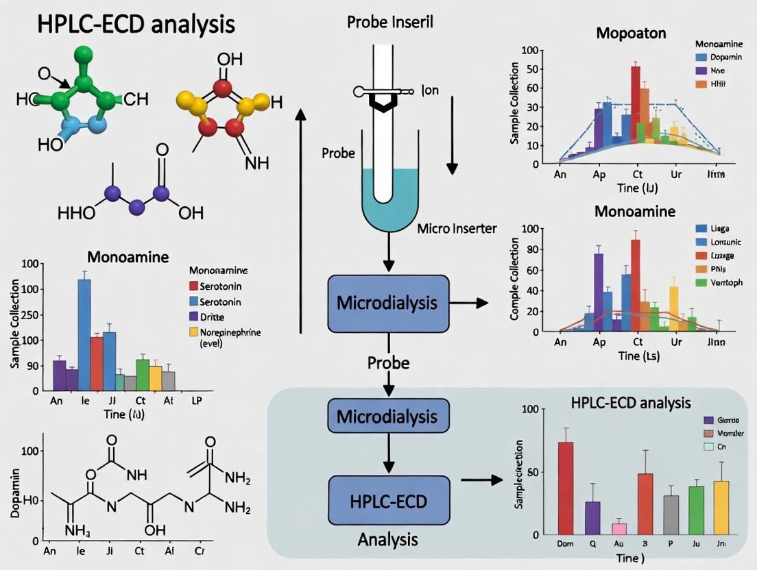
This article provides a comprehensive guide to High-Performance Liquid Chromatography with Electrochemical Detection (HPLC-ECD) for the analysis of monoamine neurotransmitters in microdialysates.
HPLC-ECD for Monoamine Analysis in Microdialysis: A Comprehensive Guide from Fundamentals to Advanced Applications
Abstract
This article provides a comprehensive guide to High-Performance Liquid Chromatography with Electrochemical Detection (HPLC-ECD) for the analysis of monoamine neurotransmitters in microdialysates. Tailored for neuroscientists, pharmacologists, and drug development professionals, it covers foundational principles, detailed methodological protocols, and advanced troubleshooting for real-time in vivo monitoring. We explore the critical role of monoamines like dopamine, serotonin, and norepinephrine in neurological function and disease. The content systematically addresses method optimization, validation against modern techniques, and practical applications in preclinical research for studying neuropharmacology, neurotoxicity, and behavioral models. This guide serves as an essential resource for achieving sensitive, selective, and reliable quantification of these crucial neurochemicals.
Understanding the Core: Why HPLC-ECD is the Gold Standard for In Vivo Monoamine Monitoring
Core Monoamines: Synthesis, Function, and Metabolites
Monoaminergic neurotransmission is critical for regulating cognition, mood, motivation, and autonomic function. HPLC-ECD coupled with in vivo microdialysis is the gold standard for monitoring real-time fluctuations of these neurotransmitters and their metabolites in the extracellular fluid of specific brain regions.
Table 1: Key Monoamines: Synthesis, Primary Functions, and Major Metabolites
| Monoamine | Biosynthetic Precursor | Key Brain Functions & Pathways | Major Metabolite(s) (via MAO/COMT) | Typical Basal ECF Concentration in Rat Striatum (nM) |
|---|---|---|---|---|
| Dopamine (DA) | Tyrosine → L-DOPA | Reward, motivation, motor control (Nigrostriatal pathway); Executive function (Mesocortical); Pleasure (Mesolimbic). | 3,4-Dihydroxyphenylacetic acid (DOPAC); Homovanillic Acid (HVA). | 0.5 - 5 nM |
| Serotonin (5-HT) | Tryptophan → 5-HTP | Mood regulation, sleep, appetite, anxiety, cognition (Raphe nuclei projections). | 5-Hydroxyindoleacetic acid (5-HIAA). | 0.1 - 2 nM |
| Norepinephrine (NE) | Dopamine (via DBH) | Arousal, alertness, stress response, attention (Locus coeruleus projections). | 3-Methoxy-4-hydroxyphenylglycol (MHPG); Normetanephrine (NMN). | 0.5 - 3 nM |
Detailed Experimental Protocols
Protocol 1:In VivoMicrodialysis Sampling for Monoamine Analysis
Objective: To collect extracellular fluid (ECF) containing monoamines and metabolites from a specific brain region (e.g., rat prefrontal cortex or striatum) for subsequent HPLC-ECD analysis.
Materials & Procedure:
- Surgical Implantation: Anesthetize the subject (e.g., rat) and stereotaxically implant a guide cannula targeting the brain region of interest.
- Probe Equilibration: On experiment day, insert a concentric-style microdialysis probe (e.g., 2-4 mm membrane length, 20kDa MWCO). Perfuse with artificial cerebrospinal fluid (aCSF: 147 mM NaCl, 2.7 mM KCl, 1.2 mM CaCl₂, 0.85 mM MgCl₂, pH 7.4) at a constant flow rate (1.0 - 1.5 µL/min) using a high-precision syringe pump.
- Equilibration Period: Allow a minimum 60-120 minute equilibration period post-insertion to stabilize basal neurotransmitter levels.
- Sample Collection: Collect dialysate into microvials prefilled with 2-5 µL of antioxidant preservation solution (0.1 M HClO₄ or 0.1 M acetic acid with 0.1 mM EDTA/0.1 mM L-cysteine). Maintain samples at 4°C (refrigerated fraction collector) and analyze immediately or store at -80°C.
Protocol 2: HPLC-ECD Analysis of Monoamines and Metabolites
Objective: To separate and quantify DA, 5-HT, NE, DOPAC, HVA, and 5-HIAA in a single microdialysis sample run.
Chromatographic Conditions:
- Column: C18 reverse-phase column (e.g., 3.0 x 100 mm, 3 µm particle size).
- Mobile Phase: 75-100 mM sodium phosphate buffer, pH 3.0-3.5, containing 1.0-1.7 mM octanesulfonic acid (ion-pair reagent), 0.1 mM EDTA, and 6-10% (v/v) methanol or acetonitrile. Degas and filter (0.22 µm).
- Flow Rate: 0.4 - 0.6 mL/min.
- Detection: Electrochemical detector with glassy carbon working electrode. Potentials: +0.7 V for analytes (oxidizing) and a secondary electrode at -0.2 V for reduction of interferents (if using dual electrode in redox mode).
- Injection Volume: 5 - 20 µL (using a low-dead-volume injector).
Procedure:
- System Preparation: Equilibrate the HPLC system with mobile phase for at least 1 hour at operational flow rate to stabilize baseline.
- Calibration: Create a 6-point calibration curve (e.g., 0.1 - 50 nM) for each analyte using external standards prepared in 0.1 M perchloric acid or aCSF.
- Sample Run: Inject dialysate samples. A typical run time is 15-25 minutes.
- Data Analysis: Identify peaks by retention time. Quantify concentrations by comparing peak area/height to the calibration curve, correcting for in vitro probe recovery (typically 10-20%).
Visualization of Pathways and Workflow
Title: Monoamine Synthesis and Metabolism Pathways
Title: Microdialysis and HPLC-ECD Analysis Workflow
The Scientist's Toolkit: Key Research Reagent Solutions
Table 2: Essential Reagents and Materials for Monoamine Microdialysis & HPLC-ECD
| Item | Function & Rationale |
|---|---|
| Artificial Cerebrospinal Fluid (aCSF) | Isotonic perfusion fluid for microdialysis probes. Mimics ionic composition of ECF to minimize osmotic stress and neuronal disturbance during sampling. |
| Antioxidant Preservation Solution (e.g., 0.1 M HClO₄ with 0.1 mM EDTA/0.1 mM L-Cysteine) | Added to collection vials to prevent oxidative degradation of easily oxidizable monoamines (especially DA and NE) prior to analysis. |
| Ion-Pair Reagent (e.g., Octanesulfonic acid sodium salt) | Added to HPLC mobile phase. Interacts with protonated amine groups, improving retention and separation of hydrophilic monoamines and metabolites on C18 columns. |
| Electrochemical Cell (Glassy Carbon Working Electrode) | The sensing surface in ECD. Applied potential oxidizes monoamines, generating a measurable current proportional to concentration. Requires regular polishing. |
| Monoamine Standard Mixture (DA, 5-HT, NE, DOPAC, HVA, 5-HIAA) | Used to create daily calibration curves for absolute quantification. Must be prepared fresh in acidic antioxidant solution from high-purity stocks. |
| Reverse-Phase C18 HPLC Column (3 µm, 100-150 mm length) | The core separation component. Small particle size provides high efficiency for resolving complex monoamine metabolite profiles in short run times. |
Within the broader thesis on HPLC-ECD analysis of microdialysis monoamines, this document details the integrated methodology that enables in vivo, real-time neurochemical monitoring. Microdialysis provides continuous, localized sampling of the brain's extracellular fluid (ECF), while High-Performance Liquid Chromatography with Electrochemical Detection (HPLC-ECD) offers the requisite sensitivity and selectivity for quantifying low concentrations of monoamines (e.g., dopamine, serotonin, norepinephrine) and their metabolites. This synergy is fundamental for studying neurochemical dynamics in response to pharmacological, behavioral, or pathological stimuli in preclinical research and drug development.
Key Advantages:
- Real-Time Pharmacodynamics: Direct measurement of drug-induced changes in neurotransmitter levels.
- High Spatial & Temporal Resolution: Probes can be targeted to specific brain regions, with sampling intervals as short as 1-10 minutes.
- Low Sample Volume Requirement: Compatible with the microliter volumes yielded by microdialysis.
- Specificity for Electroactive Analytes: ECD is inherently selective for oxidizable species like monoamines.
Table 1: Representative Basal Extracellular Levels of Monoamines in Rodent Brain
| Analyte | Brain Region | Average Basal Level (nM) | Sampling Parameters |
|---|---|---|---|
| Dopamine (DA) | Striatum | 1 - 5 | Flow: 1.0 µL/min, 10-min fraction |
| Serotonin (5-HT) | Prefrontal Cortex | 0.5 - 2 | Flow: 1.0 µL/min, 10-min fraction |
| Norepinephrine (NE) | Hippocampus | 0.5 - 3 | Flow: 1.0 µL/min, 15-min fraction |
| DOPAC (DA metabolite) | Striatum | 500 - 2000 | Flow: 1.0 µL/min, 10-min fraction |
| 5-HIAA (5-HT metabolite) | Striatum | 100 - 500 | Flow: 1.0 µL/min, 10-min fraction |
Table 2: Typical HPLC-ECD Performance Parameters for Monoamine Analysis
| Parameter | Target Specification | Typical Value |
|---|---|---|
| Lower Limit of Quantification (LLOQ) | Dopamine | 0.1 - 0.5 nM (injected) |
| Linear Dynamic Range | Dopamine | 0.1 to 1000 nM (r² > 0.995) |
| Separation Column | C18 Reverse Phase | 150 x 3.0 mm, 3 µm particle size |
| Mobile Phase | Citrate-acetate or phosphate buffer, pH 3.5-4.0, with ion-pairing reagent (e.g., OSA), organic modifier (5-10% MeOH), and electrochemical conditioning agent. | |
| Flow Rate | 0.4 - 0.6 mL/min | |
| Detection Potential | Glassy Carbon Working Electrode | +0.6 to +0.8 V vs. Ag/AgCl reference |
Experimental Protocols
Protocol 1: In Vivo Microdialysis Sampling in the Rat Striatum Objective: To collect serial dialysate samples for basal and stimulated monoamine measurement.
- Surgery & Probe Implantation: Anesthetize rat and secure in stereotaxic frame. Implant a guide cannula above the striatum (AP: +1.0 mm, ML: ±2.5 mm from bregma, DV: -3.0 mm from dura). Secure with dental cement.
- Post-Surgical Recovery: Allow animal to recover for 24-48 hours.
- Probe Insertion & Perfusion: Insert a concentric microdialysis probe (3-4 mm membrane, 20kDa MWCO) via the guide cannula. Connect to a microinfusion pump via fluorinated ethylene propylene (FEP) tubing. Perfuse with artificial cerebrospinal fluid (aCSF: 147 mM NaCl, 2.7 mM KCl, 1.2 mM CaCl₂, 0.85 mM MgCl₂, pH 7.4) at 1.0 µL/min.
- Equilibration: Allow 60-90 minutes for neurochemical equilibrium post-insertion.
- Sample Collection: Collect dialysate into microvials containing 5 µL of 0.1 M perchloric acid (to prevent oxidation) at 10-minute intervals. Keep samples on ice or at 4°C until analysis (typically within 24 hours).
- Stimulation (Optional): To evoke release, switch perfusion to aCSF containing 60-100 mM KCl for 10-20 minutes.
Protocol 2: HPLC-ECD Analysis of Dialysate Monoamines Objective: To separate and quantify monoamines in dialysate samples.
- System Preparation: Configure HPLC system with a refrigerated autosampler (set to 4°C), a degasser, a pump, and an electrochemical detector with a glassy carbon working electrode and Ag/AgCl reference cell.
- Mobile Phase: Prepare 0.1 M phosphate buffer, pH 3.0, containing 1.7 mM 1-octanesulfonic acid (OSA, ion-pair reagent), 0.1 mM EDTA (chelating agent), and 10% v/v methanol. Filter (0.22 µm) and degas thoroughly.
- Calibration: Prepare standard mixtures of analytes (DA, 5-HT, NE, DOPAC, HVA, 5-HIAA) in 0.1 M perchloric acid at concentrations spanning 0.1-100 nM. Inject 10-20 µL to generate a calibration curve.
- Sample Analysis: Inject 10-20 µL of dialysate directly. Use an isocratic elution at a flow rate of 0.5 mL/min. Typical run time is 15-20 minutes.
- Data Analysis: Quantify peaks by comparing their area under the curve (AUC) to the calibration curve. Express final ECF concentrations as nM.
Visualization: Workflows and Pathways
Title: Combined Microdialysis and HPLC-ECD Workflow
Title: Neurotransmitter Dynamics Sampled by Microdialysis
The Scientist's Toolkit: Research Reagent Solutions
Table 3: Essential Materials for Microdialysis/HPLC-ECD Experiments
| Item | Function & Critical Notes |
|---|---|
| Concentric Microdialysis Probe | Implantable device with semi-permeable membrane for molecular exchange. MWCO (e.g., 20 kDa) excludes proteins. |
| Artificial Cerebrospinal Fluid (aCSF) | Physiological perfusion fluid. Must contain Ca²⁺ for normal synaptic function. For no-Ca²⁺ experiments, replace with isotonic Mg²⁺. |
| Monoamine Standard Mixtures | Accurate quantification requires freshly prepared or certified standard solutions for calibration. |
| Ion-Pair Reagent (e.g., OSA) | Added to mobile phase to improve retention of polar monoamines on reverse-phase C18 columns. |
| Antioxidant (e.g., 0.1 M HClO₄, EDTA) | Added to collection vials to prevent oxidation of catecholamines. EDTA is also added to mobile phase. |
| HPLC Mobile Phase Buffers | Low-pH (3.0-4.0) phosphate or citrate buffers are standard for ECD to optimize oxidation efficiency and separation. |
| Glassy Carbon Working Electrode | The sensing surface in ECD. Requires periodic polishing to maintain sensitivity. |
| Sterile Guide Cannula & Obdurator | Provides a permanent guide for precise, repeatable probe insertion into the target brain region. |
Within the framework of HPLC-ECD analysis of microdialysate monoamines, understanding the precise electrochemical oxidation mechanisms is paramount. This knowledge directly informs experimental design, interpretation of chromatograms, and troubleshooting of sensitivity issues. This document details the fundamental electrochemistry of catecholamines (e.g., dopamine, norepinephrine) and indolamines (e.g., serotonin, 5-HIAA), providing application notes and protocols for their reliable detection in neuroscience and drug development research.
Electrochemical Oxidation Mechanisms
The mechanism of electrochemical oxidation at the carbon-based electrode surface (typically glassy carbon) is a two-electron, two-proton process for both classes of compounds, but the structures of the intermediates and products differ.
1.1 Catecholamines (e.g., Dopamine) Catecholamines contain an ortho-dihydroxybenzene (catechol) ring. Oxidation proceeds via a well-defined, reversible redox couple.
- Step 1: The catechol moiety is oxidized to its corresponding ortho-quinone. This is a 2e⁻/2H⁺ transfer.
- Step 2 (Post-Oxidation Chemistry): The generated ortho-quinone is highly electrophilic. It can undergo non-electrochemical chemical reactions (C), such as cyclization (for dopamine) or reaction with nucleophiles (e.g., cysteine). This EC (Electrochemical-Chemical) mechanism can be leveraged for enhanced selectivity.
1.2 Indolamines (e.g., Serotonin, 5-HIAA) Indolamines contain an indole ring system. Their oxidation is generally less reversible than catecholamines.
- Serotonin: Oxidation occurs primarily on the 5-hydroxyl group of the indole ring, generating a quinone-imine intermediate. This oxidation is often quasi-reversible.
- 5-Hydroxyindoleacetic Acid (5-HIAA): The oxidation involves the 5-hydroxyl group on the indole ring and is influenced by the carboxylic acid side chain, typically resulting in an irreversible oxidation wave.
Table 1: Key Electrochemical Parameters for Common Monoamines
| Analyte | Class | Approx. Oxidation Potential (vs. Ag/AgCl) | Reversibility | Primary Oxidation Site |
|---|---|---|---|---|
| Norepinephrine (NE) | Catecholamine | +0.15 V | Reversible | Catechol Ring |
| Epinephrine (E) | Catecholamine | +0.18 V | Reversible | Catechol Ring |
| Dopamine (DA) | Catecholamine | +0.20 V | Reversible | Catechol Ring |
| 3,4-Dihydroxyphenylacetic Acid (DOPAC) | Catecholamine Metabolite | +0.22 V | Reversible | Catechol Ring |
| Serotonin (5-HT) | Indolamine | +0.32 V | Quasi-Reversible | 5-Hydroxyl on Indole |
| 5-Hydroxyindoleacetic Acid (5-HIAA) | Indolamine Metabolite | +0.42 V | Irreversible | 5-Hydroxyl on Indole |
| Homovanillic Acid (HVA) | Catecholamine Metabolite | +0.55 V | Irreversible | Phenolic Ring |
Detailed Protocol: HPLC-ECD for Microdialysate Monoamines
2.1 Sample Preparation (Microdialysate)
- Collection: Collect microdialysate directly into vials containing 10-20 µL of antioxidant preservative solution (0.1 M HClO₄, 0.1 mM Na₂EDTA, 0.01% Na₂S₂O₅). Keep samples on ice or at 4°C.
- Storage: Immediately freeze at -80°C if not analyzed within 24 hours. Avoid freeze-thaw cycles.
- Injection: Centrifuge at 12,000 x g for 10 minutes at 4°C. Inject supernatant directly onto the HPLC. Typical injection volumes are 5-20 µL.
2.2 HPLC-ECD System Configuration and Parameters
- HPLC System: High-pressure binary or isocratic pump with pulse damper.
- Column: C18 reversed-phase column (e.g., 150 x 3.0 mm, 3 µm particle size). Maintain at 30-35°C.
- Mobile Phase: (Example formulation)
- 75 mM NaH₂PO₄
- 1.4 mM Sodium Octane Sulfonate (ion-pairing reagent)
- 10 µM Na₂EDTA (chelating agent)
- 7% (v/v) Methanol
- pH adjusted to 3.65 with H₃PO₄
- Flow Rate: 0.5 mL/min. Filter (0.2 µm) and degas continuously.
- Electrochemical Detector:
- Working Electrode: Glassy Carbon.
- Reference Electrode: Ag/AgCl.
- Guard Cell: Upstream of injector, set at +0.35 V to oxidize mobile phase contaminants.
- Analytical Cell: Dual-channel in series. Typical potentials: Channel 1: +0.15 V (catechols); Channel 2: +0.35 V (indolamines & metabolites). Apply potentials 30 minutes before analysis for stabilization.
2.3 Calibration and Quantification
- Prepare stock solutions of each analyte in 0.1 M HClO₄ with antioxidant.
- Create a calibration curve from 6-8 points spanning expected in vivo concentrations (e.g., 0.1 nM to 100 nM).
- Inject calibration standards before and after experimental samples. Use linear regression for quantification. Include a system suitability test (SST) mix daily.
The Scientist's Toolkit: Key Research Reagent Solutions
Table 2: Essential Materials for HPLC-ECD of Monoamines
| Item | Function | Critical Notes |
|---|---|---|
| Glassy Carbon Electrode | Working electrode for oxidation. | Requires daily polishing (0.05 µm alumina slurry) for reproducibility. |
| NaH₂PO₄ Buffer | Provides pH control and ionic strength for mobile phase. | pH ~3.6 maximizes separation and electrode life. |
| Ion-Pairing Reagent (e.g., Sodium Octane Sulfonate) | Modifies retention of cationic amines (DA, 5-HT) on C18 column. | Concentration is critical for resolution of early-eluting peaks. |
| Na₂EDTA | Chelates trace metal ions that catalyze analyte degradation. | Essential in both mobile phase and sample preservative. |
| Antioxidant Preservative (HClO₄/Na₂S₂O₅/EDTA) | Stabilizes easily oxidized catechols in biological samples. | Must be added immediately upon sample collection. |
| C18 Reversed-Phase Column | Separates analytes based on hydrophobicity. | Dedicated column for ECD only; guard column highly recommended. |
Visualization of Mechanisms and Workflow
Title: Dopamine Electrochemical-Chemical (EC) Oxidation Pathway
Title: HPLC-ECD Analysis Workflow for Microdialysate
Application Notes
Within a thesis investigating the role of monoamines in neuropsychiatric disorders using in vivo microdialysis, High-Performance Liquid Chromatography with Electrochemical Detection (HPLC-ECD) remains the benchmark analytical technique. Its core advantages directly address the unique challenges of microdialysate analysis:
- Sensitivity (Low Limit of Detection - LOD): HPLC-ECD achieves femtomole (fmol) to attomole (amol) on-column detection limits. This is critical as basal extracellular concentrations of monoamines (e.g., dopamine, serotonin, norepinephrine) are typically in the low nanomolar (nM) to picomolar (pM) range, with sample volumes often ≤ 10 µL.
- Selectivity (Minimal Matrix Interference): The dual selectivity of (1) reversed-phase chromatographic separation and (2) the applied electrochemical oxidation potential minimizes interference from complex brain matrix components (e.g., salts, ascorbic acid, uric acid, acidic metabolites) present in microdialysates.
- Suitability for Aqueous Samples: The aqueous nature of microdialysates (typically an artificial cerebrospinal fluid, aCSF) is perfectly compatible with common HPLC-ECD mobile phases (aqueous buffers with an organic modifier like methanol or acetonitrile), requiring minimal sample pretreatment, often just acidification and filtration.
Quantitative Performance Data for Monoamine Analysis via HPLC-ECD: Table 1: Representative Analytical Figures of Merit for Key Monoamines and Metabolites.
| Analytic | Typical Column | Mobile Phase (approx.) | Applied Potential (vs. ref.) | Limit of Detection (LOD) | Linear Range | Key Interference Resolved |
|---|---|---|---|---|---|---|
| Dopamine (DA) | C18, 3 µm, 150 x 3.2 mm | 75-100 mM NaH₂PO₄, 1.7-2.0 mM OSA, 6-10% MeOH, pH 3.6-4.0 | +0.65 - +0.75 V | 0.5 - 2 pg (3-13 fmol) | 5 pg - 100 ng | DOPAC, 5-HT, Ascorbate |
| 3,4-Dihydroxyphenylacetic Acid (DOPAC) | Same as above | Same as above | +0.65 - +0.75 V | 5 - 10 pg | 50 pg - 200 ng | DA, HVA |
| Homovanillic Acid (HVA) | Same as above | Same as above | +0.75 - +0.85 V | 10 - 20 pg | 100 pg - 200 ng | DOPAC, 5-HIAA |
| Serotonin (5-HT) | C18, 3 µm, 150 x 3.2 mm | 70-100 mM NaH₂PO₄, 0.1-0.5 mM EDTA, 8-12% MeOH, pH 4.5-5.0 | +0.60 - +0.70 V | 0.5 - 3 pg (3-17 fmol) | 5 pg - 50 ng | 5-HIAA, DA (chromatographically) |
| 5-Hydroxyindoleacetic Acid (5-HIAA) | Same as above | Same as above | +0.70 - +0.80 V | 5 - 15 pg | 50 pg - 200 ng | HVA, Uric Acid |
| Norepinephrine (NE) | C18, 3 µm, 150 x 3.2 mm | 75-100 mM NaH₂PO₄, 0.8-1.2 mM OSA, 4-6% MeOH, pH 3.1-3.5 | +0.65 - +0.75 V | 1 - 5 pg (6-30 fmol) | 10 pg - 100 ng | Epinephrine, DOPAC |
OSA: Octanesulfonic acid (ion-pairing reagent); MeOH: Methanol; Ref.: Ag/AgCl reference electrode.
Experimental Protocols
Protocol 1: Microdialysate Sample Collection and Preparation for Catecholamine Analysis Objective: To collect and stabilize striatal microdialysates for the concurrent analysis of DA, NE, DOPAC, and HVA.
- Perfusion: Implant a concentric microdialysis probe (4 mm membrane, CMA 12) into the rat striatum. Perfuse with aCSF (147 mM NaCl, 2.7 mM KCl, 1.2 mM CaCl₂, 0.85 mM MgCl₂, pH 7.4) at 1.0 µL/min.
- Baseline Collection: After a 2-hour equilibration period, collect samples every 15-20 minutes into microvials.
- Stabilization: Immediately after collection, acidify each sample by adding 1 µL of 0.1 M perchloric acid (HClO₄) or 0.1 M acetic acid to a 10 µL microdialysate aliquot. Vortex briefly.
- Cleaning: Centrifuge at 13,000 x g for 10 minutes at 4°C to precipitate proteins.
- Injection: Transfer the clear supernatant to a limited-volume HPLC vial insert. Inject 5-10 µL onto the HPLC-ECD system.
Protocol 2: HPLC-ECD System Setup and Analysis for Monoamines Objective: To establish a validated method for separating and detecting monoamines and their acidic metabolites.
- HPLC System:
- Column: Use a reverse-phase C18 column (e.g., Phenomenex Gemini, 3 µm, 150 x 3.2 mm) maintained at 30-35°C.
- Mobile Phase: Prepare 100 mM sodium phosphate buffer, pH 3.6. Add 1.7 mM octanesulfonic acid (OSA) and 7% v/v HPLC-grade methanol. Filter (0.22 µm) and degas under vacuum.
- Flow Rate: Set isocratic flow to 0.5 mL/min.
- ECD System:
- Detector: Use a dual-potential coulometric detector (e.g., ESA Coulochem III).
- Guard Cell (Upstream): Set to +350 mV to oxidize mobile phase contaminants.
- Working Electrode 1 (Screening): Set to +150 mV. This low potential oxidizes only the most easily oxidized interferents (e.g., ascorbate).
- Working Electrode 2 (Analytical): Set to +650 mV. This potential oxidizes the monoamines and metabolites of interest. The signal from Electrode 2 is subtracted from Electrode 1 for enhanced selectivity.
- Calibration: Prepare a standard mix of all analytes in aCSF acidified with 0.1 M HClO₄. Construct a 6-point calibration curve (e.g., 0.5, 2, 10, 50, 200, 1000 nM) for each compound daily.
Visualizations
Workflow for HPLC-ECD Analysis of Microdialysates
HPLC-ECD Dual Selectivity Mechanism
The Scientist's Toolkit: Essential Research Reagents & Materials
Table 2: Key Reagents and Consumables for Microdialysis HPLC-ECD.
| Item | Function & Rationale |
|---|---|
| Ion-Pairing Reagent (e.g., Octanesulfonic Acid Sodium Salt) | Adds a hydrophobic moiety to polar catecholamines and metabolites, enabling retention on reverse-phase C18 columns. Critical for separating DA from DOPAC. |
| HPLC-Grade Methanol or Acetonitrile | Organic modifier in mobile phase; adjusts retention times and peak shape. Methanol is often preferred in ECD for its lower oxidative background current. |
| Sodium Phosphate Monobasic (NaH₂PO₄) | Primary buffer salt for mobile phase. Provides stable pH in the 3.0-5.0 range, optimal for analyte stability and electrochemical oxidation. |
| Perchloric or Acetic Acid (0.1 M) | Sample acidification agent. Preserves monoamines from enzymatic degradation and oxidative loss post-collection. |
| Ethylenediaminetetraacetic Acid (EDTA) | Metal chelator added to mobile phase (esp. for 5-HT) to sequester trace metals that can catalyze analyte oxidation and degrade column performance. |
| Artificial Cerebrospinal Fluid (aCSF) | Physiological perfusion fluid matching ionic composition of brain extracellular fluid, minimizing osmotic stress during in vivo microdialysis. |
| C18 Reverse-Phase Column (3µm, 150-150mm) | The core separation component. Small particle size (3µm) provides high efficiency for resolving complex monoamine peaks in a short run time. |
| Coulometric Electrochemical Cell | Contains porous graphite working electrodes providing ~100% oxidation efficiency, yielding superior sensitivity and stability compared to amperometric detectors. |
| Microvials & Limited-Volume Inserts | Essential for handling low-volume (10-20 µL) microdialysate samples without significant loss to evaporation or vial surface adsorption. |
Historical Context and Enduring Relevance in Modern Neuroscience and Drug Discovery
The study of monoaminergic neurotransmission—dopamine (DA), norepinephrine (NE), and serotonin (5-HT)—has its roots in early 20th-century physiology and pharmacology. The discovery of these chemical messengers and their profound influence on mood, arousal, and cognition laid the foundation for modern psychopharmacology, exemplified by the development of first-generation antidepressants and antipsychotics. Today, the precise, high-resolution measurement of these monoamines via techniques like in vivo microdialysis coupled with High-Performance Liquid Chromatography and Electrochemical Detection (HPLC-ECD) remains a cornerstone of neuroscience research and CNS drug discovery. This protocol details the application of HPLC-ECD for monitoring dynamic changes in extracellular monoamines, linking historical neurochemical concepts to contemporary mechanistic studies of novel therapeutic compounds.
Table 1: Representative Basal Extracellular Monoamine Concentrations in Rat Prefrontal Cortex Microdialysate (HPLC-ECD)
| Monoamine | Average Concentration (nM) | Typical Range (nM) | Key Functional Relevance |
|---|---|---|---|
| Dopamine (DA) | 2.5 | 1.0 - 4.0 | Reward, motivation, executive function |
| Norepinephrine (NE) | 4.0 | 2.0 - 6.0 | Arousal, attention, stress response |
| Serotonin (5-HT) | 1.5 | 0.8 - 2.5 | Mood, anxiety, sleep-wake cycle |
Application Notes & Protocols
Protocol 1: In Vivo Microdialysis for Freely Moving Rodents Objective: To collect time-resolved extracellular fluid samples from a specific brain region (e.g., medial prefrontal cortex, mPFC) for subsequent monoamine analysis.
- Surgical Implantation: Anesthetize rat (e.g., isoflurane 2-5%). Stereotactically implant a guide cannula targeting the mPFC (AP: +3.2 mm, ML: ±0.8 mm, DV: -2.0 mm from bregma). Secure with dental cement.
- Post-Surgical Recovery: Allow a minimum 48-hour recovery period with analgesia.
- Microdialysis Probe Insertion & Perfusion: On experiment day, insert a concentric-style microdialysis probe (2-4 mm membrane, 20kDa MWCO) via the guide. Perfuse with artificial cerebrospinal fluid (aCSF: 147 mM NaCl, 2.7 mM KCl, 1.2 mM CaCl₂, 1.0 mM MgCl₂, pH 7.4) at 1.0 µL/min. Allow 2-3 hours for equilibration.
- Sample Collection: Collect dialysate into microvials prefilled with 5 µL of antioxidant preservative (0.1 M perchloric acid or 0.1% w/v cysteine/0.1% w/v EDTA). For baseline, collect 3-4 samples at 10-20 minute intervals. Apply pharmacological challenge (e.g., systemic drug, local reverse dialysis) and continue serial sample collection. Samples are stored at -80°C until analysis.
Protocol 2: HPLC-ECD Analysis of Monoamines in Microdialysate Objective: To separate and quantify DA, NE, and 5-HT in collected microdialysate samples.
- HPLC System Configuration: Use a reversed-phase C18 column (150 x 3.0 mm, 3 µm particle size). Mobile phase: 75 mM NaH₂PO₄, 1.7 mM 1-octanesulfonic acid, 25 µM EDTA, 10% acetonitrile, pH 3.1 (adjusted with phosphoric acid). Isocratic flow rate: 0.5 mL/min.
- Electrochemical Detection: Use a dual glassy carbon working electrode set at oxidative potentials of +450 mV (Electrode 1, for catecholamines) and +650 mV (Electrode 2, for indoleamines). Guard cell: +850 mV upstream of injector.
- Sample Run: Thaw samples on ice, inject 10 µL onto the HPLC system via an auto-sampler cooled to 4°C. Total run time: 15-20 minutes.
- Quantification: Generate a standard calibration curve daily (0.1-20 nM for each monoamine). Monoamine peaks are identified by retention time and quantified by peak area relative to standards using chromatography software.
Visualization
Diagram 1: HPLC-ECD Analysis Workflow
Diagram 2: Monoaminergic Signaling & Drug Action Pathways
The Scientist's Toolkit: Key Reagent Solutions for Microdialysis/HPLC-ECD
| Item | Function & Specification |
|---|---|
| Artificial Cerebrospinal Fluid (aCSF) | Physiological perfusion fluid for microdialysis. Must be sterile, filtered (0.2 µm), and contain specific ion concentrations (Ca²⁺, Mg²⁺, K⁺) to maintain tissue health and normal neurotransmission. |
| Antioxidant Preservative | Added to collection vials to prevent oxidation of catecholamines (DA, NE). Common: 0.1 M perchloric acid or a mixture of cysteine/EDTA. Critical for sample stability. |
| HPLC Mobile Phase | Contains ion-pairing reagent (e.g., octanesulfonic acid) to facilitate separation of hydrophilic monoamines on a reversed-phase column. Low pH (~3.1) and EDTA enhance peak resolution and prevent chelation. |
| Monoamine Standard Stock Solutions | High-purity DA, NE, and 5-HT prepared in 0.1 M perchloric acid. Used to generate daily calibration curves (e.g., 0.1, 0.5, 1, 5, 10, 20 nM) for absolute quantification. |
| Microdialysis Probe | Semi-permeable membrane (e.g., polyethersulfone) with a defined molecular weight cutoff (e.g., 20 kDa). Allows diffusion of small molecules like monoamines into the perfusate. |
| Electrode Conditioning Solution | Used to clean and polish glassy carbon working electrodes to maintain sensitivity and baseline stability (e.g., fine alumina slurry). |
Step-by-Step Protocol: Setting Up and Running a Robust HPLC-ECD Microdialysis Analysis
This application note details the optimal high-performance liquid chromatography with electrochemical detection (HPLC-ECD) system configuration for the sensitive and reliable analysis of monoamines (dopamine, norepinephrine, serotonin, and their metabolites) in microdialysis samples. The specifications are framed within a thesis investigating neurochemical dynamics in preclinical models of neurological disorders and drug action. The extreme sensitivity required for low-concentration, low-volume microdialysates dictates stringent component selection.
Optimal System Specifications & Quantitative Data
The following tables summarize the critical specifications for each module, compiled from current manufacturer data and recent methodological publications.
Table 1: Pump System Specifications
| Parameter | Optimal Specification | Rationale |
|---|---|---|
| Type | Quaternary or binary low-pressure mixing pump with degasser | Allows flexible mobile phase optimization; degasser prevents baseline noise from dissolved O₂. |
| Flow Rate Range | 0.001 to 5.0 mL/min, capable of 0.01 mL/min increments | Precise, low flow rates (0.3-0.7 mL/min) are standard for 2.0-2.1 mm ID columns to reduce solvent consumption and enhance sensitivity. |
| Pressure Limit | ≥ 6000 psi (400 bar) | Compatibility with high-efficiency, small-particle-size columns. |
| Composition Accuracy | ≤ 0.1% RSD | Critical for gradient reproducibility and retention time stability. |
| Pulsation | < 1% | Minimal pulsation is essential for stable baselines in ECD. |
Table 2: Analytical Column Specifications
| Parameter | Optimal Specification | Rationale |
|---|---|---|
| Dimensions | 150 mm x 2.0 mm or 2.1 mm internal diameter | Optimal balance between resolution, analysis time, and sensitivity for microdialysates. |
| Particle Size | 1.7 to 3.0 μm | Provides high efficiency (theoretical plates > 15,000/column). |
| Stationary Phase | C18 or C18-AQ, end-capped | Standard for monoamine separation; AQ phases offer better wettability in high-aqueous mobile phases. |
| Pore Size | 80 to 120 Å | Suitable for small molecule monoamines. |
| Temperature Control | Required (use column oven) | Maintains retention time reproducibility (typically 30-40°C). |
Table 3: Electrochemical Detector Specifications
| Parameter | Optimal Specification | Rationale |
|---|---|---|
| Type | Coulometric (dual electrode in series) or high-sensitivity amperometric | Coulometric offers superior selectivity and sensitivity (>90% oxidation efficiency). |
| Cell Design | Dual porous graphite working electrodes, in oxidation-reduction or screen mode | First electrode oxidizes analytes; second electrode reduces interferents or confirms identity via redox ratio. |
| Applied Potentials | E1: +400 to +750 mV; E2: -100 to +200 mV (vs. Pd reference) | Optimized for catecholamines and indoleamines. Exact potentials require empirical optimization. |
| Noise Level | < 1 pA peak-to-peak | Essential for detecting sub-picomole levels. |
| Data Sampling Rate | ≥ 10 Hz | Accurate peak integration for narrow HPLC peaks. |
Table 4: Autosampler Specifications
| Parameter | Optimal Specification | Rationale |
|---|---|---|
| Injection Volume | Variable, capable of 0.1 to 100 μL. 5-20 μL is typical. | Must accurately inject low volumes from limited microdialysate samples. |
| Precision | < 0.5% RSD for volume | Critical for quantitative reproducibility. |
| Carryover | < 0.05% | Avoids contamination between samples with high concentration differences. |
| Temperature Control | 4-10°C sample tray cooling | Stabilizes easily oxidized monoamines in collected dialysates. |
| Vial Capacity | ≥ 100 vials | For large batch processing (including standards, QCs, and samples). |
Detailed Experimental Protocol: HPLC-ECD Analysis of Microdialysis Monoamines
A. Reagent and Mobile Phase Preparation
- Water: Ultra-pure HPLC-grade water (18.2 MΩ·cm resistivity).
- Mobile Phase:
- Prepare 100 mM Sodium Phosphate Buffer, pH 3.0. Dissolve 13.8 g of NaH₂PO₄·H₂O in 950 mL water. Adjust to pH 3.0 with concentrated ortho-phosphoric acid. Bring to 1 L with water. Filter through a 0.22 μm nylon membrane.
- Prepare the final mobile phase daily: To 1 L of the pH 3.0 buffer, add 200 mg of Octane-1-sulfonic acid sodium salt (OSA, ion-pairing agent), 80 mg of EDTA (chelating agent), and 6% (v/v) HPLC-grade methanol. Degas under helium sparging for 10 minutes before use and maintain under a helium atmosphere during analysis.
B. System Configuration and Start-Up
- Install the specified column (e.g., 150 x 2.0 mm, 1.7 μm C18) in a column oven set to 35°C.
- Connect the electrochemical detector cell. Condition the new electrode by applying a series of increasing potentials in buffer-only mobile phase over several hours.
- Prime the pump with the degassed mobile phase at 0.2 mL/min for 30 minutes.
- Set the detector potentials. For dual-electrode coulometric mode: Electrode 1 (oxidation) = +650 mV; Electrode 2 (reduction) = -50 mV. Allow the baseline to stabilize (may take several hours).
C. Sample Preparation and Calibration
- Collect microdialysate into vials containing 2 μL of 0.1 M perchloric acid (or equivalent preservative) per 20 μL sample volume. Keep on ice or in a refrigerated autosampler at 6°C.
- Prepare a primary standard stock solution (100 μg/mL) of each analyte (DA, NE, 5-HT, DOPAC, HVA, 5-HIAA) in 0.1 M perchloric acid. Store at -80°C.
- Prepare a working composite standard mix by serial dilution in artificial cerebrospinal fluid (aCSF) with 0.01 M perchloric acid. Create a 7-point calibration curve from 0.05 to 50 ng/mL.
- Inject standards and samples (typically 10 μL) using the cooled autosampler.
D. Chromatographic Run and Data Analysis
- Run an isocratic elution at a flow rate of 0.35 mL/min. Total run time is approximately 20 minutes.
- Identify analytes by their characteristic retention times relative to standards.
- Quantify using peak area from the primary oxidation channel (E1). Use the redox ratio (peak area E2 / peak area E1) for peak purity confirmation.
- Perform linear regression on the calibration curve. Report sample concentrations in ng/mL or nM.
Diagrams
Workflow Diagram: HPLC-ECD for Microdialysis Monoamines
ECD Dual-Electrode Detection Logic
The Scientist's Toolkit: Essential Research Reagents & Materials
Table 5: Key Reagent Solutions for Microdialysis Monoamine Analysis
| Item | Function | Typical Specification/Preparation |
|---|---|---|
| Artificial CSF (aCSF) | Perfusion fluid for microdialysis; used for standard dilution. | 147 mM NaCl, 2.7 mM KCl, 1.2 mM CaCl₂, 1.0 mM MgCl₂, pH ~7.4. Filtered (0.22 μm). |
| 0.1 - 0.5 M Perchloric Acid (PCA) | Sample preservative. Prevents oxidative degradation of monoamines. | Add 2-5 μL of concentrated PCA per 100 μL sample for 0.2-0.5% final concentration. |
| Sodium Phosphate Buffer (pH 3.0) | Aqueous component of mobile phase. Low pH ensures protonation and separation. | 100 mM, adjusted with H₃PO₄. Filtered and degassed. |
| Ion-Pairing Reagent (e.g., OSA) | Added to mobile phase to improve retention of polar catecholamines on C18 column. | Octane-1-sulfonic acid sodium salt, 0.1-0.2 mM in final mobile phase. |
| EDTA Solution | Metal chelator in mobile phase. Binds trace metals that catalyze analyte oxidation. | Disodium EDTA, 50-100 μM in final mobile phase. |
| Monoamine Standard Stock Solutions | For calibration and method validation. | 100 μg/mL each in 0.1 M PCA or 0.1 M HClO₄. Aliquot and store at -80°C. |
| 3,4-Dihydroxybenzylamine (DHBA) | Internal Standard. Added to samples to correct for injection variability and recovery. | Prepare stock in 0.1 M PCA. Spike into dialysate or standard at a constant concentration (e.g., 10 ng/mL). |
In the context of a thesis focused on the HPLC-ECD analysis of monoamines (e.g., dopamine, serotonin, norepinephrine) from cerebral microdialysis samples, mobile phase optimization is the critical determinant of success. Microdialysis samples present unique challenges: ultra-low analyte concentrations (nM to pM), complex biological matrices, and the need for high sensitivity and selectivity to resolve closely related catecholamines and metabolites. This document provides application notes and detailed protocols for optimizing the four pillars of reversed-phase ion-pair HPLC-ECD mobile phase design to achieve robust, reproducible, and sensitive monoamine quantification.
The optimization of each parameter interacts with the others. The following tables summarize target ranges and effects based on current literature and practice.
Table 1: Buffer and pH Optimization Guide for Common Monoamines
| Analyte Class | Recommended Buffer | Optimal pH Range | Key Rationale |
|---|---|---|---|
| Catecholamines (DA, NE, EPI) | Citrate-Phosphate, Acetate, Phosphate | 3.0 - 3.8 | Protonates carboxyls on metabolites, stabilizes catechols from oxidation, controls silica surface charge. |
| Indoleamines (5-HT, 5-HIAA) | Citrate-Phosphate, Acetate | 4.0 - 5.0 | Balances ionization of the less acidic indoleamine group against metabolite acidity. |
| Mixed Analysis (DA, 5-HT, DOPAC, HVA, 5-HIAA) | Citrate-Phosphate with EDTA | 3.2 - 3.5 | Compromise pH for simultaneous resolution. EDTA (50-100 µM) is essential to chelate metal ions and prevent oxidation. |
Table 2: Ion-Pairing Reagents (IPRs) for Monoamine Separation
| IPR | Typical Concentration | Primary Mechanism & Effect | Application Note |
|---|---|---|---|
| Octanesulfonic Acid (OSA) | 0.5 - 2.0 mM | Pairs with protonated amines, increasing retention of cationic analytes. | Standard for catecholamines. Retention increases with chain length (e.g., heptane vs. octane). |
| Sodium Dodecyl Sulfate (SDS) | 2 - 10 mM | Strong ion-pairing, also disrupts column conditioning. Use with caution. | Can be used for very complex mixes but may require dedicated column. |
| Perfluorinated Acids (e.g., TFA, HFBA) | 0.1% v/v (TFA) | Powerful pairing agent; suppresses silanol interactions, improves peak shape. | HFBA provides greater retention for amines than TFA. Can be corrosive to ECD cells. |
Table 3: Organic Modifier Selection and Effects
| Modifier | Typical % Range (Isocratic) | Key Properties |
|---|---|---|
| Methanol | 5-12% | Lower backpressure, stronger elution strength for polar compounds, often provides better selectivity for catechols. |
| Acetonitrile | 5-10% | Lower viscosity, superior UV transparency, different selectivity. Can require higher % for similar retention time vs. MeOH. |
| Mix (MeOH/ACN) | Varies | Fine-tune selectivity and retention; used in advanced method development. |
Experimental Protocols
Protocol 1: Systematic Scouting of pH and Ion-Pair Concentration Objective: Determine the optimal pH and OSA concentration for separating a standard mix of DA, DOPAC, HVA, 5-HT, and 5-HIAA.
- Stock Solutions: Prepare 1 mM stock solutions of each analyte in 0.1 M HClO₄. Prepare separate stock solutions of 100 mM OSA and 1 M citrate-phosphate buffer (pH range 2.8, 3.2, 3.6, 4.0).
- Mobile Phase Preparation: Create a matrix of 16 test mobile phases:
- Four pH levels (2.8, 3.2, 3.6, 4.0).
- Four OSA concentrations (0.2, 0.8, 1.5, 2.5 mM).
- Hold organic constant (8% methanol) and EDTA concentration (100 µM).
- Chromatography: Use a C18 column (150 x 3.0 mm, 3 µm) at 30°C, flow rate 0.5 mL/min, ECD potential +750 mV vs. Pd reference.
- Analysis: Inject standard mix. Plot retention factor (k) and resolution (Rs) of critical pairs (e.g., DOPAC vs. 5-HIAA) against pH and [OSA]. Select condition offering baseline resolution (Rs >1.5) and total run time <15 minutes.
Protocol 2: Organic Modifier Optimization for Speed and Resolution Objective: Fine-tune the organic percentage to balance analysis time and resolution.
- Baseline Mobile Phase: Use the optimal buffer pH and OSA concentration from Protocol 1.
- Gradient Scouting: Perform a fast linear gradient from 5% to 20% methanol over 10 minutes. Note the elution window for all analytes.
- Isocratic Fine-Tuning: Prepare isocratic mobile phases at methanol percentages bracketing the midpoint of the elution window (e.g., 6%, 7%, 8%, 9%).
- Evaluation: Inject standards. Select the lowest organic percentage that provides resolution of all critical pairs with a run time under 12 minutes. Monitor backpressure.
Visualizations
Title: Mobile Phase Optimization Workflow for HPLC-ECD
Title: Ion-Pairing Mechanism on C18 Column
The Scientist's Toolkit: Key Reagent Solutions
| Reagent/Material | Function in Mobile Phase Optimization |
|---|---|
| Citric Acid & Sodium Phosphate Dibasic | Forms a versatile, biologically compatible citrate-phosphate buffer system with excellent buffering capacity in the pH 2.5-5.5 range. |
| Octanesulfonic Acid Sodium Salt | The standard ion-pairing reagent for retaining cationic monoamines. Stock solutions (e.g., 100 mM in water) are stable at 4°C for months. |
| Ethylenediaminetetraacetic Acid (EDTA) Disodium Salt | Essential antioxidant. Chelates trace metal ions (Fe²⁺, Cu²⁺) in buffers that catalyze the oxidation of catecholamines. Use at 50-200 µM. |
| HPLC-Grade Methanol & Acetonitrile | Organic modifiers. Methanol often preferred for ECD due to its different selectivity and lower cost. Both must be low in UV absorbance and purity >99.9%. |
| Perchloric Acid (0.1 M) | Standard solution for preparing and stabilizing monoamine stock standards and for acidifying microdialysis samples to prevent degradation. |
| C18 Reverse-Phase Column (150 x 3.0 mm, 3 µm) | Optimal column geometry for microdialysis: narrow bore increases sensitivity, 3 µm particles offer good efficiency at moderate pressure. |
Within the context of HPLC-ECD analysis for microdialysis monoamines research, the integrity of neurochemical measurements is paramount. Monoamines like dopamine, serotonin, and norepinephrine are inherently prone to enzymatic and oxidative degradation. Microdialysates present unique challenges due to their low volume, low analyte concentration, and continuous collection over extended periods. This application note details evidence-based protocols to preserve sample integrity from the point of collection to instrumental analysis.
Key Degradation Pathways & Stabilization Strategies
The primary mechanisms of monoamine loss in microdialysates are enzymatic breakdown by monoamine oxidases and non-enzymatic oxidation in aqueous solutions. Stabilization requires a multi-pronged approach targeting pH, oxidation, and enzymatic activity.
Title: Monoamine Degradation Pathways and Stabilization Strategies
Quantitative Impact of Handling Conditions
The following table summarizes key quantitative findings on factors affecting monoamine stability in microdialysates.
Table 1: Impact of Handling Conditions on Monoamine Stability
| Condition Variable | Analyte | Stability Outcome (vs. Optimal) | Key Measurement | Reference Context |
|---|---|---|---|---|
| pH 7.4, 4°C | Dopamine | ~40% loss after 6 hrs | Peak area, HPLC-ECD | Basal aCSF, no additives |
| pH 2.0, 4°C | Dopamine | >95% retained after 24 hrs | Peak area, HPLC-ECD | Acidified with 0.1M HClO₄ |
| + 0.1mM Na₂EDTA | Serotonin | ~85% retained after 12 hrs | Recovery (%) | Minimizes metal-catalyzed oxidation |
| + 0.1% Ascorbic Acid | Norepinephrine | >90% retained after 6 hrs | Recovery (%) | Antioxidant in collection vial |
| -20°C Storage | DOPAC, HVA | >90% stable for 1 month | Concentration vs. baseline | Acidified samples |
| Room Temp Collection | 5-HIAA | ~30% loss after 2 hrs | Peak height reduction | No cooling of collection vial |
Detailed Experimental Protocols
Protocol 1: Collection Vial Preparation forIn VivoMicrodialysis
Objective: To prepare antioxidant-fortified, low-adhesion vials for continuous sample collection.
Materials: See "The Scientist's Toolkit" below. Procedure:
- Pipette 5 µL of the Antioxidant/Chelator Solution into the bottom of a low-volume polypropylene vial.
- Gently swirl the vial to coat the bottom surface. Leave uncapped in a laminar flow hood to evaporate the methanol (∼10 minutes). This deposits a thin, stabilized film.
- Cap vials and store at 4°C until use (up to 1 week).
- Immediately prior to starting microdialysis, add the required volume of Acidified Perfusion Solution (e.g., 20-50 µL) to the vial to reconstitute the stabilizers. Place vial in the refrigerated fraction collector (set to 4-6°C).
Protocol 2: Immediate Post-Collection Processing
Objective: To further stabilize and prepare samples for short-term storage or analysis.
Procedure:
- Following collection, immediately remove vials from the fraction collector.
- Centrifuge at 4°C, 5000 x g for 5 minutes to pellet any potential particulate matter.
- Using a low-adhesion pipette tip, carefully transfer the clarified supernatant to a fresh, labeled microcentrifuge tube prefilled with 2 µL of Internal Standard Solution.
- Vortex the tube gently for 10 seconds.
- For Analysis within 24 hrs: Store tubes at -80°C until HPLC-ECD injection.
- For Immediate Analysis: Keep tubes in a chilled (4°C) autosampler.
Protocol 3: HPLC-ECD System Suitability Test for Stability Assessment
Objective: To validate that the HPLC-ECD system can detect degradation products and confirm analyte stability.
Materials: Mobile phase (e.g., 75 mM NaH₂PO₄, 1.4 mM OSA, 25 µM EDTA, 10% methanol, pH 3.1), C18 reversed-phase column (2.1 x 100 mm, 3 µm), dual glassy carbon working electrodes. Procedure:
- Prepare a fresh standard mix of analytes (e.g., DA, 5-HT, NE) and their primary oxidation products (e.g., isoproterenol for NE) and metabolites (DOPAC, HVA, 5-HIAA).
- Inject 5 µL of the standard. Use a gradient or isocratic method to separate all compounds.
- Optimize ECD potentials (typically +300 to +800 mV vs. Pd reference) for maximum analyte signal and minimal baseline noise.
- The system is suitable if baseline resolution (R > 1.5) is achieved between the monoamine and its nearest neighbor/metabolite, and the signal-to-noise ratio for a 1 nM standard is > 5:1.
- Process experimental samples. Monitor chromatograms for the emergence of unexpected peaks (potential degradation products) and a decrease in parent monoamine peak area.
The Scientist's Toolkit: Essential Research Reagents & Materials
Table 2: Key Reagents for Microdialysate Stabilization
| Item | Function & Rationale |
|---|---|
| Perchloric Acid (0.1-0.2 M) | Function: Primary acidifying agent. Rationale: Lowers pH to 2-3.5, instantly denatures enzymes (MAO), and protonates monoamines, reducing oxidative susceptibility. |
| Sodium Metabisulfite (0.1% w/v) | Function: Antioxidant. Rationale: Effective oxygen scavenger, more stable than ascorbate in acidic solutions for long-term storage. |
| Na₂EDTA (0.1-0.3 mM) | Function: Chelating agent. Rationale: Binds trace metal ions (Fe²⁺, Cu²⁺) that catalyze the oxidation of catecholamines. |
| Low-Adhesion Polypropylene Vials/Tubes | Function: Sample collection and storage. Rationale: Minimizes analyte adsorption to container walls, crucial for low-concentration samples. |
| Refrigerated Fraction Collector | Function: Sample collection hardware. Rationale: Maintains samples at 4-6°C immediately upon emergence from the probe, slowing all kinetic degradation processes. |
| Internal Standard (e.g., Dihydroxybenzylamine - DHBA) | Function: Analytical control. Rationale: Added immediately post-collection, it corrects for volumetric errors and any proportional losses during subsequent handling/injection. |
| Antioxidant/Chelator Stock in MeOH | Function: Stabilizer film. Rationale: Methanol allows for even coating and rapid evaporation in collection vials, leaving a precise, non-diluting stabilizer film. |
Title: Microdialysate Handling Workflow for HPLC-ECD
Rigorous sample handling is the critical first step in generating reliable data for HPLC-ECD analysis of monoamines. The synergistic application of immediate acidification, antioxidant/chelator use, consistent cooling, and low-adhesion labware can preserve >90% of labile monoamines. Integrating these protocols into a standardized workflow, as part of a thesis on microdialysis monoamine research, ensures that observed neurochemical fluctuations reflect in vivo physiology rather than ex vivo artifact.
Within the scope of a doctoral thesis investigating neurochemical dynamics via in vivo microdialysis coupled with High-Performance Liquid Chromatography with Electrochemical Detection (HPLC-ECD), the development of a robust chromatographic method is paramount. Accurate quantification of monoamine neurotransmitters (e.g., dopamine, serotonin, norepinephrine) and their metabolites (e.g., DOPAC, HVA, 5-HIAA) from cerebral microdialysates presents a significant analytical challenge. The complex biological matrix, extremely low analyte concentrations (picomolar to nanomolar), and the structural similarity of target molecules necessitate a method achieving baseline resolution (Rs ≥ 1.5). This application note details a validated protocol for the simultaneous separation and detection of a complex monoamine panel, forming the analytical cornerstone for hypothesis-driven research in neuropharmacology and drug development.
Experimental Protocols
Protocol 2.1: Mobile Phase Preparation and Degassing
Objective: Prepare a consistent, oxygen-free, buffered mobile phase to ensure stable baselines and reproducible retention times in ECD. Procedure:
- In a 2L glass vessel, add 1.9L of HPLC-grade water.
- Weigh and add the following reagents:
- Citric Acid Monohydrate: 3.42 g (80 mM final concentration)
- Sodium Phosphate Dibasic (Anhydrous): 6.40 g (40 mM final concentration)
- Sodium Octanesulfonic Acid (SOS): 0.432 g (2 mM final concentration)
- Ethylenediaminetetraacetic Acid Disodium Salt (Na2EDTA): 0.0744 g (0.1 mM final concentration)
- Adjust pH to 3.65 ± 0.02 using concentrated ortho-phosphoric acid.
- Add 80 mL of HPLC-grade acetonitrile (4% v/v final) and 20 mL of HPLC-grade tetrahydrofuran (1% v/v final).
- QS to a final volume of 2.0L with HPLC-grade water. Mix thoroughly.
- Filter through a 0.22 µm nylon membrane filter under vacuum.
- Degas continuously via sparging with high-purity helium (≥99.999%) at a rate of 50-100 mL/min for 20 minutes prior to use. Maintain a slight helium blanket during system operation.
Protocol 2.2: Chromatographic System Configuration and Conditions
Objective: Establish optimal hardware and runtime parameters for peak resolution and detection sensitivity. Materials: HPLC pump with pulse damper, refrigerated autosampler (set to 6°C), column oven, C18 reverse-phase analytical column (150 x 3.0 mm, 3 µm particle size), guard column, electrochemical detector with glassy carbon working electrode and Ag/AgCl reference electrode. Procedure:
- Install and condition the guard and analytical columns at a flow rate of 0.40 mL/min for at least 60 minutes.
- Set the column oven temperature to 30°C.
- Set the electrochemical detector parameters:
- Working Electrode Potential: +750 mV vs. Ag/AgCl reference.
- Filter Constant: 0.1 Hz.
- Data Collection Rate: 2 Hz.
- Set autosampler injection volume to 10 µL (using partial loop fill mode).
- Set the mobile phase flow rate to 0.40 mL/min. Total run time: 35 minutes.
- Perform a minimum of 5-10 injections of a standard mixture to equilibrate the system and stabilize the electrode response before data collection.
Protocol 2.3: Standard and Sample Preparation
Objective: Prepare calibration standards and process microdialysate samples for reliable quantification. Procedure for External Calibration Standards:
- Prepare individual 1 mM stock solutions of each analyte (DA, 5-HT, NE, DOPAC, HVA, 5-HIAA) in 0.1 M perchloric acid (PCA) containing 0.1 mM Na2EDTA. Store at -80°C.
- Create a composite working standard by serial dilution in "artificial cerebrospinal fluid" (aCSF: 147 mM NaCl, 2.7 mM KCl, 1.2 mM CaCl2, 1.0 mM MgCl2) to span a concentration range of 0.5 nM to 200 nM.
- Immediately prior to injection, mix 20 µL of standard with 5 µL of antioxidant/internal standard mix (0.1 M PCA, 0.1 mM Na2EDTA, 50 nM 3,4-Dihydroxybenzylamine, DHBA). Procedure for Microdialysate Samples:
- Collect microdialysate fractions directly into microvials containing 5 µL of antioxidant solution (0.1 M PCA, 0.1 mM Na2EDTA) on ice.
- Centrifuge samples at 10,000 x g for 5 minutes at 4°C to pellet any particulate matter.
- Transfer 20 µL of the clear supernatant to a HPLC vial insert and add 5 µL of the internal standard mix (containing DHBA).
- Inject immediately or store at -80°C (single freeze-thaw cycle recommended).
Data Presentation: Quantitative Method Performance
Table 1: Chromatographic Performance Parameters for Baseline Separation
| Analytic | Abbr. | Retention Time (min) ± RSD% (n=10) | Resolution (Rs) from Previous Peak | Theoretical Plates (N) | LOD (pM, S/N=3) |
|---|---|---|---|---|---|
| Norepinephrine | NE | 8.2 ± 0.4 | - | 12500 | 85 |
| 3,4-Dihydroxyphenylacetic Acid | DOPAC | 12.5 ± 0.3 | 4.8 | 14200 | 120 |
| Dopamine | DA | 16.8 ± 0.2 | 6.1 | 13800 | 50 |
| 3,4-Dihydroxybenzylamine (IS) | DHBA | 20.1 ± 0.2 | 3.9 | 14500 | - |
| 5-Hydroxyindoleacetic Acid | 5-HIAA | 24.7 ± 0.3 | 5.2 | 13500 | 200 |
| Homovanillic Acid | HVA | 28.9 ± 0.3 | 4.1 | 14000 | 250 |
| Serotonin | 5-HT | 32.5 ± 0.4 | 4.5 | 12800 | 65 |
Table 2: Validation Results for Quantification in aCSF Matrix
| Analytic | Linearity Range (nM) | R² | Intra-day Accuracy (% Nominal) | Intra-day Precision (% RSD) | Inter-day Precision (% RSD) |
|---|---|---|---|---|---|
| NE | 1 - 200 | 0.9992 | 98.5 - 102.1 | 1.8 | 3.5 |
| DOPAC | 2 - 200 | 0.9985 | 97.8 - 103.5 | 2.2 | 4.1 |
| DA | 0.5 - 200 | 0.9995 | 99.2 - 101.8 | 1.5 | 2.9 |
| 5-HIAA | 5 - 200 | 0.9980 | 96.9 - 104.2 | 2.8 | 4.8 |
| HVA | 10 - 200 | 0.9978 | 97.5 - 103.0 | 3.0 | 5.2 |
| 5-HT | 1 - 200 | 0.9990 | 98.1 - 102.5 | 2.0 | 3.8 |
The Scientist's Toolkit: Essential Research Reagent Solutions
Table 3: Key Reagents and Materials for HPLC-ECD of Monoamines
| Item | Function & Critical Specification |
|---|---|
| C18 Reverse-Phase Column (150 x 3.0 mm, 3µm) | Core separation medium. A narrow bore (3.0 mm) increases mass sensitivity for microdialysis volumes. |
| Ion-Pairing Reagent (e.g., Sodium Octanesulfonate, SOS) | Interacts with protonated amine groups, increasing retention and resolution of cationic analytes (DA, NE, 5-HT) on the C18 phase. |
| Chelating Agent (Na₂EDTA) | Binds trace metal ions in mobile phase and samples, preventing catalytic oxidation of catechols and stabilizing baseline. |
| Anti-Oxidant Solution (0.1 M Perchloric Acid + EDTA) | Acidifies and stabilizes microdialysate samples immediately upon collection, minimizing analyte degradation. |
| Internal Standard (e.g., DHBA) | A structurally similar, non-endogenous compound added to all samples and standards to correct for injection variability and detection drift. |
| HPLC-Grade Acetonitrile & THF | Organic modifiers. THF, in low percentages (1-2%), uniquely improves peak shape for indoles like 5-HIAA and 5-HT. |
| High-Purity Helium Gas (≥99.999%) | Used for mobile phase degassing. Removal of dissolved oxygen is critical for low-noise ECD operation. |
| pH-Adjusting Acid (Ortho-Phosphoric Acid) | Provides precise pH control of phosphate-citrate buffer. pH is the most critical variable for retention time reproducibility. |
Visualized Workflows and Pathways
Title: Workflow for Monoamine Analysis from Microdialysis
Title: Monoamine Signaling and Microdialysis Sampling Pathway
Application Note AN-101: Acute SSRI Effects on Extracellular 5-HT in the Prefrontal Cortex
Thesis Context: This protocol demonstrates the core HPLC-ECD methodology for quantifying pharmacologically-induced changes in extracellular serotonin (5-HT) via in vivo microdialysis, supporting the thesis that optimized analyte separation is critical for interpreting neuropharmacological efficacy.
Background: Selective serotonin reuptake inhibitors (SSRIs) elevate extracellular 5-HT by blocking SERT. This application note details the quantification of acute escitalopram effects in rat medial prefrontal cortex (mPFC) dialysate.
Key Quantitative Data: Table 1: Extracellular 5-HT in mPFC Following Acute Systemic Escitalopram Administration (Mean ± SEM, n=8 rats/group).
| Treatment (Dose, s.c.) | Baseline 5-HT (nM) | Peak % Change from Baseline | Time to Peak (min post-inj.) | AUC (0-180 min) |
|---|---|---|---|---|
| Vehicle (1 mL/kg saline) | 0.52 ± 0.07 | +5.2 ± 3.1% | N/A | 98.5 ± 8.2 |
| Escitalopram (5 mg/kg) | 0.49 ± 0.05 | +285.4 ± 22.7%* | 80 | 352.7 ± 24.6* |
| Escitalopram (10 mg/kg) | 0.51 ± 0.06 | +412.8 ± 31.5%* | 90 | 498.4 ± 33.1* |
p < 0.01 vs. Vehicle (Two-way ANOVA, Tukey's post-hoc).
Protocol 1: In Vivo Microdialysis and HPLC-ECD Analysis of Acute SSRI Response.
- Surgery: Anesthetize adult Sprague-Dawley rat (isoflurane, 2-3% in O2). Implant guide cannula (CMA 12) targeting mPFC (AP: +3.2 mm, ML: -0.8 mm, DV: -3.0 mm from dura). Secure with dental cement.
- Microdialysis: 24-48h post-surgery, insert microdialysis probe (CMA 12, 2mm membrane, 20kDa MWCO). Perfuse with artificial cerebrospinal fluid (aCSF: 147mM NaCl, 2.7mM KCl, 1.2mM CaCl2, 0.85mM MgCl2, pH 7.4) at 1.0 µL/min. After 2h equilibration, collect baseline samples every 20min for 1h.
- Drug Administration: Administer escitalopram oxalate (5 or 10 mg/kg, s.c.) or vehicle. Continue sample collection for 3h.
- HPLC-ECD Analysis:
- System: ESA Coulochem III with analytical cell (5014B; E1: +150 mV, E2: -220 mV).
- Column: C18 reverse-phase column (3.2 x 150 mm, 3 µm particle size).
- Mobile Phase: 75 mM NaH2PO4, 1.4 mM octanesulfonic acid, 10 µM EDTA, 7% acetonitrile (v/v), pH 3.0. Flow rate: 0.5 mL/min.
- Sample Injection: 10 µL of dialysate, undiluted.
- Data Quantification: Calculate 5-HT concentrations using external standard curves (0.1-20 nM) run daily. Normalize data as % of mean baseline.
Research Reagent Solutions:
| Item | Function |
|---|---|
| Escitalopram Oxalate | Selective serotonin reuptake inhibitor (SSRI); test compound. |
| HPLC Mobile Phase (with OSA) | Ion-pairing reagent (OSA) enhances retention/separation of monoamines on C18 column. |
| aCSF Perfusate | Maintains ionic homeostasis and minimizes tissue damage during microdialysis. |
| 5-HT Creatinine Sulfate Standard | Primary standard for calibration curve generation in HPLC-ECD. |
| EDTA in Mobile Phase & aCSF | Chelating agent that prevents oxidation of monoamines by metal ions. |
Diagram 1: SSRI action and 5-HT microdialysis sampling.
Application Note AN-102: MPTP-Induced Dopaminergic Neurotoxicity in the Striatum
Thesis Context: This case study validates the HPLC-ECD protocol's sensitivity for measuring catastrophic depletions in striatal dopamine (DA) and its metabolites, a key model for Parkinsonian neurotoxicity.
Background: 1-Methyl-4-phenyl-1,2,3,6-tetrahydropyridine (MPTP) is metabolized to MPP+, which selectively destroys nigrostriatal DA neurons, enabling the study of neuroprotective agents.
Key Quantitative Data: Table 2: Striatal Tissue Monoamine Content 7 Days Post-MPTP Administration (Mean ± SEM, ng/mg tissue, n=10 mice/group).
| Treatment | Dopamine (DA) | DOPAC | HVA | DA Metabolite Ratio (DOPAC+HVA/DA) |
|---|---|---|---|---|
| Saline Control | 12.45 ± 0.91 | 1.22 ± 0.09 | 1.05 ± 0.08 | 0.18 |
| MPTP (30 mg/kg, i.p., x2) | 1.89 ± 0.31* | 0.28 ± 0.05* | 0.31 ± 0.06* | 0.31* |
p < 0.001 vs. Control (Student's t-test).
Protocol 2: Post-Mortem Tissue Analysis of MPTP Neurotoxicity.
- Neurotoxin Model: C57BL/6 mice receive two intraperitoneal injections of MPTP-HCl (15 mg/kg free base) in saline, 6h apart. Control mice receive saline.
- Tissue Dissection: 7 days post-injection, euthanize mice, rapidly remove brains, and dissect striatum on an ice-cold plate. Snap-freeze in liquid N2. Store at -80°C.
- Tissue Homogenization: Homogenize striatal tissue in 0.1 M perchloric acid (with 0.1 mM EDTA) (10 µL/mg tissue). Centrifuge at 15,000 g for 15 min at 4°C.
- HPLC-ECD Analysis:
- System: As in Protocol 1.
- Column: As in Protocol 1.
- Mobile Phase: 75 mM NaH2PO4, 1.7 mM octanesulfonic acid, 25 µM EDTA, 10% acetonitrile (v/v), pH 3.0. Flow rate: 0.6 mL/min.
- Sample Injection: 20 µL of filtered (0.2 µm) supernatant.
- Data Quantification: Calculate tissue content (ng/mg) using external standards for DA, DOPAC, and HVA.
Diagram 2: MPTP neurotoxicity mechanism and analysis.
Application Note AN-103: Monoamine Correlates of Sucrose Preference Test (SPT)
Thesis Context: This integrated behavioral and neurochemical protocol directly links HPLC-ECD data from nucleus accumbens (NAc) microdialysis to a key behavioral phenotype (anhedonia), a cornerstone for translational research.
Background: The Sucrose Preference Test (SPT) measures anhedonia, a core depressive symptom. This protocol correlates reduced sucrose preference with attenuated NAc DA response to a palatable stimulus.
Key Quantitative Data: Table 3: Sucrose Preference and Associated NAc DA Response (Mean ± SEM, n=12 rats/group).
| Animal Model (4 weeks) | Sucrose Preference (%) | Baseline NAc DA (nM) | DA Peak Post-Sucrose (% Baseline) |
|---|---|---|---|
| Control (Group-housed) | 72.5 ± 3.2 | 0.21 ± 0.03 | +155.2 ± 12.7 |
| Chronic Mild Stress (CMS) | 38.4 ± 5.1* | 0.18 ± 0.04 | +62.8 ± 9.4* |
p < 0.01 vs. Control.
Protocol 3: Concurrent Microdialysis and Sucrose Preference Behavioral Testing.
- Chronic Mild Stress (CMS): Expose rats to unpredictable mild stressors (e.g., damp bedding, white noise, restraint) for 4 weeks. Control rats are housed normally.
- SPT Procedure: At week 4, house rats individually. Present two bottles for 24h: one with 1% sucrose solution, one with tap water. Measure consumption. Sucrose Preference = [Sucrose intake/(Sucrose + Water intake)] * 100%.
- On-Board Microdialysis During Sucrose Exposure: Implant microdialysis guide cannula targeting NAc. 48h later, perform microdialysis as in Protocol 1. After stable baseline collection, present a 1% sucrose solution to the rat in the dialysis chamber. Collect dialysate for 2h post-presentation.
- HPLC-ECD Analysis: Use Protocol 1, but with mobile phase optimized for DA separation (acetonitrile increased to 9%).
- Correlation Analysis: Perform linear regression between individual animal's sucrose preference score and its peak DA response (% baseline).
Research Reagent Solutions:
| Item | Function |
|---|---|
| Sucrose Solution (1%) | Palatable stimulus to evoke hedonic response and associated DA release. |
| CMS Regimen Protocol | Standardized stressor schedule to induce anhedonia-like behavioral state. |
| NAc-Targeted Dialysis Probe | Precisely samples extracellular fluid from key reward circuitry node. |
| DA/5-HT Multi-Analyte HPLC Column | Allows simultaneous measurement of DA and 5-HT to study interplay in reward. |
Diagram 3: Integrating behavioral models with neurochemical analysis.
Solving Common HPLC-ECD Challenges: A Troubleshooting Guide for Reliable Data
Diagnosing and Fixing Baseline Noise, Drift, and Poor Peak Shape
Application Notes & Protocols for HPLC-ECD Analysis of Microdialysis Monoamines
Within the broader thesis on the high-performance liquid chromatography with electrochemical detection (HPLC-ECD) analysis of monoamines from cerebral microdialysis, achieving a stable baseline and optimal peak shape is critical for reliable quantification of dopamine, serotonin, norepinephrine, and their metabolites. Baseline noise, drift, and poor peak shape directly compromise detection limits, precision, and data integrity, particularly in the context of low-concentration, in vivo neurochemical monitoring for drug development research.
Table 1: Common Sources of Baseline Disturbances and Their Characteristics
| Issue | Typical Cause | Observed Symptom | Quantitative Impact on Signal |
|---|---|---|---|
| High-Frequency Noise | Electrical interference, pump pulsation, dirty electrode. | Rapid, jagged signal variations. | Increases baseline standard deviation (> 5 pA). |
| Low-Frequency Noise / Drift | Temperature fluctuations, mobile phase degassing failure, column contamination. | Slow, wandering baseline over minutes/hours. | Baseline slope > 0.1 nA/hr at constant potential. |
| Cyclical Drift | Inadequate thermostatting, HPLC pump mixer inefficiency. | Regular, repeating baseline waves. | Amplitude often correlates with room temperature cycles. |
| Poor Peak Shape (Tailing) | Secondary interactions on column, void at column inlet, incorrect mobile phase pH. | Asymmetry factor (As) > 1.5. | Reduces resolution, increases integration error. |
| Poor Peak Shape (Fronting) | Column overloading, channeling in column bed. | Asymmetry factor (As) < 0.8. | Leads to co-elution and inaccurate quantification. |
Table 2: Troubleshooting Protocol Outcomes for Monoamine Analysis
| Corrective Action | Target Parameter | Expected Improvement (Typical Values Pre- vs. Post-Fix) |
|---|---|---|
| Electrode Polishing/Reassembly | Signal-to-Noise (S/N) for DA | S/N improves from <10:1 to >50:1 for 10 fmol standard. |
| Mobile Phase Filtration & Degassing | Baseline Drift | Drift reduced from >2 nA/hr to <0.5 nA/hr. |
| Guard Column Replacement | Peak Asymmetry (As) | As for 5-HIAA returns from 1.8 to 1.1. |
| pH Adjustment of Mobile Phase | Peak Resolution (Rs) | Rs between DOPAC and HVA increases from 1.0 to >1.5. |
| Reference Electrode Maintenance | Baseline Stability (RMS Noise) | Noise decreases from ~3-4 pA to ~1-2 pA. |
Experimental Protocols
Protocol 1: Systematic Diagnosis of Baseline Issues
- Isolate Components: Run system with mobile phase flowing directly to detector (bypass column). Observe baseline.
- Noise persists? Issue is detector or mobile phase related. Proceed to step 2.
- Noise eliminated? Issue is column or injector related. Proceed to step 5.
- Assess Mobile Phase/Detector:
- Replace mobile phase with fresh, freshly degassed (via helium sparging for 15 min) batch.
- If noise continues, disconnect detector from data system and measure voltage output with a voltmeter to rule out software/data acquisition issues.
- Evaluate Electrochemical Cell:
- Disassemble and inspect working electrode surface for scratches or deposits.
- Polish electrode with sequential 0.3 µm and 0.05 µm alumina slurry on a microcloth, following manufacturer's instructions.
- Sonicate electrode in water and isopropanol for 5 minutes each.
- Reassemble cell, ensuring correct gasket alignment and torque specification.
- Check Grounding & Electrical Integrity: Ensure all instrument components share a common, dedicated ground. Use shielded cables and ensure no high-frequency electrical devices (e.g., centrifuges, dimmer switches) are on the same circuit.
- Assess Column/Injector:
- Reconnect column. If noise returns, condition column with 20 column volumes of mobile phase.
- If drift/poor peaks persist, install a new guard column. If issue resolves, the guard column was saturated with matrix components from microdialysates.
- Perform a blank injection (e.g., artificial cerebrospinal fluid). If anomalous peaks appear, clean or replace injector rotor seal.
Protocol 2: Optimization of Peak Shape for Monoamines
- Mobile Phase pH Adjustment:
- For catecholamines (DA, NE, DOPAC, HVA), the optimal mobile phase pH is typically 3.0-3.2 (using citrate-phosphate or acetate buffers). For serotonin and 5-HIAA, pH can be adjusted up to 4.0 for better shape.
- Prepare a series of buffers at 0.1 pH unit intervals around the target. Analyze a standard mix and calculate asymmetry factor (As at 10% peak height) for each analyte.
- Column Temperature Stabilization:
- Place analytical column in a dedicated, active HPLC column heater. Set temperature to 30-35°C for C18 columns. Monitor baseline stability over 2 hours.
- Sample Composition Matching:
- Ensure the injection solvent (typically 0.1M Perchloric acid or a weak acid for microdialysates) is no stronger in elution power than the mobile phase. Dilute samples with mobile phase if tailing is observed.
Visualizations
Diagram Title: HPLC-ECD Troubleshooting Decision Tree
Diagram Title: Microdialysis Monoamine Analysis & QC Workflow
The Scientist's Toolkit: Research Reagent & Material Solutions
Table 3: Essential Materials for Reliable HPLC-ECD of Monoamines
| Item | Function & Rationale |
|---|---|
| Alumina Slurry (0.05 µm & 0.3 µm) | For periodic polishing of the glassy carbon working electrode to restore a pristine, electroactive surface and minimize noise. |
| In-Line Degasser (Helium Sparging Kit) | To remove dissolved oxygen from the mobile phase, which causes significant baseline drift and noise in ECD. |
| Citrate-Phosphate Buffer Salts | The standard buffer system for monoamine separation; provides optimal pH control (∼pH 3.1) and chelates metal ions. |
| Octadecylsilane (C18) Guard Columns | Protects the expensive analytical column from irreversible adsorption of proteins and other matrix components present in microdialysates. |
| Electrochemical Cell Gasket/Spacer Set | Proper assembly with new seals ensures no leakage or diffusion limitations, critical for peak shape and sensitivity. |
| Monoamine Standard Mixture (DA, 5-HT, NE, DOPAC, HVA, 5-HIAA) | For daily system suitability testing, calibration, and diagnosing changes in retention time or peak shape. |
| Perchloric Acid (0.1M) with EDTA/Sodium Metabisulfite | Standard sample preservation solution; acid stabilizes amines, EDTA chelates metals, metabisulfite prevents oxidation. |
| Active Column Heater/Chiller | Maintains constant column temperature to ensure reproducible retention times and minimize cyclical baseline drift. |
Within the context of HPLC-ECD analysis of monoamines from microdialysis samples, maintaining optimal electrode performance is paramount. Sensitivity loss and increased noise are frequently attributed to electrode fouling, a process where adsorption of sample components (e.g., proteins, metabolites, oxidation by-products) onto the working electrode surface diminishes its catalytic activity. This application note provides detailed protocols for diagnosing fouling and restoring electrode performance through systematic cleaning and re-polishing, essential for ensuring the reproducibility and accuracy of long-term neurochemical monitoring in drug development research.
Diagnosis of Electrode Fouling
A consistent decline in analytical performance indicates potential fouling. Key diagnostic metrics are summarized in Table 1.
Table 1: Diagnostic Metrics for HPLC-ECD Electrode Fouling
| Metric | Optimal Range | Indication of Fouling | Typical Measurement |
|---|---|---|---|
| Signal Response | Stable, high nA/pmol | >20% decrease from baseline | Injection of standard (e.g., 10 nM DA, 5-HT) |
| Background Current | Stable, low nA | Gradual or sudden increase | Current at mobile phase baseline |
| Noise Level | <1-2% of signal | Significant increase (>5%) | Peak-to-peak baseline variation |
| Peak Shape | Symmetric, sharp | Tailing, broadening, loss of resolution | Asymmetry factor (Tf), plate count (N) |
| Retention Time | Stable (± 0.1 min) | Drift may accompany fouling | Time for primary analyte peak |
Protocols for Cleaning and Re-polishing
Preliminary Cleaning Protocol (Daily/Maintenance)
This non-invasive protocol is recommended as a first-line response and for routine maintenance.
Materials & Reagents:
- HPLC system with ECD cell disconnected.
- Sonicator bath.
- Nitric Acid Solution (1.0 M): Dilute 6.25 mL of 70% HNO₃ to 100 mL with HPLC-grade water. CAUTION: Corrosive.
- Ammonium Hydroxide Solution (1.0 M): Dilute 6.8 mL of 29% NH₄OH to 100 mL with HPLC-grade water. CAUTION: Corrosive, fumes.
- HPLC-grade water and methanol.
- Appropriate safety PPE (gloves, goggles, lab coat).
Procedure:
- System Shutdown: Power down the potentiostat and disconnect the ECD cell from the HPLC system.
- Cell Disassembly: Carefully disassemble the working electrode (typically glassy carbon) from the cell body according to the manufacturer's instructions.
- Sonication: Place the working electrode in a vial containing ~10 mL of 1.0 M nitric acid. Sonicate for 10 minutes.
- Rinse: Thoroughly rinse the electrode with copious amounts of HPLC-grade water.
- Second Sonication: Place the electrode in a vial containing ~10 mL of 1.0 M ammonium hydroxide. Sonicate for 10 minutes.
- Final Rinse: Rinse sequentially with HPLC-grade water and methanol. Dry with a gentle stream of inert gas (N₂ or Ar).
- Reassembly & Equilibration: Reassemble the cell, reconnect to the HPLC system, and re-equilibrate with mobile phase under applied potential until a stable baseline is achieved (typically 30-60 min).
Electrode Re-polishing Protocol (When Cleaning Fails)
If the cleaning protocol does not restore performance (>80% of original response), mechanical re-polishing of the glassy carbon surface is required.
Materials & Reagents:
- Polishing Kit: Alumina micropolishing powders (1.0 µm, 0.3 µm, and 0.05 µm).
- Polishing Pads (microporous cloth or specialized electrode polishing pads).
- HPLC-grade water.
- Lens cleaning tissue or lint-free wipes.
- Ultrasonic bath.
Procedure:
- Prepare Polishing Slurries: On separate, clean polishing pads, create fine slurries of each alumina powder using HPLC-grade water.
- Coarse Polish (1.0 µm): Using a figure-eight pattern, gently polish the electrode face on the 1.0 µm slurry for 30-60 seconds. Apply minimal, even pressure.
- Rinse: Rinse the electrode thoroughly with HPLC-grade water to remove all 1.0 µm alumina particles.
- Intermediate Polish (0.3 µm): Repeat the polishing process on the 0.3 µm slurry for 60 seconds.
- Rinse: Rinse thoroughly.
- Fine Polish (0.05 µm): Repeat the polishing process on the 0.05 µm slurry for 90-120 seconds. This step is critical for achieving a mirror-finish, electroactive surface.
- Final Ultrasonic Cleaning: Place the electrode in a vial of HPLC-grade water and sonicate for 5 minutes to remove any embedded alumina particles.
- Dry & Reassemble: Dry with inert gas and reassemble the cell.
- Re-conditioning: Reconnect to the HPLC, apply the operating potential, and equilibrate with mobile phase for at least 2 hours or until optimal response to standards is achieved. Multiple injections of standard may be needed to "break-in" the newly polished surface.
The Scientist's Toolkit: Key Research Reagent Solutions
Table 2: Essential Materials for ECD Maintenance in Monoamine Analysis
| Item | Function/Application | Critical Notes |
|---|---|---|
| Alumina Polishing Slurries (1.0, 0.3, 0.05 µm) | Mechanical re-polishing of glassy carbon working electrode to restore a pristine, reproducible surface. | Use sequential grades. Final 0.05 µm polish is essential for low noise. |
| High-Purity Nitric Acid (HNO₃) | Acidic cleaning agent for sonication; removes inorganic deposits and some organic contaminants. | Use at 0.5-1.0 M concentration. Always add acid to water. |
| Ammonium Hydroxide (NH₄OH) | Basic cleaning agent for sonication; effective for removing organic/polymeric fouling layers. | Use at 0.5-1.0 M concentration. Use in a fume hood. |
| HPLC-Grade Methanol & Water | Rinsing and final cleaning solvents; prevent water spots and facilitate drying. | Low electrochemical background is crucial. |
| Microporous Polishing Pads | Provide a flat, slightly yielding surface for uniform abrasive polishing. | Dedicate a separate pad for each alumina grade to avoid cross-contamination. |
| Electrode Standard Solution | Contains target monoamines (DA, 5-HT, NE) at known concentrations in preservative-free artificial CSF or mobile phase. | Used for daily performance validation and post-maintenance calibration. |
Workflow and Pathway Diagrams
Diagram 1: Decision Workflow for ECD Maintenance
Diagram 2: Impact of Fouling on Data Quality
Resolving Retention Time Shift and Resolution Degradation.
1. Introduction In the context of a broader thesis on the HPLC-ECD analysis of microdialysis monoamines (e.g., dopamine, serotonin, norepinephrine), method robustness is paramount for longitudinal in vivo studies. Retention time (RT) shift and resolution (Rs) degradation are critical failures, leading to misidentification, inaccurate quantification, and increased data variability. This document details the primary causes and provides application notes and protocols for systematic diagnosis and resolution.
2. Primary Causes and Diagnostic Data The following table summarizes root causes, diagnostic observations, and quantitative impact on RT and Rs.
| Root Cause Category | Specific Issue | Diagnostic Observation (HPLC-ECD) | Typical Impact on RT | Typical Impact on Rs |
|---|---|---|---|---|
| Mobile Phase | Buffer Concentration / pH Drift | Consistent drift in RT for all analytes; change in system pressure. | High (minutes) | Moderate to High |
| Organic Solvent Degradation/Evaporation | Change in elution strength; RT shift for late-eluting compounds. | Moderate | High | |
| Microbial Growth in Aqueous Phase | Increased backpressure; noisy baseline; peak broadening. | Low to Moderate | High | |
| Stationary Phase | Column Degradation (Silanol Activity, C18 Loss) | Peak tailing, especially for amines; loss of efficiency (N). | Low to Moderate | High |
| Column Clogging (Frit) | High system pressure; peak broadening. | Low | High | |
| System | Temperature Fluctuation | RT variability correlates with ambient temperature changes. | Moderate | Low to Moderate |
| Pump Inaccuracy (Flow Rate) | Proportional RT shift; change in system pressure. | High | Moderate | |
| EC Cell Contamination | Baseline noise/drift; loss of sensitivity; minimal RT effect. | None | Low (if noise obscures) | |
| Samples | Matrix Effects (Microdialysis) | Build-up on guard/column; progressive changes over runs. | Progressive Shift | Progressive Degradation |
3. Experimental Protocols for Diagnosis and Resolution
Protocol 3.1: Systematic Diagnosis of RT Shift Objective: Isolate the component causing RT instability. Materials: HPLC-ECD system, fresh mobile phase (pH verified), standard monoamine mixture (e.g., DA, 5-HT, NE, DOPAC, 5-HIAA at 10 nM each). Procedure:
- Establish Baseline: Equilibrate system with fresh mobile phase (e.g., 75-100 mM phosphate/acetate buffer, pH 3.0-3.8, 6-10% methanol, 1-2 mM OSA, 0.5-1 mL/min) for 1 hour. Inject standard. Record RTs, efficiency (N), and asymmetry (As).
- Test Mobile Phase Stability: Using the same bottled mobile phase, run 5-6 standard injections over 8 hours. Plot RT vs. injection number. A linear drift indicates mobile phase instability (evaporation, pH change).
- Test Temperature Control: Place column in thermostat-controlled oven (set to 30-35°C). Repeat step 2. Compare RT variance with and without temperature control.
- Test Pump Flow Accuracy: Use a calibrated flowmeter or gravimetric method to measure actual flow rate at the column outlet.
- Test Column/Guard: Replace guard column. If issue persists, replace/switch analytical column with a fresh, certified column. Re-run baseline test.
Protocol 3.2: Restoration of Degraded Resolution Objective: Recover Rs between critical monoamine pairs (e.g., NE and EPI). Materials: Degraded C18 column (3 μm, 150 x 3.2 mm), guard column, HPLC pumps, wash solvents. Procedure for Column Cleaning:
- Backflush Column: Disconnect column from detector. Reverse column direction.
- Gradient Wash: At 0.2 mL/min, flush with: 100% Water (20 min), 100% Acetonitrile (30 min), 100% Isopropanol (60 min), 100% Acetonitrile (30 min), 100% Water (20 min).
- Equilibrate: Reconnect in normal orientation. Equilibrate with analytical mobile phase for 2 hours. Test with standard.
- If Rs Remains Poor: Implement a mobile phase re-optimization test. Prepare three variants: a) Original pH, b) pH ± 0.2 units, c) Organic % ± 1-2%. Run standard to identify conditions restoring Rs and peak shape.
Protocol 3.3: Preventing Microdialysis Matrix Effects Objective: Extend column lifetime when analyzing complex microdialysates. Materials: On-line injector with dual-loop, in-line filter (0.5 μm), strong cation exchange (SCX) guard cartridge. Procedure:
- Sample Cleanup: Use an SCX guard cartridge prior to the analytical column to capture monoamines and exclude hydrophilic interferents. Elute with analytical mobile phase.
- In-line Filtration: Install a low-volume in-line filter between injector and guard column.
- Scheduled Flush: After every 5-10 microdialysate injections, program a "wash injection" of 10-20 μL of 5-10% acetic acid or a low-concentration EDTA solution to chelate metals, followed by equilibration.
4. The Scientist's Toolkit: Research Reagent Solutions
| Item | Function in HPLC-ECD Monoamine Analysis |
|---|---|
| Sodium Octane Sulfonate (OSA) | Ion-pairing reagent. Imparts negative charge to pair with protonated amines, controlling retention on C18. |
| Citric Acid / Phosphate Buffer | Maintains low pH (3.0-3.8). Suppresses silanol activity and ensures amines are fully protonated. |
| EDTA (Disodium Salt) | Metal chelator. Added to mobile phase (50-100 μM) to bind trace metals that catalyze oxidation and degrade analytes. |
| Methanol (HPLC Grade, Low Peroxide) | Organic modifier. Lower UV cut-off than ACN, compatible with ECD. Must be fresh to avoid aldehyde formation. |
| Antioxidant (e.g., Ascorbic Acid) | Added to microdialysate collection vials (50-100 μM) to prevent oxidative degradation of catecholamines. |
| SCX Guard Cartridge | Pre-concentrates cationic analytes and excludes anionic/neutral matrix components from microdialysate. |
| Electrode Cleaning Kit (Alumina Slurry) | For polishing the working electrode of the ECD cell to restore sensitivity and signal-to-noise. |
5. Visualizations
Title: Diagnostic & Resolution Workflow for HPLC-ECD Issues
Title: Monoamine Analysis Workflow from Microdialysis to HPLC-ECD
Optimizing Signal-to-Noise Ratio and Lowering Limits of Detection (LOD/LOQ)
Within HPLC-ECD analysis of monoamines from microdialysate, the matrix is complex and analyte concentrations are exceptionally low (often low nM to pM). The core thesis of advancing this field hinges on rigorous optimization of Signal-to-Noise Ratio (S/N) and the systematic lowering of Limits of Detection (LOD) and Quantification (LOQ). This document provides detailed application notes and protocols to achieve these goals.
Theoretical Foundations & Key Parameters
Signal-to-Noise Ratio (S/N): Calculated as S/N = H / h, where H is the peak height of the analyte and h is the peak-to-peak noise of the baseline. A S/N ≥ 3 is typically required for LOD, and ≥ 10 for LOQ.
Limits of Detection & Quantification:
- LOD = (3.3 * σ) / S where σ is the standard deviation of the response (noise) and S is the slope of the calibration curve.
- LOQ = (10 * σ) / S
Key factors influencing these parameters are summarized in Table 1.
Table 1: Key Optimization Parameters for HPLC-ECD of Monoamines
| Parameter | Impact on S/N & LOD/LOQ | Optimization Goal |
|---|---|---|
| Electrochemical Potential | Specific oxidation potential maximizes signal for target analyte (e.g., DA, 5-HT) while minimizing co-oxidation of interferents. | Apply optimal working electrode potential (e.g., +0.7 V vs. Pd reference for DA). |
| Mobile Phase Composition | pH affects analyte ionization and retention; ionic strength and organic modifier affect efficiency and peak shape. | Use citrate-acetate buffers (pH 3.5-4.0), low % methanol/ACN, and ion-pairing reagents (e.g., OSA). |
| Flow Rate & Column Temp. | Lower flow rates (~0.5-1.0 mL/min) improve reaction time at electrode; controlled temperature (25-40°C) enhances reproducibility. | Balance separation efficiency with detection sensitivity and run time. |
| Working Electrode State | Polishing removes adsorbed contaminants, restoring responsive electrode surface. | Regular manual polishing (alumina slurry) or use of replaceable cartridge electrodes. |
| System & Electronic Noise | Pulsations, temperature fluctuations, and electrical interference create baseline instability. | Use pulse-dampeners, thermal insulation, Faraday cages, and high-quality grounding. |
| Sample Pre-treatment | Removes salts, proteins, and interferents that elevate baseline noise and cause electrode fouling. | Online or offline purification (e.g., SPE, on-column focusing). |
Detailed Experimental Protocols
Protocol 3.1: Systematic Optimization of ECD Parameters
Objective: To determine the optimal working electrode potential for maximizing S/N for dopamine (DA) and serotonin (5-HT). Materials: HPLC-ECD system with glassy carbon working electrode, Ag/AgCl reference electrode; Standard solutions of DA and 5-HT (100 nM in 0.1M perchloric acid). Procedure:
- Set initial chromatographic conditions: C18 column (150 x 4.6 mm, 5 µm), mobile phase: 75 mM phosphate buffer, pH 3.0, 1.7 mM OSA, 10% methanol, flow rate 1.0 mL/min.
- Set detector potential to +0.6 V. Inject 20 µL of standard mixture.
- Record peak heights (H) for DA and 5-HT. Measure peak-to-peak baseline noise (h) over a 1-minute window near the analytes.
- Increase potential in +0.05 V increments up to +0.9 V, repeating injection and S/N calculation at each step.
- Plot S/N vs. Applied Potential for each analyte. The potential yielding the highest S/N for the target analyte with acceptable selectivity is optimal.
Protocol 3.2: On-Column Focusing for Lowering LOD
Objective: To concentrate a large-volume, low-concentration microdialysate sample on the head of the analytical column. Materials: HPLC system with autosampler and switching valve; Hypersil GOLD aQ or similar aqueous-compatible C18 column (50 x 4.6 mm, 5 µm); Loading pump (optional). Procedure:
- Conditioning: Flush and equilibrate the focusing column with the initial, weak mobile phase (e.g., 100% aqueous buffer, pH 4.0) at 1.0 mL/min for 10 min.
- Loading: Using the autosampler or a loading pump, inject a large volume (e.g., 50-100 µL) of filtered, acidified microdialysate sample onto the focusing column at a high flow rate (e.g., 1.5 mL/min). The hydrophobic analytes (DA, NE, 5-HT) will be retained, while polar salts and matrix components elute to waste.
- Elution & Analysis: After loading, actuate the switching valve to place the focusing column in-line with the analytical column and gradient pump. Start the analytical gradient (increasing organic modifier) to elute the focused analytes onto the analytical column for separation and ECD detection. This method can lower practical LOD by 5-10x.
Protocol 3.3: Determination of Method LOD and LOQ
Objective: To empirically calculate the LOD and LOQ for an optimized HPLC-ECD method. Materials: A minimum of 10 independent blank microdialysate matrix samples (analyte-free); Low-concentration standard in matrix. Procedure:
- Analyze the 10 blank matrix samples using the final method.
- Measure the baseline response (peak area/height) at the exact retention time of the target analyte for each blank.
- Calculate the standard deviation (σ) of these blank responses.
- Prepare a calibration curve in matrix covering a low range (e.g., 0.05 - 5 nM). Ensure linearity (R² > 0.99).
- Determine the slope (S) of the calibration curve.
- Calculate: LOD = (3.3 * σ) / S and LOQ = (10 * σ) / S. Verify by analyzing samples at these calculated concentrations; S/N should be ~3 for LOD and ~10 for LOQ.
Visualized Workflows & Pathways
HPLC-ECD Optimization Workflow
Noise Source to Solution Pathway
The Scientist's Toolkit: Research Reagent Solutions
Table 2: Essential Materials for HPLC-ECD Monoamine Analysis
| Item | Function & Importance |
|---|---|
| Citrate-Acetate Buffer Salts | Provides optimal pH (3.5-4.0) for analyte protonation and stable electrochemical oxidation. Lowers background current. |
| Octanesulfonic Acid (OSA) Sodium Salt | Ion-pairing reagent. Interacts with protonated amine groups, improving retention of cationic monoamines on C18 columns. |
| Methanol & Acetonitrile (HPLC Grade) | Organic modifiers. Fine-tune retention and selectivity. Acetonitrile often provides lower background current. |
| Alumina Slurry (0.05 µm & 0.3 µm) | For sequential polishing of glassy carbon working electrode to a mirror finish, restoring sensitivity and peak shape. |
| Perchloric Acid (0.1 - 0.5 M) | Sample preservative and deproteinizing agent. Stabilizes monoamines in microdialysate by preventing enzymatic degradation. |
| Solid-Phase Extraction (SPE) Cartridges (e.g., SDB-RPS) | For offline sample cleanup. Remove interfering ions and molecules, concentrating analytes and protecting the column/electrode. |
| Pd/H2 Reference Electrode | Maintains a stable reference potential. Preferred over Ag/AgCl for biological samples due to chloride ion leakage. |
| Aqueous-Compatible C18 Columns (e.g., Hypersil GOLD aQ) | Allows for high-aqueous mobile phases and on-column focusing with 100% aqueous injection, crucial for retaining polar analytes. |
Within the context of HPLC-ECD analysis of monoamines from microdialysis samples, preventing analytical artifacts is paramount for data integrity. Electrochemical (EC) detection, while exquisitely sensitive for catecholamines and indoleamines, is susceptible to interferences from chemical contaminants and physical system carryover. These artifacts can lead to false positives, inflated concentrations, and compromised pharmacokinetic or pharmacodynamic interpretations in neuroscience research and drug development. This application note details protocols for identifying, managing, and preventing these critical sources of error.
Electrochemical Interferences
Interferents are electroactive compounds co-eluting with analytes of interest (e.g., dopamine, serotonin, metabolites). Common sources include:
- System-Dependent Contaminants: Mobile phase impurities, leaching from tubing/pump seals, and oxidized electrode products.
- Sample-Dependent Contaminants: Metabolites from drug candidates, ascorbate, uric acid, and compounds leaching from the microdialysis membrane or guide cannula.
System Carryover
Carryover refers to the partial transfer of a analyte from one injection to subsequent injections, often due to adsorption/desorption processes within the autosampler, injection valve, or column.
Table 1: Quantitative Impact of Common Artifacts in Monoamine Analysis
| Artifact Source | Typical Manifestation | Potential Impact on Peak Area | Commonly Affected Analytes |
|---|---|---|---|
| Mobile Phase Contamination | Elevated baseline noise, ghost peaks. | +5% to +25% variability | All, particularly early eluting peaks. |
| Ascorbic Acid Interference | Co-elution or baseline shift. | Can obscure or falsely increase DA, 5-HT. | Dopamine (DA), Serotonin (5-HT). |
| Autosampler Adsorptive Carryover | Peak in subsequent blank injection. | 0.1% - 2.0% of previous peak. | All, particularly less hydrophilic metabolites. |
| Column Contamination (Biofouling) | Peak broadening, retention time drift. | -10% to -50% signal loss over time. | All. |
Experimental Protocols for Diagnosis and Mitigation
Protocol 3.1: Comprehensive System Carryover Test
Objective: Quantify carryover contributed by the autosampler, injection valve, and column.
- Stabilization: Equilibrate HPLC-ECD system with mobile phase for ≥1 hour.
- Blank Baseline: Inject 5-10 consecutive blank matrix injections (e.g., artificial cerebrospinal fluid).
- High Concentration Sample: Inject a standard containing all target analytes at a concentration 10x the upper limit of quantification (ULOQ).
- Post-High Blank Series: Immediately inject at least 3 consecutive blank matrix injections.
- Low Concentration Sample: Inject a standard at the lower limit of quantification (LLOQ).
- Analysis: Calculate carryover percentage for each analyte:
% Carryover = (Mean Peak Area in Post-High Blanks / Peak Area of High Sample) * 100Acceptance Criterion: Carryover should be <0.5% of the ULOQ peak area and not interfere with the LLOQ accuracy.
Protocol 3.2: Identification of Electrochemical Interferents
Objective: Distinguish analyte peaks from co-eluting interferents.
- Multiple Electrode Potential Confirmation:
- Analyze the sample using the standard optimal oxidation potential (e.g., +650 mV for DA).
- Re-analyze the same sample at a second, lower potential (e.g., +450 mV).
- Calculate the current ratio (Response at High Potential / Response at Low Potential) for the suspect peak.
- Compare this ratio to the ratio obtained for a pure analyte standard. A mismatch suggests an interference.
- Standard Addition: Spike the microdialysate sample with a known amount of analyte standard. A non-quantitative recovery (>±15% of expected) indicates interference affecting the peak.
Key Mitigation Strategies and Workflow
The Scientist's Toolkit: Research Reagent Solutions
Table 2: Essential Materials for Preventing HPLC-ECD Artifacts in Microdialysis Research
| Item | Function & Rationale |
|---|---|
| PEEKsil or Fused Silica Capillary Tubing | Inert tubing minimizes adsorption of analytes and reduces chemical leaching compared to standard stainless steel. |
| High-Purity Solvents & Salts (HPLC/MS Grade) | Minimizes baseline noise and ghost peaks from mobile phase contaminants. |
| Mobile Phase Additives: Octanesulfonic Acid or Heptanesulfonic Acid | Ion-pairing agents crucial for retaining and separating cationic monoamines. Must be of high purity. |
| Mobile Phase Additives: EDTA or Citric Acid | Chelating agents that bind metal ions, preventing catalytic oxidation of analytes and reducing interferent formation. |
| Antioxidants in Sample Vial (e.g., 0.1M HClO₄, 0.1% Cysteine/EDTA) | Preserves monoamines in collected microdialysate by preventing enzymatic and non-enzymatic degradation. |
| In-Line Mobile Phase Degasser | Removes dissolved oxygen, which causes baseline drift and noise in ECD. |
| Autosampler Wash Solvents (e.g., 20% Methanol in Weak Acid) | Strong wash solvent reduces adsorptive carryover in the needle and injection port. |
| Guard Column | Protects the expensive analytical column from particulates and irreversible contamination (biofouling). |
Benchmarking HPLC-ECD: Validation, Comparison to MS, and Ensuring Data Integrity
Application Notes for HPLC-ECD Analysis of Microdialysis Monoamines
Within the context of a thesis on HPLC-ECD analysis of monoamines (e.g., dopamine, serotonin, norepinephrine) from cerebral microdialysates, rigorous method validation is paramount. This complex biological matrix demands validated protocols to ensure data integrity for neuropharmacological and drug development research. The following application notes detail the validation parameters and experimental protocols essential for establishing a reliable analytical method.
Specificity
Definition: The ability to assess unequivocally the analyte in the presence of components that may be expected to be present, such as matrix constituents, metabolites, or degradation products.
Protocol:
- Samples: Prepare and analyze:
- Blank artificial cerebrospinal fluid (aCSF).
- aCSF spiked with target monoamines (DA, 5-HT, NE) at low (e.g., 1 nM) and high (e.g., 50 nM) concentrations.
- aCSF spiked with likely interferents (e.g., DOPAC, HVA, 5-HIAA, ascorbic acid, uric acid) at physiologically relevant high levels.
- Authentic microdialysate samples (from in vivo experiments).
- Analysis: Inject all samples onto the HPLC-ECD system. Chromatographic conditions (column, mobile phase pH/ion-pairing agent, and electrochemical potential) must be optimized to achieve baseline separation of all analytes from each other and from endogenous interferents. Peak purity can be assessed by comparing voltammetric ratios at multiple electrode potentials.
- Acceptance Criterion: No significant co-eluting peaks (interference < 20% of analyte peak area at LLOQ) in the retention window of each analyte.
Linearity & Range
Definition: The ability of the method to obtain test results proportional to analyte concentration within a given range.
Protocol:
- Calibration Standards: Prepare a minimum of six non-zero calibration standards in aCSF, covering the expected in vivo concentration range (e.g., 0.1 nM to 100 nM). A quadratic or weighted linear (1/x or 1/x²) regression model is often required for ECD due to detector response characteristics.
- Analysis: Analyze each standard in triplicate. Plot mean peak area (or height) vs. nominal concentration.
- Acceptance Criterion: The correlation coefficient (r) should be >0.995. The residual for each calibration point should be within ±15% (±20% at LLOQ).
Table 1: Representative Linearity Data for Monoamines
| Analyte | Range (nM) | Regression Model | Correlation Coefficient (r) | % Residuals (Mean ± SD) |
|---|---|---|---|---|
| Dopamine | 0.2 - 75 | y = 45.2x + 12.1 (1/x² weighted) | 0.9987 | 3.1 ± 2.8 |
| Norepinephrine | 0.5 - 100 | y = 38.7x + 8.5 (1/x² weighted) | 0.9982 | 4.2 ± 3.5 |
| Serotonin | 0.1 - 50 | y = 60.1x + 5.3 (1/x² weighted) | 0.9991 | 2.8 ± 2.1 |
Accuracy
Definition: The closeness of agreement between the test result and an accepted reference value (spiked value).
Protocol (Recovery Experiment):
- Samples: Prepare quality control (QC) samples at three concentration levels (Low, Mid, High) in triplicate within the calibration range in aCSF.
- Analysis: Analyze QC samples against a freshly prepared calibration curve. Calculate the measured concentration.
- Calculation: % Accuracy = (Mean Measured Concentration / Nominal Spiked Concentration) x 100.
- Acceptance Criterion: Accuracy should be within 85-115% (80-120% at LLOQ).
Table 2: Accuracy and Intra-day Precision (Repeatability)
| Analyte | QC Level (nM) | Accuracy (%) | Precision (RSD, %) |
|---|---|---|---|
| Dopamine | 0.6 (LLOQ) | 92.5 | 6.8 |
| 10 (Low) | 102.3 | 4.1 | |
| 40 (High) | 98.7 | 3.2 | |
| Serotonin | 0.3 (LLOQ) | 88.4 | 7.9 |
| 8 (Low) | 104.1 | 5.0 | |
| 30 (High) | 96.9 | 3.8 |
Precision
Definition: The closeness of agreement between a series of measurements under specified conditions. Includes repeatability (intra-day) and intermediate precision (inter-day, inter-operator).
Protocol:
- Repeatability: Analyze the three-level QC samples (n=6 each) in a single run by one analyst.
- Intermediate Precision: Analyze the same QC levels across three different days, with two different analysts if possible.
- Calculation: Express as % Relative Standard Deviation (%RSD).
- Acceptance Criterion: RSD ≤ 15% (≤20% at LLOQ) for both repeatability and intermediate precision.
Robustness
Definition: A measure of the method's capacity to remain unaffected by small, deliberate variations in procedural parameters.
Protocol (Deliberate Variation):
- Parameters Varied: Systematically vary one parameter at a time and analyze mid-level QC samples (n=3).
- Variations: Mobile phase pH (±0.2 units), organic content (±2%), flow rate (±5%), column temperature (±2°C), and electrochemical detector potential (±10 mV).
- Assessment: Monitor impact on chromatographic resolution (Rs > 2.0 for critical pairs), retention time (RT stability), and QC accuracy/precision.
- Acceptance Criterion: All results for the varied parameters should meet system suitability and accuracy/precision criteria.
Table 3: Robustness Test Results (Mid-Level QC)
| Varied Parameter | Condition | Dopamine Recovery (%) | Critical Resolution (Rs) |
|---|---|---|---|
| Mobile Phase pH | 3.1 (Nominal: 3.3) | 94.2 | 2.5 |
| 3.5 | 101.8 | 2.1 | |
| Flow Rate | 0.48 mL/min (Nominal: 0.5) | 103.5 | 2.3 |
| 0.52 mL/min | 97.1 | 2.2 | |
| Detector Potential | +740 mV (Nominal: +750) | 89.4* | 2.4 |
| +760 mV | 105.6* | 2.3 |
*Indicates parameter is highly critical; potential must be tightly controlled.
The Scientist's Toolkit: Key Reagents & Materials
| Item | Function in HPLC-ECD of Monoamines |
|---|---|
| Octadecylsilane (C18) Column | Reverse-phase chromatography column (e.g., 150 x 3.2 mm, 3 µm) for separating monoamines based on hydrophobicity. |
| Ion-Pairing Reagent (e.g., OSA) | Octanesulfonic acid sodium salt. Added to mobile phase to improve retention and separation of polar, charged catecholamines. |
| Electrochemical Detector | Equipped with glassy carbon working electrode and Ag/AgCl reference electrode. Selectively oxidizes monoamines at applied potential. |
| Artificial CSF (aCSF) | Buffer matching ionic composition of brain extracellular fluid (e.g., NaCl, KCl, CaCl₂, MgCl₂). Used for calibration standards and perfusate. |
| Antioxidants (e.g., AA/AcA) | Ascorbic Acid (0.1 mM) and Acetic Acid (0.01 M). Added to sample vials to prevent oxidation of monoamines prior to injection. |
| Monoamine Standards | High-purity dopamine HCl, serotonin HCl, norepinephrine bitartrate for preparing calibration and QC solutions. |
Experimental Workflows and Relationships
Title: Method Validation Sequential Workflow
Title: HPLC-ECD Analysis of Microdialysate Workflow
Title: Relationship of Validation Parameters to Research Goal
Application Notes
This document provides a comparative analysis of High-Performance Liquid Chromatography with Electrochemical Detection (HPLC-ECD) and Liquid Chromatography-Tandem Mass Spectrometry (LC-MS/MS) for the quantification of monoamines in microdialysis samples. The context is a thesis focused on advancing in vivo neurochemical monitoring via HPLC-ECD.
Core Analytical Comparison
Table 1: Instrumental & Performance Comparison
| Parameter | HPLC-ECD | LC-MS/MS (Triple Quadrupole) |
|---|---|---|
| Detection Principle | Redox reaction at electrode surface | Mass-to-charge ratio & fragmentation |
| Typical Sensitivity (LOD) | 50-500 fg on-column (e.g., Dopamine) | 5-50 fg on-column (e.g., Dopamine) |
| Selectivity | Moderate (co-eluting redox-active species interfere) | Very High (mass & fragmentation fingerprint) |
| Dynamic Range | 2-3 orders of magnitude | 4-6 orders of magnitude |
| Sample Volume Requirement | 5-20 µL (microdialysis friendly) | 1-10 µL (ideal for low volume) |
| Run Time per Sample | 8-15 minutes | 5-10 minutes (with fast LC) |
| Assay Development Complexity | Low to Moderate | High (requires optimization of MS parameters) |
| Instrument Cost (Capital) | $30,000 - $80,000 | $150,000 - $350,000+ |
| Annual Operational Cost | $5,000 - $15,000 | $20,000 - $50,000+ |
| Ease of Use & Maintenance | High (robust, less downtime) | Moderate to Low (requires specialist expertise) |
| Primary Best-Fit Application | Targeted, high-throughput monoamine analysis in dedicated labs. | Multiplexed panels, metabolite profiling, unknown ID. |
Table 2: Application-Specific Fit for Microdialysis Monoamines
| Research Scenario | Recommended Platform | Rationale |
|---|---|---|
| High-throughput, single-lab studies of dopamine/serotonin kinetics. | HPLC-ECD | Superior cost-effectiveness and operational simplicity for focused targets. |
| Simultaneous quantification of monoamines & their metabolites (e.g., HVA, DOPAC, 5-HIAA). | LC-MS/MS | Unmatched selectivity in complex matrices without long run times for baseline separation. |
| Studies with limited sample volume (e.g., mouse basal ganglia). | LC-MS/MS | Higher sensitivity allows for reliable measurement from sub-microliter injections. |
| Labs with budget constraints or lacking dedicated MS staff. | HPLC-ECD | Lower barrier to entry, easier maintenance, and stable performance. |
| Discovery-phase work probing novel neurotransmitters or unexpected metabolites. | LC-MS/MS | Capability for untargeted screening and structural confirmation. |
Experimental Protocols
Protocol 1: HPLC-ECD Analysis of Rat Prefrontal Cortex Dialysate for Dopamine and Serotonin
Principle: Microdialysate is directly injected onto a reverse-phase C18 column. Analytes are separated isocratically and detected via oxidation at a glassy carbon working electrode.
Reagents & Solutions:
- Mobile Phase: 75 mM NaH₂PO₄, 1.4 mM sodium octanesulfonate (ion-pair reagent), 10 µM EDTA, 7% (v/v) methanol, pH adjusted to 3.6 with phosphoric acid. Filter (0.22 µm) and degas.
- Standards: Stock solutions (1 mg/mL in 0.1 M HClO₄) of dopamine (DA), serotonin (5-HT), and internal standard (e.g., Dihydroxybenzylamine, DHBA). Prepare working standards in artificial cerebrospinal fluid (aCSF).
Procedure:
- System Setup: Assemble HPLC-ECD system with a degasser, isocratic pump, pulse damper, manual injector with 20 µL loop, C18 column (150 x 3.0 mm, 3 µm), and electrochemical detector with glassy carbon working electrode and Ag/AgCl reference electrode.
- Electrode Conditioning: Before first use, apply a cyclic voltammetry profile (+1.0 V to -1.0 V) in mobile phase for 30 minutes. Set operating potential to +0.7 V vs. ref.
- Chromatography: Equilibrate column with mobile phase at 0.4 mL/min for 1 hour. System backpressure should stabilize.
- Calibration: Inject 20 µL of standard mixtures (e.g., 0.1, 0.5, 1, 5, 10 nM DA & 5-HT) in triplicate. Plot peak area (analyte/IS ratio) vs. concentration.
- Sample Analysis: Thaw microdialysate samples on ice. Centrifuge at 10,000 x g for 5 min (4°C). Inject 20 µL of clear supernatant directly.
- Data Analysis: Quantify using the internal standard method (DHBA). Peaks are identified by retention time.
Protocol 2: LC-MS/MS Analysis of Monoamines and Metabolites in Mouse Striatal Microdialysate
Principle: Analytes are separated on a HILIC or charged surface hybrid column and detected via Multiple Reaction Monitoring (MRM) for maximum specificity and sensitivity.
Reagents & Solutions:
- Mobile Phase A: 0.1% (v/v) Formic acid in water.
- Mobile Phase B: 0.1% (v/v) Formic acid in acetonitrile.
- Standards: Prepare stock and working standards for DA, 5-HT, NE, DOPAC, HVA, 5-HIAA, and deuterated internal standards (e.g., DA-d₄, 5-HT-d₄) in 0.1 M formic acid.
Procedure:
- Sample Preparation: Mix 5 µL of microdialysate with 5 µL of ice-cold internal standard solution (in 0.1 M FA). Centrifuge at 14,000 x g for 10 min (4°C). Transfer supernatant to a low-volume autosampler vial.
- LC Conditions:
- Column: HILIC column (e.g., 100 x 2.1 mm, 1.7 µm).
- Gradient: 95% B at 0 min -> 60% B at 3.0 min -> 95% B at 3.1-5.0 min.
- Flow Rate: 0.4 mL/min. Column Temp: 40°C. Injection Volume: 2 µL.
- MS/MS Conditions:
- Ion Source: Electrospray Ionization (ESI), Positive mode.
- Source Parameters: Capillary Voltage 3.0 kV, Source Temp 150°C, Desolvation Temp 500°C, Cone/Desolvation Gas optimized.
- MRM Transitions: Optimize for each analyte (e.g., DA: 154 > 137; 5-HT: 177 > 160). Use deuterated IS for corresponding analytes.
- Calibration & Analysis: Prepare calibration standards in aCSF. Inject and acquire data in MRM mode. Use the peak area ratio (analyte/IS) for quantification.
Visualization
Diagram Title: Microdialysis Monoamine Analysis Workflow: HPLC-ECD vs LC-MS/MS
Diagram Title: Key Monoamine Synthesis and Degradation Pathways
The Scientist's Toolkit: Research Reagent Solutions
Table 3: Essential Materials for Monoamine Microdialysis Analysis
| Item | Function | Key Consideration |
|---|---|---|
| Microdialysis Probe (e.g., CMA 12) | In vivo sampling of extracellular fluid. | Molecular weight cut-off (MWCO 20 kDa) and membrane length must be matched to target brain region. |
| Artificial Cerebrospinal Fluid (aCSF) | Perfusion fluid mimicking extracellular ionic composition. | Must contain ions (Na+, K+, Ca2+, Mg2+, Cl-) and be pH-adjusted (~7.4). For monoamines, often includes ascorbate (antioxidant). |
| Ion-Pair Reagent (e.g., Sodium Octanesulfonate) | Modifies stationary phase to retain and separate cationic analytes (DA, NE, 5-HT) on C18 columns for HPLC-ECD. | Concentration and chain length critically impact retention time and peak shape. |
| Deuterated Internal Standards (e.g., DA-d₄, 5-HT-d₄) | Added to samples prior to LC-MS/MS analysis to correct for ionization variability and sample preparation losses. | Essential for achieving high precision and accuracy in quantitative MS. |
| Perchloric Acid (0.1 M) or Formic Acid | Sample stabilization and protein precipitation agent. Prevents oxidative degradation of catecholamines. | Required for making standard stocks and often for diluting microdialysate. |
| Electrochemical Working Electrode (Glassy Carbon) | Surface for analyte oxidation in HPLC-ECD. Signal is proportional to concentration. | Requires periodic polishing and conditioning to maintain stable response. |
| HILIC Chromatography Column | Provides retention for polar monoamines and metabolites in LC-MS/MS, compatible with high organic mobile phases for superior ESI sensitivity. | Alternative to traditional reverse-phase ion-pair methods, offering different selectivity. |
This document presents application notes and protocols for the cross-validation of High-Performance Liquid Chromatography with Electrochemical Detection (HPLC-ECD) data, the core methodology of the broader thesis on in vivo microdialysis monoamine analysis. While HPLC-ECD offers excellent sensitivity and selectivity for basal monoamine levels, cross-validation with complementary techniques is critical for confirming analyte identity, assessing temporal resolution, and verifying findings in dynamic pharmacological studies. Fast-scan cyclic voltammetry (FSCV) provides sub-second temporal resolution for monitoring transient neurotransmitter release events. Microplate fluorometry offers a high-throughput, cost-effective method for validating relative concentration changes from a large number of microdialysis samples. This integrated approach strengthens the overall conclusions of the research.
Table 1: Comparison of Analytical Techniques for Monoamine Detection
| Parameter | HPLC-ECD (Thesis Core) | Fast-Scan Cyclic Voltammetry (FSCV) | Microplate Fluorometry |
|---|---|---|---|
| Primary Analytes | DA, 5-HT, NE, and metabolites (DOPAC, HVA, 5-HIAA) | Primarily DA, sometimes 5-HT | DA, 5-HT (via derivatization) |
| Temporal Resolution | 5-20 minutes (per fraction) | < 0.1 seconds (real-time) | 5-20 minutes (per sample, batch) |
| Sensitivity | Low pico-mole to femtomole range | Nano-molar to low micromolar | Nano-molar range |
| In Vivo Applicability | Yes (via microdialysis) | Yes (via implanted carbon fiber microelectrode) | No (ex vivo sample analysis) |
| Selectivity | High (chromatographic separation + electrochemical signature) | Moderate (based on electrochemical signature) | Moderate to Low (requires specific derivatization) |
| Throughput | Low (serial analysis) | Very High (continuous real-time) | Very High (parallel, 96-well plate) |
| Key Strength | Quantitative, multi-analyte, gold standard for basal levels | Real-time kinetics of neurotransmitter release | High-throughput, cost-effective validation |
Table 2: Example Cross-Validation Data from a Pharmacological Challenge (Hypothetical Data from Recent Literature)
| Experimental Condition | HPLC-ECD (DA pmol/fraction) | FSCV (DA nM, peak) | Fluorometry (Relative Fluorescence Units, RFU) |
|---|---|---|---|
| Baseline | 2.1 ± 0.3 | 25 ± 5 | 1050 ± 120 |
| Post-Amphetamine | 12.5 ± 1.8 | 450 ± 75 | 8950 ± 980 |
| Post-RA (Uptake Inhibitor) | 8.3 ± 1.1 | 220 ± 40 | 5210 ± 610 |
Experimental Protocols
Protocol 3.1: Cross-Validation using Fast-Scan Cyclic Voltammetry (FSCV)
Objective: To validate electrically evoked dopamine release events measured by HPLC-ECD from dialysate with real-time, spatially resolved FSCV in vivo.
Materials: Triple-barrel carbon fiber microelectrode, FSCV potentiostat (e.g., Pine WaveNeuro), stereotaxic frame, rat with indwelling microdialysis guide cannula and voltammetry electrode implant, artificial cerebrospinal fluid (aCSF).
Methodology:
- Electrode Preparation: Fabricate a carbon fiber microelectrode. Calibrate in vitro in a flow injection system with known concentrations of dopamine (1 µM) in aCSF. Apply a triangular waveform (e.g., -0.4 V to +1.3 V and back vs. Ag/AgCl, 400 V/s, 10 Hz).
- In Vivo Measurement: In an anesthetized or behaving rat, position the microelectrode in the same brain region (e.g., striatum) as the microdialysis probe.
- Stimulation: Deliver a brief electrical stimulation (e.g., 60 Hz, 24 pulses, 120 µA) to the medial forebrain bundle to evoke dopamine release.
- Data Acquisition: Record the electrochemical current in real-time. Use principal component analysis (PCA) with standard training sets (DA, pH, metabolites) to resolve the dopamine signal.
- Cross-Validation: Following the experiment, perform a microdialysis experiment in the same subject/cohort. Apply an identical stimulation protocol and collect dialysate fractions around the time of stimulation for later HPLC-ECD analysis. Compare the temporal profile and relative magnitude of the evoked response.
Protocol 3.2: Cross-Validation using Microplate Fluorometry
Objective: To validate relative concentration changes of serotonin (5-HT) across a large set of microdialysis samples from a pharmacological time-course study.
Materials: Collected microdialysis fractions, black 96-well microplate, plate reader with fluorescence capabilities (λex ~345 nm, λem ~485 nm), O-phthalaldehyde (OPA) derivatization reagent, borate buffer (pH ~10.5).
Methodology:
- Derivatization: Mix 20 µL of each microdialysis fraction with 10 µL of OPA reagent (containing β-mercaptoethanol) directly in a microplate well.
- Incubation: Allow the reaction to proceed at room temperature for 2-5 minutes. The OPA reacts with the primary amine of 5-HT to form a fluorescent isoindole.
- Measurement: Immediately read the fluorescence intensity of each well.
- Data Analysis: Generate a standard curve of fluorescence intensity versus 5-HT concentration (0-500 nM) run in parallel on the same plate. Use this curve to convert the sample RFUs to estimated concentrations.
- Cross-Validation: Plot the fluorometry-derived 5-HT time-course against the absolute concentrations obtained from HPLC-ECD analysis of a subset of key fractions. Assess the correlation (R²) between the two datasets.
Diagrams
Diagram 1: Cross-Validation Workflow for Microdialysis Monoamines
Diagram 2: Complementary Detection of Evoked Dopamine Release
The Scientist's Toolkit: Essential Research Reagents & Materials
Table 3: Key Reagents and Materials for Cross-Validation Experiments
| Item | Function in Cross-Validation | Specific Example / Note |
|---|---|---|
| Carbon Fiber (for FSCV) | The working electrode material. Provides a small, sensitive, and fast-responding surface for electrochemical oxidation/reduction of neurotransmitters. | 7 µm diameter T-650 fiber, sealed in a pulled glass capillary. |
| OPA Derivatization Reagent | Reacts with primary amines (e.g., in serotonin, dopamine) to form highly fluorescent products for fluorometric detection. | Must contain a thiol (β-mercaptoethanol or 2-mercaptoethanol) and be prepared fresh or stored in aliquots at -20°C. |
| Artificial CSF (aCSF) | Physiological perfusion fluid for in vivo microdialysis and in vitro FSCV calibration. Maintains ionic balance and pH. | Typical composition: 145 mM NaCl, 2.8 mM KCl, 1.2 mM MgCl2, 1.2 mM CaCl2, 5.4 mM D-glucose, 0.25 mM ascorbic acid, pH 7.4. |
| Monoamine Standards | Essential for creating calibration curves for HPLC-ECD, FSCV, and fluorometry to convert signal to concentration. | High-purity DA, 5-HT, NE, DOPAC, HVA, 5-HIAA. Prepare daily from stock solutions in 0.1M HClO4 or aCSF. |
| Tetrodotoxin (TTX) | Sodium channel blocker. Used in control experiments to confirm neurotransmitter release is dependent on neuronal activity (action potentials). | Apply via microdialysis perfusion (1 µM) to abolish vesicular release. A key control for both HPLC and FSCV. |
| Nomifensine or Cocaine | Dopamine transporter (DAT) inhibitors. Used pharmacologically to increase extracellular DA. A common challenge to validate system sensitivity across techniques. | Administer systemically or locally via reverse dialysis. |
| Black 96-Well Plates | For fluorometric assays. Minimizes light scattering and crosstalk between wells during fluorescence measurement. | Use clear bottom for top-reading instruments, solid black for bottom-reading. |
Establishing System Suitability Tests for Daily Quality Control
Application Notes
System Suitability Tests (SSTs) are a critical component of quality control (QC) in High-Performance Liquid Chromatography with Electrochemical Detection (HPLC-ECD) for microdialysis monoamine analysis. Within a research thesis focused on the neurochemical dynamics of monoamines (e.g., dopamine, serotonin, norepinephrine), robust daily SSTs ensure that the analytical system's sensitivity, resolution, and reproducibility are maintained, guaranteeing the integrity of time-series data from in vivo experiments. Consistent SST performance directly validates the detection of subtle, physiologically relevant fluctuations in monoamine levels.
Core SST Parameters for HPLC-ECD in Microdialysis Research:
- Retention Time (tR) Stability: Critical for accurate peak identification in complex biological matrices.
- Peak Area/Height Precision (%RSD): Measures detector (ECD) response stability, directly impacting quantitative accuracy for low-concentration analytes.
- Theoretical Plates (N): Indicates column efficiency and performance degradation.
- Tailing Factor (Tf): Assesses peak shape, which affects integration accuracy and resolution.
- Signal-to-Noise Ratio (S/N): Directly confirms the lower limit of quantification (LLOQ) capability of the ECD system for basal monoamine levels.
- Resolution (Rs): Ensures separation of monoamines and their metabolites (e.g., DOPAC, HVA, 5-HIAA).
SST Protocol for HPLC-ECD of Monoamines
I. Preparation of System Suitability Test Solution
- Analytes: Prepare a fresh, aqueous standard mixture containing representative monoamines and metabolites relevant to your study (e.g., Dopamine, Serotonin, Norepinephrine, DOPAC, 5-HIAA).
- Concentration: The concentration should be at the high end of the expected physiological range (typically 10-50 nM) to ensure a clear signal above baseline noise.
- Preservative: Include 0.1 M Perchloric or Phosphoric Acid (0.01% final concentration) and 0.1 mM EDTA to prevent oxidation.
- Matrix: Dissolve in the same perfusion solution used for microdialysis (e.g., artificial cerebrospinal fluid, aCSF) filtered through a 0.1 µm membrane.
II. Chromatographic Conditions (Example)
- Column: C18, 3.0 µm particle size, 150 x 2.1 mm.
- Mobile Phase: 75-100 mM Sodium Phosphate buffer, pH 3.0-3.8, 1-2 mM Octanesulfonic acid (ion-pair reagent), 6-10% Methanol.
- Flow Rate: 0.2 - 0.5 mL/min.
- Temperature: 30-35°C.
- ECD Settings: Glassy carbon working electrode, +0.7 V vs. Ag/AgCl reference electrode.
III. SST Execution and Acceptance Criteria
- System Equilibration: Pump mobile phase for at least 30-60 minutes until a stable baseline is achieved (< 5 pA drift over 10 min).
- Injection: Perform six (6) replicate injections of the SST standard solution.
- Data Analysis: Calculate the following parameters from the chromatograms of the six replicates.
Table 1: SST Parameters and Acceptance Criteria for Monoamine Analysis
| Parameter | Formula/Target | Acceptance Criterion (Typical) | Rationale for Monoamine Research |
|---|---|---|---|
| Retention Time RSD | (SD of tR / Mean tR) x 100 | ≤ 1.0% for each analyte | Ensures consistent identification in serial dialysate samples. |
| Peak Area RSD | (SD of Area / Mean Area) x 100 | ≤ 5.0% for each analyte | Validates quantitative precision for concentration changes. |
| Theoretical Plates (N) | 16 * (tR / Peak Width)^2 | > 5000 per column | Monitors column performance degradation over time. |
| Tailing Factor (Tf) | (Width at 5% height) / (2 * Front half-width) | ≤ 2.0 | Ensures symmetrical peaks for accurate integration. |
| Signal-to-Noise (S/N) | 2 * (Peak Height / Peak-to-Peak Noise) | ≥ 10 (for LLOQ conc.) | Confirms system sensitivity for basal monoamine levels. |
| Resolution (Rs) | 2*(tR2 - tR1) / (Peak Width1 + Width2) | > 1.5 between critical pairs | Guarantes separation of analytes from interfering peaks. |
IV. Data Logging and Corrective Action
- Maintain a daily SST log sheet.
- If SST fails, cease experimental sample analysis. Investigate: mobile phase freshness, electrode condition (requires periodic polishing), column temperature, or pump pulsation.
Title: Daily HPLC-ECD System Suitability Test Workflow
Title: Electrochemical Detection (ECD) Principle for Monoamines
The Scientist's Toolkit: Key Reagent Solutions for HPLC-ECD SST
| Reagent/Material | Function in SST & Monoamine Analysis |
|---|---|
| Certified Reference Standards | High-purity dopamine, serotonin, norepinephrine, and metabolites for preparing the definitive SST calibration solution. |
| HPLC-Grade Water & Methanol | Essential for mobile phase preparation; impurities cause high baseline noise and electrode fouling in ECD. |
| Sodium Phosphate, HPLC Grade | Primary buffer for mobile phase; precise pH (3.0-3.8) is critical for retention time stability and ECD response. |
| Alkyl Sulfonate Ion-Pair Reagent | (e.g., Octanesulfonic acid sodium salt). Adds charge to cations, enabling retention of polar monoamines on C18 columns. |
| EDTA (Disodium Salt) | Chelating agent added to standards and dialysates to bind metal ions that catalyze monoamine oxidation. |
| Perchloric or Phosphoric Acid | Added to SST standards (low concentration) to mimic acidified microdialysis samples and prevent analyte degradation. |
| Artificial Cerebrospinal Fluid (aCSF) | The physiologically relevant matrix for preparing SST standards, ensuring similar matrix effects to real samples. |
| Glassy Carbon Electrode Polishing Kit | (Alumina slurry, polishing pads). Essential maintenance for restoring ECD sensitivity and peak shape. |
Best Practices for Data Calibration, Integration, and Reporting in Preclinical Studies
Application Notes for HPLC-ECD Analysis of Microdialysis Monoamines
Reliable quantification of extracellular monoamines (dopamine, norepinephrine, serotonin) via in vivo microdialysis coupled to High-Performance Liquid Chromatography with Electrochemical Detection (HPLC-ECD) is critical for neuropharmacological and preclinical drug development studies. The following notes outline best practices to ensure data integrity from acquisition to reporting.
Core Principles:
- Pre-analytical Calibration: System performance must be validated daily using external standards. A minimum of a five-point calibration curve (e.g., 0.1, 0.5, 1.0, 5.0, 10.0 nM) for each analyte is required. The correlation coefficient (R²) should be ≥0.995.
- Internal Standardization: For microdialysis, an internal standard (e.g., dihydroxybenzylamine, DHBA) is added to all samples, standards, and dialysate collection vials to correct for recovery variability, injector precision, and detector drift.
- Integration Consistency: Chromatogram peak integration parameters (baseline start/end, peak detection threshold) must be defined in a Standard Operating Procedure (SOP) and applied uniformly across all samples in a study.
- Normalization & Reporting: Data should be reported as a percentage of baseline (% Baseline) following established preclinical reporting standards (e.g., MIABIE guidelines). Raw concentration values (corrected for in vitro recovery) must also be archived.
Table 1: Representative HPLC-ECD System Calibration & Performance Metrics
| Parameter | Target Specification | Typical Value | Acceptance Criteria |
|---|---|---|---|
| Calibration Curve R² | ≥ 0.995 | 0.998 - 0.999 | Must meet target for study validity. |
| Retention Time Stability | < 2% RSD | 0.5 - 1.0% RSD | Ensures correct peak identification. |
| Limit of Detection (LOD) | Sub-picomole | 0.02 - 0.05 pmol (on-column) | Signal-to-Noise ratio > 3:1. |
| Limit of Quantification (LOQ) | Defined by curve | 0.05 - 0.1 pmol (on-column) | Signal-to-Noise ratio > 10:1; accuracy 80-120%. |
| Intra-day Precision (%RSD) | < 5% | 2 - 4% | Measured from replicate (n=6) standard injections. |
| Inter-day Precision (%RSD) | < 8% | 3 - 6% | Measured from daily calibration standards. |
| In Vitro Recovery (%) | Compound-specific | 10-20% (for typical probe) | Must be determined for each probe/batch. |
Detailed Experimental Protocols
Protocol 2.1: Daily HPLC-ECD System Calibration and Quality Control
Objective: To verify detector linearity, sensitivity, and chromatographic performance prior to sample analysis. Materials: Monoamine standard stock solutions (1 mM in 0.1M HClO₄), DHBA internal standard stock (100 µM), artificial cerebrospinal fluid (aCSF), mobile phase (e.g., 75 mM NaH₂PO₄, 1.7 mM octanesulfonic acid, 25 µM EDTA, 10% v/v acetonitrile, pH 3.7). Procedure:
- Prepare a serial dilution of monoamine standards in aCSF to create concentrations spanning the expected in vivo range (e.g., 0.1 nM to 10 nM).
- Spike each calibration standard and a blank (aCSF) with a fixed, final concentration of DHBA (e.g., 5 nM).
- Inject each standard in duplicate, from lowest to highest concentration.
- Generate calibration curves by plotting the peak area ratio (Analyte Peak Area / DHBA Peak Area) against the known analyte concentration.
- Inject a mid-level QC standard (e.g., 1 nM) after every 6-8 unknown samples to monitor system drift.
Protocol 2.2:In VivoMicrodialysis Sampling and Data Normalization
Objective: To collect brain dialysate and calculate monoamine release as a percentage of stable baseline. Materials: Stereotaxic apparatus, guide cannula, concentric microdialysis probe (2-4 mm membrane), perfusion pump, liquid swivel, fraction collector, HPLC-ECD system. Procedure:
- Surgery & Recovery: Implant guide cannula targeting brain region of interest (e.g., striatum, PFC). Allow 24-48 hours for recovery.
- Probe Equilibration: Insert microdialysis probe and perfuse with aCSF at 1.0 µL/min. Allow 60-90 min for equilibration.
- Baseline Collection: Collect dialysate samples every 10-20 minutes for a minimum of 3 samples to establish a stable baseline. Immediately add internal standard (DHBA) to collection vials.
- Intervention: Administer drug or vehicle. Continue sample collection for the experimental duration.
- HPLC-ECD Analysis: Inject samples and quantify concentrations using the daily calibration curve.
- Normalization: For each animal, calculate the mean baseline concentration from the 3 pre-treatment samples. Express all data points as % Baseline = (Sample Concentration / Mean Baseline Concentration) * 100.
Visualizations
Experimental Workflow for Microdialysis Monoamine Analysis
Data Flow from Chromatogram to Final Report
The Scientist's Toolkit: Essential Research Reagent Solutions
Table 2: Key Reagents and Materials for Reliable Microdialysis-HPLC-ECD
| Item | Function & Specification | Critical Notes |
|---|---|---|
| Monoamine Standard Solutions | Primary reference for calibration. >95% purity, prepared in 0.1M HClO₄ or 0.1N HCl. | Aliquot and store at -80°C. Avoid freeze-thaw cycles (>3). |
| Internal Standard (e.g., DHBA) | Corrects for analytical variability. Should elute near analytes but be fully resolved. | Must be absent in biological samples. Use consistent concentration across all vials. |
| Artificial Cerebrospinal Fluid (aCSF) | Perfusate for microdialysis. Typically: 147mM NaCl, 2.7mM KCl, 1.2mM CaCl₂, 1.0mM MgCl₂, pH 7.4. | Must be sterile-filtered (0.2 µm) and prepared daily or aliquoted and frozen. |
| HPLC Mobile Phase | Chromatographic separation. Low-UV grade salts, HPLC-grade water and organic solvent (e.g., MeCN). | Degas thoroughly. Use inert (PEEK) tubing. pH is critical for retention time stability. |
| Electrode Conditioning Solution | Maintains ECD electrode performance. Typically 1:1 (v/v) isopropanol to mobile phase or specialized solutions. | Apply according to manufacturer SOP when noise increases or sensitivity drops. |
| Microdialysis Probes | Semi-permeable membrane for in vivo sampling. Defined membrane length and molecular weight cutoff. | Determine in vitro recovery prior to use. Handle carefully to avoid membrane damage. |
Conclusion
HPLC-ECD remains a fundamentally powerful and accessible technique for the sensitive, selective analysis of monoamines in microdialysis, offering unparalleled direct detection of redox-active neurochemicals in complex biological matrices. Mastery of its foundational principles, coupled with rigorous methodological execution and proactive troubleshooting, is essential for generating high-fidelity, translational neurochemical data. While advanced techniques like LC-MS/MS offer complementary capabilities, the cost-effectiveness and specific electrochemical sensitivity of HPLC-ECD secure its ongoing vital role in neuroscience research and CNS drug development. Future directions involve further integration with automated sampling, miniaturized systems for freely moving animals, and expanded metabolite panels to provide deeper, more dynamic insights into brain chemistry, paving the way for novel therapeutic interventions in psychiatric and neurodegenerative disorders.