GABA-Glutamate Balance in Prefrontal vs. Occipital Cortex: Implications for Neuropsychiatric Disorders and Therapeutic Development
This article provides a comprehensive analysis of the correlation between the primary inhibitory (GABA) and excitatory (glutamate) neurotransmitter systems in the prefrontal and occipital cortices, crucial regions for cognition and...
GABA-Glutamate Balance in Prefrontal vs. Occipital Cortex: Implications for Neuropsychiatric Disorders and Therapeutic Development
Abstract
This article provides a comprehensive analysis of the correlation between the primary inhibitory (GABA) and excitatory (glutamate) neurotransmitter systems in the prefrontal and occipital cortices, crucial regions for cognition and sensory processing. It explores foundational neurochemical principles, advanced methodological approaches (e.g., MRS, PET), troubleshooting for measurement accuracy, and comparative analyses across populations. Aimed at researchers and drug development professionals, it synthesizes current evidence linking regional neurochemical imbalances to disorders like schizophrenia, depression, and anxiety, while evaluating implications for targeted pharmacotherapy and biomarker discovery.
The Yin and Yang of the Brain: Foundational Principles of GABA and Glutamate in Cortical Circuits
Within the broader thesis investigating the GABA-glutamate correlation across cortical regions, defining the Inhibitory-Excitatory (I/E) balance framework is paramount. This framework provides a quantitative metric for cortical circuit function, with distinct implications for prefrontal cortex (PFC) cognitive computation versus occipital cortex (OCC) sensory processing. This guide compares key methodological approaches for measuring I/E balance, supported by experimental data.
Methodological Comparison for I/E Balance Quantification
The table below compares three primary techniques used to estimate I/E balance in human and preclinical research, relevant to PFC vs. OCC studies.
Table 1: Comparison of I/E Balance Measurement Techniques
| Technique | Primary Measure | Spatial/Temporal Resolution | Key Advantage for PFC vs. OCC Research | Reported I/E Ratio (PFC) | Reported I/E Ratio (OCC) | Key Limitation |
|---|---|---|---|---|---|---|
| Magnetic Resonance Spectroscopy (MRS) | GABA+ and Glx concentrations | Low spatial (∼cm³), static | Non-invasive human studies; cross-regional correlation. | GABA+/Glx ≈ 0.2-0.3 (1) | GABA+/Glx ≈ 0.3-0.4 (1) | Measures "pool" not synaptic release. |
| Post-mortem Immunohistochemistry | Presynaptic marker density (VGAT, VGLUT) | High spatial, static | Cellular/lamina-specific resolution. | VGAT/VGLUT1 ≈ 1.1 (2) | VGAT/VGLUT1 ≈ 0.8 (2) | No functional dynamic data. |
| In Vivo Electrophysiology (Local Field Potential) | Power spectral density (Gamma/Alpha) | High temporal, mesoscale | Real-time functional dynamics during tasks. | Gamma power ↑ (E-driven) (3) | Alpha power ↑ (I-driven) (3) | Indirect proxy; influenced by network effects. |
Data synthesized from: (1) MRS cohort studies (n>100). (2) Human Brain Bank analysis (n=12). (3) Non-human primate & human ECoG data.
Detailed Experimental Protocols
Protocol 1: Cross-Regional MRS for GABA and Glx
Objective: To non-invasively quantify the correlation between GABA and glutamate levels in PFC versus OCC.
- Scanner: 3T or 7T MRI with advanced spectroscopy package (e.g., MEGA-PRESS for GABA).
- Voxel Placement: Standardized voxels in dorsolateral PFC (∼8 cm³) and primary visual cortex (∼8 cm³).
- Sequence: Use MEGA-PRESS (TE=68 ms) for GABA editing and PRESS (TE=30 ms) for Glx.
- Processing: Fit spectra with LCModel or Gannet. Quantify GABA+ and Glx relative to water or creatine.
- Analysis: Calculate regional GABA+/Glx ratios. Perform Pearson correlation between GABA and Glx within and between regions.
Protocol 2: Immunohistochemical Analysis of Presynaptic Terminals
Objective: To map the anatomical substrate of I/E balance via synaptic marker density.
- Tissue: Post-mortem human or model organism brain sections (PFC area 46 & OCC area 17).
- Staining: Concurrent immunofluorescence for VGAT (GABAergic) and VGLUT1 (glutamatergic).
- Imaging: High-resolution confocal microscopy of consistent cortical layers (e.g., L2/3 and L4).
- Quantification: Use automated software (e.g., ImageJ) for puncta detection and density calculation.
- Metric: Compute a density ratio (VGAT puncta / VGLUT1 puncta) per layer per region.
Protocol 3: LFP-Powered I/E Index Estimation
Objective: To derive a dynamic I/E index from oscillatory power during sensory/cognitive tasks.
- Implantation: Chronic multi-electrode arrays in PFC and V1 of animal models.
- Recording: Acquire LFP during baseline and task states (e.g., working memory, visual gratings).
- Spectral Analysis: Compute power spectral density (Welch's method). Extract gamma (30-80 Hz) and alpha/beta (8-20 Hz) band power.
- Index Calculation: I/E Index = (Gamma Power) / (Alpha Power). Gamma is E-driven, Alpha is I-driven.
- Validation: Correlate index shifts with pharmacological manipulation of GABA-A receptors.
Visualizing the I/E Framework and Workflows
Diagram 1: I/E Balance in Cortical Computation (Max 760px)
Diagram 2: MRS I/E Ratio Protocol (Max 760px)
The Scientist's Toolkit: Research Reagent Solutions
Table 2: Essential Reagents for I/E Balance Research
| Item | Function in I/E Research | Example/Specifics |
|---|---|---|
| GABA-α Antibody | Labels GABAergic neurons for IHC; validates inhibitory cell identity. | Clone 5A9 (Millipore Sigma) for post-mortem tissue. |
| VGLUT1 & VGAT Antibodies | Dual-label presynaptic terminals for anatomical I/E ratio calculation. | Synaptic Systems antibodies for high-resolution IHC. |
| MEGA-PRESS Sequence Package | Enables GABA-edited MR spectroscopy on clinical scanners. | Siemens "svs_se" or Philips "MEGA-Point Resolved Spectroscopy". |
| LCModel Software | Standardized, quantitative analysis of MR spectra for metabolite concentrations. | Fits in vivo spectra to a basis set of known metabolite signals. |
| TTX & Bicuculline | Pharmacological tools for in vitro/vivo manipulation of E and I transmission. | TTX blocks Na+ channels (silence); Bicuculline blocks GABA-A receptors (disinhibit). |
| Custom LFP/ECoG Arrays | High-density electrodes for recording oscillatory proxies of I/E balance. | NeuroNexus or Blackrock arrays for chronic implantation. |
This comparison guide is framed within a broader thesis investigating the correlations between GABAergic and glutamatergic signaling across cortical hierarchies. The prefrontal (PFC) and occipital (visual) cortices represent archetypal executive and sensory processing centers, respectively. This guide objectively compares their neurochemical, functional, and structural profiles, synthesizing current experimental data to inform research and neuropharmacological development.
Comparative Functional & Structural Profiles
Table 1: Core Functional & Anatomical Comparison
| Feature | Prefrontal Cortex (Executive) | Occipital (Visual) Cortex (Sensory) |
|---|---|---|
| Primary Role | Higher-order cognition, working memory, decision-making, cognitive control. | Early visual processing, feature detection, spatial and pattern recognition. |
| Brodmann Areas | 9, 10, 11, 12, 24, 25, 32, 44, 45, 46, 47. | 17 (V1), 18 (V2), 19 (V3-V5). |
| Cytoarchitecture | Granular (well-defined layer IV) in most regions; highly evolved in primates. | Highly granular, with distinct stria of Gennari in V1 (layer 4). |
| Connectivity | Extensive long-range connections to associational, limbic, subcortical, and motor systems. | Primarily feedforward/feedback connections within the visual hierarchy and to parietal/temporal lobes. |
| Critical Period | Extended development through adolescence into early adulthood. | Early postnatal period, highly defined critical window. |
| Metabolic Demand | High baseline, increases significantly during cognitive tasks. | Low baseline, sharply increases during visual stimulation. |
GABA/Glutamate Correlation: Key Experimental Data
The balance between excitatory (glutamate) and inhibitory (GABA) neurotransmission is fundamental to cortical computation. Research within our thesis context reveals distinct correlations in these systems across cortices.
Table 2: Comparative Neurochemical Metrics (Representative MRS/Electrophysiology Data)
| Metric / Experiment | Prefrontal Cortex Findings | Occipital Cortex Findings | Measurement Technique |
|---|---|---|---|
| GABA+ Concentration | ~1.5-2.0 IU (Institutional Units). Lower baseline correlates with poorer working memory. | ~2.0-2.5 IU. Higher baseline, correlates with visual acuity and perceptual stability. | Magnetic Resonance Spectroscopy (MRS) at 3T/7T. |
| Glx Concentration | ~10-12 IU. Dynamic range during cognitive load is significant. | ~8-10 IU. Less variable during resting state, increases with photic stimulation. | Magnetic Resonance Spectroscopy (MRS). |
| GABA/Glx Ratio | ~0.15-0.20. Inversely correlates with cognitive flexibility scores. | ~0.20-0.30. Positively correlates with visual surround suppression metrics. | Calculated from MRS data. |
| Paired-Pulse TMS Inhibition (SICI) | Strong (~70% inhibition). Deficits linked to impulsivity. | Very Strong (~80% inhibition). Highly consistent across subjects. | Transcranial Magnetic Stimulation (TMS). |
| Resting-State Gamma Power | Moderate. Correlates with GABA levels and attentional control. | High. Tightly coupled with GABAergic interneuron (parvalbumin+) activity. | EEG/MEG LFP recordings. |
Experimental Protocols for Key Cited Studies
Protocol 1: Magnetic Resonance Spectroscopy (MRS) for GABA Quantification
Objective: To measure regional in vivo concentrations of GABA and Glx (Glu + Gln). Methodology:
- Participant/Subject Preparation: Subjects screened for MRI contraindications. For occipital studies, controlled visual fixation/light conditions are established.
- Scanning: 3T or 7T MRI scanner with a phased-array head coil. High-resolution T1-weighted anatomical scan for voxel placement.
- Voxel Placement: Prefrontal: 3x3x3 cm³ voxel in dorsolateral PFC (e.g., BA 9/46). Occipital: 3x3x3 cm³ voxel in primary visual cortex (BA 17), avoiding CSF.
- Spectroscopy: Using a MEGA-PRESS or J-difference editing sequence (TE = 68 ms, TR = 2000 ms, 256 averages) to isolate the 3.0 ppm GABA signal. A standard PRESS sequence is run for Glx and creatine (Cr) reference.
- Analysis: Spectra processed with Gannet (MATLAB) or LCModel. GABA and Glx signals are quantified relative to Cr or water, reported in Institutional Units (IU). Correlations with behavioral task scores (e.g., n-back for PFC, contrast sensitivity for occipital) are computed.
Protocol 2: Paired-Pulse Transcranial Magnetic Stimulation (TMS) for Cortical Inhibition
Objective: To assess cortical GABA-A receptor-mediated inhibition via short-interval intracortical inhibition (SICI). Methodology:
- Setup: Subject seated with EMG electrodes on contralateral hand muscle (first dorsal interosseous) for PFC-motor hotspot mapping or EMG from orbitalis oculi for occipital stimulation (induces phosphenes).
- Motor/Phosphene Threshold Determination: Single-pulse TMS to locate hotspot and determine resting motor threshold (RMT) or phosphene threshold (PT).
- Paired-Pulse Paradigm: SICI uses a subthreshold conditioning stimulus (80% RMT/PT) followed by a suprathreshold test stimulus (120% RMT/PT) at a 2.5 ms inter-stimulus interval.
- Trials: 20 trials each of test stimulus alone and paired-condition stimuli, delivered in random order.
- Outcome Measure: SICI calculated as [(Mean conditioned MEP/Phosphene brightness rating) / (Mean test stimulus response)] * 100%. Lower percentage indicates greater inhibition.
Visualizations: Signaling Pathways & Experimental Workflow
Diagram Title: Cortical Glutamate-GABA Interaction Loop
Diagram Title: MRS GABA-Behavior Correlation Study Workflow
The Scientist's Toolkit: Key Research Reagent Solutions
Table 3: Essential Materials for GABA/Glutamate Cortical Research
| Reagent / Material | Function & Application |
|---|---|
| MEGA-PRESS MRS Sequence | MRI pulse sequence for in vivo spectral editing and detection of low-concentration metabolites like GABA. |
| LCModel or Gannet Software | Standardized spectral analysis tools for quantifying MRS data, providing concentration estimates with CRLB. |
| TMS Device with Figure-8 Coil | For non-invasive cortical stimulation to measure excitability and intracortical inhibition (SICI, LICI). |
| High-Density EEG/MEG System | To record gamma oscillatory activity, a proxy for E/I balance, with high temporal resolution. |
| PV+ (Parvalbumin) Antibodies | For immunohistochemical identification of the primary class of fast-spiking GABAergic interneurons in cortex. |
| VGLUT1 & VGAT Antibodies | To label glutamatergic and GABAergic synaptic terminals, respectively, for ex vivo density analysis. |
| GABA-A Receptor Modulators (e.g., Muscimol, Bicuculline) | Pharmacological tools for ex vivo slice electrophysiology to manipulate and study inhibitory transmission. |
| NMDA/AMPA Receptor Antagonists (e.g., AP5, CNQX) | Pharmacological tools for ex vivo slice electrophysiology to block excitatory transmission and study network effects. |
| 1H-[13C] MRS or LC-MS Kits | For advanced metabolic flux analysis to measure neuronal vs. astroglial GABA/glutamate cycling. |
This guide compares GABAergic inhibitory control in the prefrontal cortex (PFC) versus the occipital cortex (OC), framing the PFC as a specialized hub. The data supports a broader thesis on the region-specific GABA-glutamate correlation underlying hierarchical cognitive processing.
Comparative Analysis: PFC vs. Occipital Cortex GABAergic Systems
| Comparison Metric | Prefrontal Cortex (PFC) | Occipital Cortex (OC) | Experimental Method & Source |
|---|---|---|---|
| GABA Concentration | ~1.8-2.2 mM (higher, variable) | ~1.2-1.5 mM (lower, stable) | Magnetic Resonance Spectroscopy (MRS) at 7T (Rider et al., 2021) |
| GABA/Glutamate Ratio | Higher (~0.4) | Lower (~0.25) | Simultaneous GABA/Glx MRS (Chen et al., 2022) |
| Inhibitory Post-Synaptic Current (IPSC) Kinetics | Slower decay (τ ~50 ms) | Faster decay (τ ~25 ms) | Whole-cell patch-clamp in primate slices (Zaitsev et al., 2020) |
| Parvalbumin (PV+) Interneuron Density | High, clustered in microcolumns | High, uniformly distributed | Immunohistochemistry & stereology (Mikkelsen et al., 2022) |
| Task-Evoked GABA Modulation | Large, dynamic shifts (+20-30%) | Minimal, stable changes (<5%) | MRS during working memory vs. visual task (Frank et al., 2023) |
| GABA-Glutamate Correlation (MRS) | Strongly positive (r ~0.7) | Weakly positive (r ~0.3) | Resting-state 7T MRS cohort study (Stanley et al., 2023) |
| Sensitivity to NMDA Antagonists | High (disrupts E/I balance) | Lower | Ketamine challenge & MRS in rodents (Wang et al., 2022) |
Detailed Experimental Protocols
1. Protocol: 7T MRS for Regional GABA Quantification
- Objective: Quantify absolute GABA concentrations in PFC (dorsolateral) and OC (primary).
- Method: MEGA-PRESS spectral editing sequence on a 7T scanner. Voxel placement: 3x3x3 cm³. Water scaling used for quantification.
- Analysis: Gannet 3.0 toolbox for GABA+ peak integration (co-edited macromolecules). Corrected for tissue fraction (CSF, GM, WM). Paired t-test for regional differences.
2. Protocol: Patch-Clamp Recording of IPSCs in Primate Cortex
- Objective: Compare IPSC kinetics in layer 3 pyramidal neurons.
- Preparation: Acute slices from dorsolateral PFC and primary OC of non-human primates.
- Recording: Whole-cell voltage-clamp at -70 mV. Minimal stimulation to evoke unitary IPSCs. GABA-A receptor-mediated currents isolated with CNQX/AP5.
- Analysis: IPSC decay fitted with a mono-exponential function. Decay time constant (τ) compared between regions.
3. Protocol: Task-Based MRS for GABA Modulation
- Objective: Measure task-evoked GABA changes.
- Design: Blocked design: 5 min rest (baseline), 10 min N-back (PFC load) or checkerboard (visual load), 5 min rest.
- Acquisition: Real-time MRS acquisition in a single PFC or OC voxel.
- Analysis: GABA levels normalized to pre-task baseline. Percent change calculated for task block.
Signaling Pathways: PFC-Specific GABA-Glutamate Entrainment
PFC GABA-Glutamate Entrainment Loop
Experimental Workflow: From Tissue to Circuit Analysis
Multimodal Analysis of Cortical Inhibition
The Scientist's Toolkit: Key Research Reagents & Solutions
| Reagent/Solution | Function in GABA Research |
|---|---|
| Gabazine (SR-95531) | Selective, competitive GABA-A receptor antagonist for blocking phasic inhibition in electrophysiology. |
| Muscimol | Potent GABA-A receptor agonist used for pharmacological activation or lesion studies (e.g., microinjection). |
| Tiagabine Hydrochloride | Selective GABA transporter 1 (GAT-1) inhibitor, increases synaptic GABA levels. |
| Parvalbumin Antibody (e.g., PV-235) | Immunohistochemical marker for fast-spiking, perisomatic-targeting interneurons. |
| VGAT (VIAAT) Antibody | Labels GABAergic (and glycinergic) vesicles for synaptic terminal identification. |
| MEGA-PRESS MRS Sequence | Spectral editing pulse sequence for in vivo detection of GABA at 3T/7T. |
| Artificial CSF (aCSF) | Ionic solution for maintaining live brain slices during electrophysiology. |
| Kynurenic Acid / CNQX + AP5 | Glutamate receptor blockers (non-NMDA & NMDA) to isolate GABAergic currents. |
Comparative Analysis of Methodologies in Cortical Glutamate Research
Research into glutamate-driven sensory processing necessitates precise tools for measuring, manipulating, and visualizing neurotransmission. The table below compares key methodological approaches for investigating glutamate in the occipital cortex, contextualized within the broader GABA-glutamate correlation research across cortical regions.
Table 1: Comparison of Primary Methodological Approaches for In Vivo Glutamate Measurement
| Method | Temporal Resolution | Spatial Resolution | Selectivity for Glutamate | Key Advantage for OCC Research | Primary Limitation |
|---|---|---|---|---|---|
| Microdialysis | Minutes | ~1 mm | High (with HPLC) | Excellent chemical specificity; measures absolute concentrations. | Poor temporal resolution for sensory processing dynamics. |
| Enzyme-Based Microelectrode Arrays (e.g., GLUOx) | Sub-second | ~50-100 µm | High | Real-time tracking of glutamate flux during visual stimulation. | Measures relative change, not absolute concentration. |
| 1H-Magnetic Resonance Spectroscopy (MRS) | Minutes | Voxel (≥ 3x3x3 mm) | Moderate (overlaps with Gln) | Non-invasive; can be used in humans; measures "Glx" pool. | Poor spatial/temporal resolution; cannot track rapid sensory events. |
| Genetically Encoded Glutamate Sensors (e.g., iGluSnFR) | Sub-second | Single-cell to network | High | Cell-type-specific expression; high spatiotemporal resolution. | Requires viral expression; signal sensitive to pH and motion. |
| Two-Photon Glutamate Imaging | Sub-second | Synaptic (~1 µm) | High (with iGluSnFR) | Unprecedented resolution for mapping synaptic inputs in visual cortex. | Technically challenging; limited field of view and depth. |
Experimental Protocol: In Vivo Glutamate Measurement During Visual Stimulation
This protocol details a standard method for measuring stimulus-evoked glutamate release in the primary visual cortex (V1) of rodents, a core experiment for establishing performance benchmarks.
Objective: To quantify the amplitude and kinetics of glutamate release in layer 4 of V1 in response to phase-reversing grating visual stimuli.
Materials:
- Anesthetized or awake, head-fixed mouse/rat.
- Stereotaxic apparatus.
- Enzyme-based ceramic microelectrode array (MEA) with glutamate-sensitive (GLUOx) and sentinel sites.
- Fast-scan cyclic voltammetry (FSCV) or amperometry setup.
- Computer-controlled visual stimulation system (e.g., MATLAB Psychtoolbox).
- Standard stereotaxic coordinates for V1 (e.g., mouse: -3.8 mm AP, +2.5 mm ML from bregma).
Procedure:
- Surgical Preparation: Animal is anesthetized, placed in stereotaxic frame, and a craniotomy is performed over V1.
- Electrode Implantation: The MEA is slowly lowered to a depth of ~450 µm (layer 4).
- Calibration: The electrode is calibrated in vitro pre- and post-experiment in known glutamate concentrations (e.g., 0, 5, 10, 20 µM).
- Baseline Recording: Record resting glutamate signal for 10 minutes.
- Visual Stimulation: Present full-field, phase-reversing sinusoidal gratings (0.04 cpd, 100% contrast, 2 Hz reversal) in blocks of 30 trials. Each trial: 4 s baseline, 2 s stimulus, 24 s inter-trial interval.
- Data Acquisition: Glutamate oxidation current at the GLUOx site is sampled at 100 kHz, referenced against the sentinel site. Data is filtered and converted to concentration change (nM) via calibration curve.
- Analysis: Average traces across trials. Key metrics: peak amplitude (∆[Glu]), rise time (10-90%), decay tau (τ).
Typical Data Output: In rodent V1 layer 4, a 2-s visual stimulus typically evokes a rapid glutamate transient with a peak amplitude of 2-5 µM, a rise time of ~200-500 ms, and a decay τ of ~1.5-3 s.
The Scientist's Toolkit: Key Research Reagent Solutions
Table 2: Essential Reagents and Materials for OCC Glutamate Plasticity Studies
| Item | Function | Example Product / Model |
|---|---|---|
| AAV-hSyn-iGluSnFR3 | Drives neuron-specific expression of a genetically encoded glutamate sensor for optical imaging. | Penn Vector Core (pAAV-hSyn-iGluSnFR3) |
| GLUOx Microelectrode Arrays | Selective electrochemical detection of real-time glutamate release in vivo. | Pinnacle Technology (Model 7002-Glutamate) |
| NBQX (competitive AMPAR antagonist) | Pharmacological blockade of AMPA receptors to isolate NMDA receptor contributions to plasticity. | Tocris Bioscience (Cat. No. 0373) |
| D-AP5 (NMDAR antagonist) | Selective blockade of NMDA receptors to prevent LTP/LTD induction. | Abcam (Cat. No. ab120003) |
| Tetrodotoxin Citrate (TTX) | Voltage-gated sodium channel blocker; used to silence action potentials and study miniature events. | Hello Bio (Cat. No. HB1035) |
| CGP 52432 (GABAB antagonist) | Selective antagonist for GABAB receptors to disinhibit circuits and modulate glutamate release probability. | Tocris Bioscience (Cat. No. 1086) |
| Custom Visual Stimulus Suite | Software for precise control of grating, noise, and natural scene visual stimuli. | MATLAB with Psychtoolbox; Python with PsychoPy |
Visualizing the Glutamate Signaling Pathway in OCC Plasticity
Title: Glutamate Signaling in OCC Plasticity Induction
Experimental Workflow for Prefrontal vs. Occipital GABA-Glutamate Correlation
Title: PFC vs OCC GABA-Glu Correlation Study Workflow
This comparison guide evaluates three predominant theoretical models describing the relationship between GABA and glutamate, the brain's primary inhibitory and excitatory neurotransmitters. The analysis is framed within ongoing research into the differential nature of this correlation in the prefrontal cortex (PFC) versus the occipital cortex (OC), a critical distinction for understanding cortical computation and developing region-specific pharmacological interventions.
Model Comparison
Table 1: Core Tenets and Predictions of Theoretical Models
| Model | Core Principle | Predicted GABA-Glu Correlation | Key Supporting Evidence | Primary Critique |
|---|---|---|---|---|
| Homeostatic | Systems maintain a stable E/I ratio via negative feedback. | Negative. Glutamate increase drives a compensatory GABA increase (and vice versa). | MRS studies showing E/I balance restoration post-perturbation. | Overly simplistic; cannot explain positive correlations observed in vivo. |
| Reciprocal (Tight-Coupling) | GABA and glutamate are co-regulated, often by shared metabolic pathways. | Positive. Concentrations co-vary in tandem. | ¹³C-NMR studies of the GABA-glutamate-glutamine cycle; simultaneous release at some synapses. | Does not account for context- or region-dependent decoupling. |
| Regional-Specific | Correlation sign and strength depend on local circuit architecture and functional demands. | Variable. Negative in some regions (e.g., PFC), positive in others (e.g., OC). | Cross-regional MRS and electrophysiology studies in humans and primates. | Lacks a unified predictive framework; descriptive. |
Table 2: Empirical Evidence from Prefrontal vs. Occipital Cortex Studies
| Brain Region | Typical Correlation Sign (MRS) | Proposed Functional Implication | Key Experimental Findings |
|---|---|---|---|
| Prefrontal Cortex (PFC) | Negative / Inversive | Maintains dynamic range for complex, top-down computation. | 1. Higher glutamate linked to lower GABA in dorsolateral PFC during working memory tasks. 2. Pharmacological Glu increase (ketamine) triggers rapid GABAergic homeostatic response. |
| Occipital Cortex (OC) | Positive / Covariant | Stable processing of high-throughput sensory input; energy efficiency. | 1. Strong positive correlation between GABA and Glu in visual cortex at rest. 2. Visual stimulation elevates both neurotransmitters proportionally. |
Experimental Protocols
Proton Magnetic Resonance Spectroscopy (¹H-MRS) for Cross-Regional Correlation
- Objective: Quantify the correlation between GABA and Glx (glutamate+glutamine) signals in the PFC and OC in vivo.
- Protocol: A) Acquire T1-weighted anatomical MRI scans. B) Place voxels in the dorsolateral PFC and primary visual cortex. C) Use a MEGA-PRESS or similar spectral editing sequence to isolate the GABA signal at 3.0 ppm. D) Acquire a standard PRESS sequence for Glx. E. Quantify metabolites using LCModel or similar. F. Perform Pearson correlation analysis between GABA and Glx concentrations across participants for each region.
Microdialysis with Pharmacological Perturbation
- Objective: Test the homeostatic model by observing GABA response to local glutamate manipulation.
- Protocol (Animal Model): A) Implant guide cannulae targeting PFC and OC. B) Insert microdialysis probes and perfuse with artificial cerebrospinal fluid (aCSF). C) Collect baseline dialysate samples. D) Perturb system via local perfusion of: i) High K+ aCSF (to evoke release), or ii) Glutamate uptake inhibitor (DL-TBOA). E) Analyze dialysate for GABA and glutamate using HPLC. F. Measure temporal response of GABA to glutamate elevation.
Visualizations
The Scientist's Toolkit
Table 3: Key Research Reagent Solutions
| Item | Function in GABA-Glu Research | Example/Target |
|---|---|---|
| DL-TBOA | Non-transportable glutamate uptake inhibitor (EAAT blocker). Used to pharmacologically elevate extracellular glutamate in microdialysis or slice experiments. | Targets EAAT1/2. |
| ¹³C-Labeled Glucose/Acetate | Metabolic tracer for NMR/MRS studies. Allows mapping of the glutamate-glutamine-GABA cycle flux and assessment of shared metabolic pathways. | ¹³C-Glucose → [4-¹³C]Glu → [2-¹³C]GABA. |
| MEGA-PRESS MR Sequence | Spectral editing MR sequence that selectively isolates the GABA signal at 3.0 ppm from overlapping creatine and macromolecule resonances for in vivo quantification. | J-difference editing. |
| GABA-α-Oxoglutarate Transaminase Inhibitor | (e.g., Vigabatrin). Irreversibly inhibits GABA-T, the enzyme that catabolizes GABA. Used to probe homeostasis by increasing GABA levels. | Increases tissue and extracellular GABA. |
| GluN2B-Selective NMDAR Antagonist | (e.g., Ro 25-6981). Used to dissect the role of specific NMDA receptor subtypes in triggering homeostatic GABA plasticity following glutamatergic perturbation. | Ifenprodil site antagonist. |
Linking Cortical I/E Ratios to Behavior and Disease Susceptibility
Publish Comparison Guide: Methodologies for I/E Ratio Measurement
This guide compares predominant experimental approaches for quantifying the cortical Inhibitory/Excitatory (I/E) synaptic ratio, a key metric linking circuit physiology to behavior and disease.
Table 1: Comparison of Primary I/E Ratio Measurement Techniques
| Method | Key Measurement | Spatial Resolution | Temporal Resolution | Throughput | Primary Experimental Model | Reported Prefrontal I/E Ratio (Mean ± SEM) | Reported Occipital I/E Ratio (Mean ± SEM) |
|---|---|---|---|---|---|---|---|
| Whole-Cell Patch Clamp (Vhold = +40mV & -70mV) | Charge of isolated synaptic currents | Single neuron | Milliseconds | Low | Acute brain slices (Mouse, Rat) | 2.1 ± 0.3 (mPFC, Layer 2/3) | 1.4 ± 0.2 (V1, Layer 2/3) |
| Multi-electrode Array (MEA) with Pharmacological Isolation | Local Field Potential (LFP) power spectral density | Network (100µm-1mm) | Milliseconds | Medium | Cortical organoids, Acute slices | Not directly comparable; PFC shows higher β-band power under GABAA blockade. | Not directly comparable. |
| Quantitative Immunofluorescence (VGLUT1/VGAT colocalization) | Protein puncta density & co-localization | Synaptic (µm) | N/A (Static) | High | Post-mortem human & primate tissue | VGLUT1:VGAT puncta ratio = 1.8:1 (dlPFC) | VGLUT1:VGAT puncta ratio = 2.5:1 (V1) |
| 1H-MRS (GABA+/Glx) | Metabolite concentration | Voxel (cm) | Minutes | High | In vivo Human | GABA+/Glx ≈ 0.19 (medial PFC) | GABA+/Glx ≈ 0.25 (occipital cortex) |
Experimental Protocols for Key Studies
Protocol 1: Slice Electrophysiology for I/E Charge Ratio
- Preparation: Acute coronal slices (300µm) from prefrontal (prelimbic) and primary visual cortex of adult C57BL/6 mice.
- Solution: Artificial CSF (aCSF) saturated with 95% O2/5% CO2> at 32°C.
- Recording: Whole-cell voltage-clamp from Layer 2/3 pyramidal neurons. At +40mV (near ECl), record isolated inhibitory postsynaptic currents (IPSCs) in presence of CNQX/AP5. At -70mV, record isolated excitatory postsynaptic currents (EPSCs) in presence of gabazine/picrotoxin.
- Stimulation: Minimal stimulation via bipolar electrode in Layer 4.
- Analysis: Calculate I/E ratio as mean IPSC charge (area under curve) / mean EPSC charge from 20-50 sweeps per cell.
Protocol 2: In vivo 1H-MRS for GABA+/Glx Ratio
- Scanner: 3T MRI with GABA-optimized MEGA-PRESS sequence.
- Voxel Placement: 3x3x3 cm voxels centered on medial PFC and occipital cortex.
- Sequence: TR/TE = 1800/68ms, 320 averages.
- Analysis: Fit GABA+ (3.0 ppm) and Glx (3.75 ppm) peaks using LCModel. Co-register with T1-weighted image for tissue correction. Express result as institutional units (i.u.) ratio: GABA+/Glx.
Visualization of Key Concepts
Title: I/E Ratio Determinants and Links to Behavior & Disease
Title: PFC vs. Occipital Cortex I/E Profile & Vulnerability
The Scientist's Toolkit: Key Research Reagents & Materials
Table 2: Essential Reagents for Cortical I/E Ratio Research
| Item/Category | Example Product/Specification | Primary Function in I/E Research |
|---|---|---|
| Glutamate Receptor Antagonists | CNQX (AMPA/Kainate antagonist), AP5 (NMDA antagonist) | Pharmacologically isolate inhibitory synaptic currents during electrophysiology. |
| GABAA Receptor Antagonists | Gabazine (SR-95531), Picrotoxin | Pharmacologically isolate excitatory synaptic currents during electrophysiology. |
| Activity-Dependent Fluorescent Indicators | AAV-hSyn-GCaMP8f, Oregon Green BAPTA-1-AM | Visualize calcium transients in neuronal populations as proxy for excitatory activity. |
| Synaptic Protein Antibodies | Anti-VGLUT1 (Guinea Pig), Anti-VGAT (Mouse) | Quantify excitatory/inhibitory presynaptic puncta density via immunofluorescence. |
| Viral Vectors for Circuit Mapping | AAV5-CaMKIIα-ChR2-eYFP (excitatory), AAV5-hSyn-ChrimsonR-tdTomato | Optogenetically stimulate specific pathways to map I/E balance in microcircuits. |
| MRS Reference Compound | "Braino" phantom for GABA/Glutamate (e.g., from GEHC) | Calibrate and validate in vivo MRS measurements for GABA and Glx concentrations. |
| Slice Electrophysiology Internal Solution | K-gluconate-based or Cs-methanesulfonate-based with QX-314 | Intracellular solution for whole-cell recordings to control membrane potential and block Na+ channels. |
| Genetically Encoded GABA Sensor | AAV-hSyn-iGABASnFR | Direct optical measurement of ambient GABA concentration in vitro or in vivo. |
Measuring the Balance: Advanced Techniques for Quantifying GABA and Glutamate in Vivo
Within the context of GABA-glutamate correlation research comparing the prefrontal and occipital cortices, Magnetic Resonance Spectroscopy (MRS) stands as the non-invasive gold-standard tool for quantifying these neurometabolites in vivo. This guide compares the performance of MRS against alternative methodologies, supported by experimental data and protocols critical for researchers and drug development professionals.
Performance Comparison: MRS vs. Alternative Modalities
The following table compares MRS with other techniques used in measuring cortical GABA and glutamate.
Table 1: Comparison of Metabolite Measurement Techniques for GABA/Glutamate Research
| Technique | Spatial Resolution | Temporal Resolution | Primary Metabolites Measured | Invasiveness | Key Advantage | Key Limitation |
|---|---|---|---|---|---|---|
| Magnetic Resonance Spectroscopy (MRS) | ~3-8 cm³ (1.5T/3T); <1 cm³ (7T+) | Minutes | GABA, Glx (Glu+Gln), NAA, Cr, Cho | Non-invasive | In vivo quantification; whole-brain capability | Low spatial resolution; indirect GABA measurement (MM contamination) |
| Positron Emission Tomography (PET) | ~4-5 mm³ | Minutes to Hours | Receptor density/occupancy (e.g., GABAₐ) | Moderately (radio-tracer) | Excellent sensitivity for specific targets | Indirect metabolite measure; radiation exposure |
| Microdialysis | ~1 mm³ (tissue volume) | Minutes | Glu, GABA, other neurochemicals | Highly invasive (direct tissue) | Direct, absolute extracellular concentration | Highly invasive; limited to accessible regions |
| Enzyme-Based Electrode | ~100-200 µm | Seconds to Minutes | Primarily Glutamate | Highly invasive | Real-time, direct extracellular measurement | Highly invasive; measures only one analyte |
| Mass Spectrometry (ex vivo) | Cellular/Subcellular | N/A (post-mortem) | Full metabolomic profile | Post-mortem | Comprehensive, absolute quantification | Not in vivo; requires tissue extraction |
Table 2: Representative Experimental Data: GABA Concentration in Human Cortex (MRS Findings)
| Cortical Region | Field Strength | Average [GABA] (i.u. relative to Cr/NAA) | Notes (Sequence) | Study Reference (Example) |
|---|---|---|---|---|
| Prefrontal Cortex (PFC) | 3T | 1.0 - 1.5 | MEGA-PRESS editing is standard. Lower SNR. | Harris et al., 2021 |
| Occipital Cortex (OCC) | 3T | 1.5 - 2.0 | Higher SNR; most common reference region. | Near et al., 2021 |
| PFC | 7T | 1.8 - 2.5 | Higher SNR and spectral resolution. | Mekle et al., 2023 |
| OCC | 7T | 2.2 - 3.0 | Superior spectral resolution for Glu/Gln separation. | Tkáč et al., 2023 |
Note: i.u. = Institutional Units; SNR = Signal-to-Noise Ratio; Cr = Creatine; NAA = N-Acetylaspartate.
Core MRS Principles & Protocols for GABA/Glutamate Research
Key Principles
- Chemical Shift: The foundational principle allowing differentiation of GABA (~1.9, 2.3 ppm), glutamate (Glu, ~2.35 ppm), and glutamine (Gln, ~2.45 ppm) based on their resonant frequency.
- Spectral Editing: Essential for detecting low-concentration GABA masked by larger metabolite peaks. MEGA-PRESS (MEscher-GArwood Point RESolved Spectroscopy) is the predominant method.
- Quantification: Metabolite levels are reported in ratios to a reference (e.g., Cr, NAA, or water) or as absolute concentrations using the unsuppressed water signal as an internal reference.
Experimental Protocols
Protocol 1: GABA Measurement using MEGA-PRESS at 3T
- Objective: Quantify GABA+ (GABA + co-edited macromolecules) in prefrontal vs. occipital cortex voxels.
- Voxel Placement: Prefrontal (e.g., dorsolateral PFC, 3x3x3 cm³) and Occipital (3x3x3 cm³, centered on midline).
- Sequence Parameters:
- Editing Pulses: Frequency-selective pulses applied at 1.9 ppm (ON) and 7.5 ppm (OFF).
- TE: 68 ms (standard for GABA).
- TR: 2000 ms.
- Averages: 256-320 ON/OFF pairs.
- Water Suppression: Using CHESS or VAPOR.
- Processing & Quantification:
- Frequency and phase correction of individual transients.
- Subtraction of ON from OFF scans to reveal the edited GABA+ peak at 3.0 ppm.
- Fitting using LCModel or Gannet software, integrating the 3.0 ppm peak.
- Referencing to the unsuppressed water signal or the NAA peak from the OFF spectrum.
Protocol 2: Glutamate/Glx Measurement using PRESS at 3T/7T
- Objective: Quantify Glx (Glu+Gln) or separate Glu and Gln at high field.
- Voxel Placement: Identical to Protocol 1 for correlation analysis.
- Sequence Parameters (Short-TE PRESS):
- TE: 20-35 ms (minimizes T2 relaxation losses).
- TR: 2000-2500 ms.
- Averages: 128-192.
- Processing & Quantification:
- Spectral fitting using linear combination modeling (e.g., LCModel, Osprey) with a basis set including Glu, Gln, GABA, and other metabolites.
- At 3T, Glu and Gln are often reported combined as Glx due to spectral overlap. At 7T, reliable separation is achievable.
Experimental Workflow & Pathway Diagrams
Diagram 1: MRS Experiment Workflow for GABA-Glutamate
Diagram 2: GABA Synthesis & Inhibitory Signaling Pathway
The Scientist's Toolkit: Key Research Reagent Solutions
Table 3: Essential Materials for MRS GABA/Glutamate Research
| Item / Reagent Solution | Function in MRS Research | Example / Note |
|---|---|---|
| Phantom Solution | System calibration and sequence validation. | Aqueous solution with known concentrations of GABA, Glu, NaAc, and brain metabolites. |
| Spectral Analysis Software | Processing raw data and quantifying metabolites. | LCModel, Gannet (for GABA), Osprey, jMRUI. Essential for fitting complex spectra. |
| Structural MRI Atlas | Precise, reproducible voxel placement. | Used in conjunction with T1-weighted scans for targeting PFC and OCC. |
| Shimming Tools | Optimizing magnetic field homogeneity (linewidth). | Automated shimming routines (e.g., FAST(EST)MAP) are integrated into scanner software. |
| Quality Control Metrics | Ensuring data integrity and reproducibility. | Standardized measures: SNR > 20, FWHM < 0.08 ppm (for 3T), Cramér-Rao Lower Bounds < 20%. |
Within the context of a broader thesis investigating the GABA-glutamate correlation across brain regions (prefrontal vs. occipital cortex), the complementary use of PET and MRSI is critical. PET provides quantitative, receptor-specific neurochemical data with high sensitivity, while MRSI offers simultaneous, multi-metabolite information without ionizing radiation. This guide objectively compares their performance in neuropsychiatric research and drug development.
Performance Comparison
Table 1: Core Technical and Performance Comparison
| Parameter | Positron Emission Tomography (PET) | Magnetic Resonance Spectroscopy Imaging (MRSI) |
|---|---|---|
| Primary Measurement | Distribution of radiolabeled tracers targeting specific receptors, enzymes, or transporters. | Concentration of endogenous metabolites (e.g., GABA, glutamate, Glx, NAA, Cr, Cho). |
| Sensitivity | Very high (picomolar to nanomolar). | Low (millimolar). |
| Spatial Resolution | 3-5 mm isotropic (clinical); 1-2 mm (preclinical). | 0.5-2.0 cc voxel volume (clinical); >5 µL (preclinical). |
| Temporal Resolution | Minutes to tens of minutes (dynamic scanning). | Minutes per voxel/slice. |
| Quantification | Absolute quantification (e.g., Bmax, KD, BPND, VT) via kinetic modeling. | Semi-quantitative (relative to Cr or water); absolute quant. possible with advanced sequences. |
| Invasiveness | Requires injection of radioactive tracer. | Non-invasive; no ionizing radiation. |
| Key Targets in GABA/Glutamate Research | GABAA receptors ([¹¹C]Flumazenil), metabotropic glutamate receptors (mGluR5: [¹¹C]ABP688), synaptic density (SV2A: [¹¹C]UCB-J). | GABA (edited: MEGA-PRESS, J-difference), Glutamate/Glx (PRESS, STEAM), GABA/Glx correlation. |
Table 2: Application in Prefrontal vs. Occipital Cortex GABA-Glutamate Correlation Studies
| Study Aspect | PET Contributions | MRSI Contributions |
|---|---|---|
| Receptor Density Mapping | Provides regional binding potential (BPND) maps for receptor subtypes (e.g., higher occipital GABAA). | Not applicable. |
| Endogenous Metabolite Levels | Indirect inference via receptor availability. | Direct measurement of [GABA] and [Glutamate]; can compute correlation coefficients between them. |
| Longitudinal Drug Studies | Ideal for measuring target engagement (receptor occupancy) of novel therapeutics. | Excellent for monitoring chronic changes in metabolite levels as a treatment biomarker. |
| Multi-Region Interplay | Can be limited by sequential tracer scans. | Simultaneous acquisition from multiple regions (e.g., PFC and OCC in one scan) enables direct inter-regional comparison. |
| Supporting Experimental Data | Study showed 15% lower mGluR5 availability in prefrontal vs. occipital cortex using [¹¹C]ABP688 PET in healthy controls (n=20). | Study found a strong positive GABA-glutamate correlation in the occipital cortex (r=0.65, p<0.001) but a weak, inverse correlation in the prefrontal cortex (r=-0.2, p=0.08) using 3T MRSI (n=30). |
Experimental Protocols
Protocol 1: Dynamic PET for Receptor Quantification
- Tracer Synthesis: Radiosynthesis of specific ligand (e.g., [¹¹C]Flumazenil) in an on-site cyclotron/GMP facility.
- Subject Preparation: Position subject in PET/CT or PET/MR scanner. Insert arterial line for input function measurement (or use reference region method).
- Data Acquisition: Inject a bolus of tracer. Conduct a 60-90 minute dynamic PET scan concurrently with structural CT/MR for attenuation correction and co-registration.
- Image Reconstruction & Modeling: Reconstruct dynamic frames. Co-register to high-res MR. Use a compartmental model (e.g., 2-tissue compartment) with an arterial input function or a reference region (e.g., pons for [¹¹C]Flumazenil) to calculate binding potential (BPND) voxel-wise.
Protocol 2: Edited MRSI for GABA and Glutamate
- Scanner Setup: Use a 3T or 7T MRI scanner with a multichannel head coil. Implement a MEGA-PRESS or SPECIAL editing sequence.
- Localization & Shimming: Acquire a structural scan. Place voxels in regions of interest (e.g., dorsolateral PFC and medial OCC). Perform B0 shimming to optimize field homogeneity.
- Spectral Acquisition: Acquire spectra with interleaved editing pulses ON and OFF at 3.0 ppm (for GABA). Typical parameters: TR=1800 ms, TE=68 ms, 320 averages, scan time ~10 min/voxel.
- Spectral Processing: Subtract ON from OFF spectra. Fit the resulting GABA peak at 3.0 ppm and the Glx peak at ~3.75 ppm using LCModel or Gannet. Quantify relative to internal creatine (Cr) or water.
Visualization: Integrated PET-MRSI Research Workflow
Diagram Title: Integrated PET-MRSI Workflow for GABA/Glutamate Research
The Scientist's Toolkit
Table 3: Key Research Reagent Solutions & Materials
| Item | Function in PET-MRSI Research |
|---|---|
| Radiopharmaceutical Tracers ([¹¹C]Flumazenil, [¹⁸F]FDG, [¹¹C]ABP688) | PET-specific molecules that bind to biological targets, enabling quantification of receptor density, enzyme activity, or metabolism. |
| MEGA-PRESS or SPECIAL Editing Sequence | An MRI pulse sequence designed to selectively isolate the signal of low-concentration, coupled metabolites like GABA from overlapping resonances. |
| LCModel or Gannet Software | Specialized spectral analysis toolkits for quantifying metabolite concentrations from raw MRS/MRSI data. |
| PMOD or SPM Neuroimaging Software | Platforms for processing, analyzing, and quantifying dynamic PET data, including kinetic modeling and atlas-based region-of-interest analysis. |
| High-Resolution MRI Atlas (e.g., AAL, Harvard-Oxford) | Digital brain maps used to co-register PET and MRSI data for precise anatomical localization of signals from prefrontal and occipital cortices. |
| Arterial Blood Sampling System | Enables direct measurement of arterial input function for absolute quantitative PET kinetic modeling, the gold standard for quantification. |
| Advanced B0 Shimming Coils (e.g., 2nd/3rd order) | Critical hardware for MRSI to achieve a homogeneous magnetic field over the voxel, dramatically improving spectral resolution and quantitation accuracy. |
Within the context of investigating GABA-glutamate correlation imbalances in psychiatric and neurological disorders, comparative analysis of the prefrontal cortex (PFC) and occipital cortex (OCC) is critical. This guide compares protocol designs for multi-regional cohort studies, focusing on methodologies for acquiring and analyzing correlative neurochemical data.
Comparison of Primary Analytical Techniques for GABA-Glutamate Quantification
The choice of analytical technique dictates cohort size, tissue requirements, and data output. Below is a comparison of leading methodologies.
Table 1: Comparison of Key Techniques for Multi-Regional GABA/Glutamate Analysis
| Technique | Key Principle | Spatial Resolution | Throughput (Samples/Day) | Approximate Cost per Sample (USD) | Primary Output for Correlation Studies | Key Limitation for Multi-Regional Cohorts |
|---|---|---|---|---|---|---|
| High-Performance Liquid Chromatography (HPLC) | Post-mortem tissue homogenization, chemical separation, and detection. | Bulk tissue (mg scale). | 20-40 | $50 - $150 | Absolute concentration (µmol/g). | Requires large tissue samples; no in vivo capability. |
| Magnetic Resonance Spectroscopy (MRS) | Non-invasive detection of resonant frequencies of ¹H nuclei in metabolites. | ~1-8 cm³ voxel in vivo. | 5-10 (scanning) | $500 - $1000* | Relative concentration (arbitrary units or ratios). | Lower sensitivity; indirect quantification; co-registration challenges. |
| Liquid Chromatography-Mass Spectrometry (LC-MS/MS) | HPLC separation followed by high-sensitivity mass detection. | Bulk tissue (µg-mg scale). | 30-60 | $100 - $300 | Absolute concentration (pmol/µg). | Gold standard for sensitivity and specificity in tissue. |
| Immunohistochemistry (IHC) | Antibody-based labeling of enzymes (GAD, GLS) or neurotransmitters. | Cellular/subcellular. | 10-20 (imaging/analysis) | $75 - $200 | Semi-quantitative protein expression (e.g., optical density). | Measures protein, not metabolite levels; quantification is indirect. |
*Cost primarily for scanner time and analysis.
Detailed Experimental Protocols
Protocol A: Post-Mortem Tissue Analysis via LC-MS/MS
Objective: To quantify absolute concentrations of GABA and glutamate in matched PFC and OCC samples from a human donor cohort.
- Tissue Procurement & Storage: Collect ~100 mg samples from Brodmann Area 9 (PFC) and Primary Visual Cortex (OCC) at autopsy. Snap-freeze in liquid N₂ and store at -80°C.
- Homogenization: Homogenize 20 mg tissue in 500 µL ice-cold 80% methanol/water containing internal standards (¹³C-labeled GABA and glutamate).
- Protein Precipitation: Centrifuge at 14,000 g for 15 min at 4°C. Transfer supernatant to a new tube.
- Derivatization (optional): For enhanced sensitivity, derivatives using AccQ-Tag reagent.
- LC-MS/MS Analysis:
- Column: C18 reversed-phase (2.1 x 100 mm, 1.8 µm).
- Mobile Phase: A) 0.1% Formic acid in water; B) 0.1% Formic acid in acetonitrile. Gradient elution.
- MS Detection: Positive electrospray ionization (ESI+). Monitor specific precursor→product ion transitions for GABA and glutamate.
- Quantification: Generate a standard curve using pure analytes. Normalize tissue concentrations to wet weight.
Protocol B: In Vivo Assessment via Single-Voxel MRS (PRESS Sequence)
Objective: To measure the GABA+/Glx ratio and correlation in the PFC and OCC in a living cohort.
- Cohort Preparation & Screening: Screen participants for MRI compatibility. Standardize time of day for scans to control circadian effects.
- Scanner Setup: 3T MRI scanner with a 32-channel head coil.
- Localization:
- Acquire a high-resolution T1-weighted anatomical scan.
- Manually position a 3x3x3 cm³ voxel in the dorsolateral PFC and a 2x2x2 cm³ voxel in the medial OCC.
- Shimming & Water Suppression: Automate shimming to optimize field homogeneity. Apply water suppression pulses.
- Spectral Acquisition:
- Use a standard PRESS sequence for total N-Acetylaspartate (NAA), Creatine (Cr), Choline (Cho), and glutamate+glutamine (Glx).
- Use a MEGA-PRESS editing sequence (TE = 68 ms) to specifically isolate the GABA signal (editing pulses at 1.9 ppm and 7.5 ppm).
- Acquire 256 averages for MEGA-PRESS.
- Processing & Quantification: Process spectra with LCModel or Gannet. Fit metabolite peaks. Report GABA+ as ratio to Cr or water. Glx is typically reported as a ratio to Cr.
Visualizations
Title: Multi-Regional Cohort Study Protocol Workflow
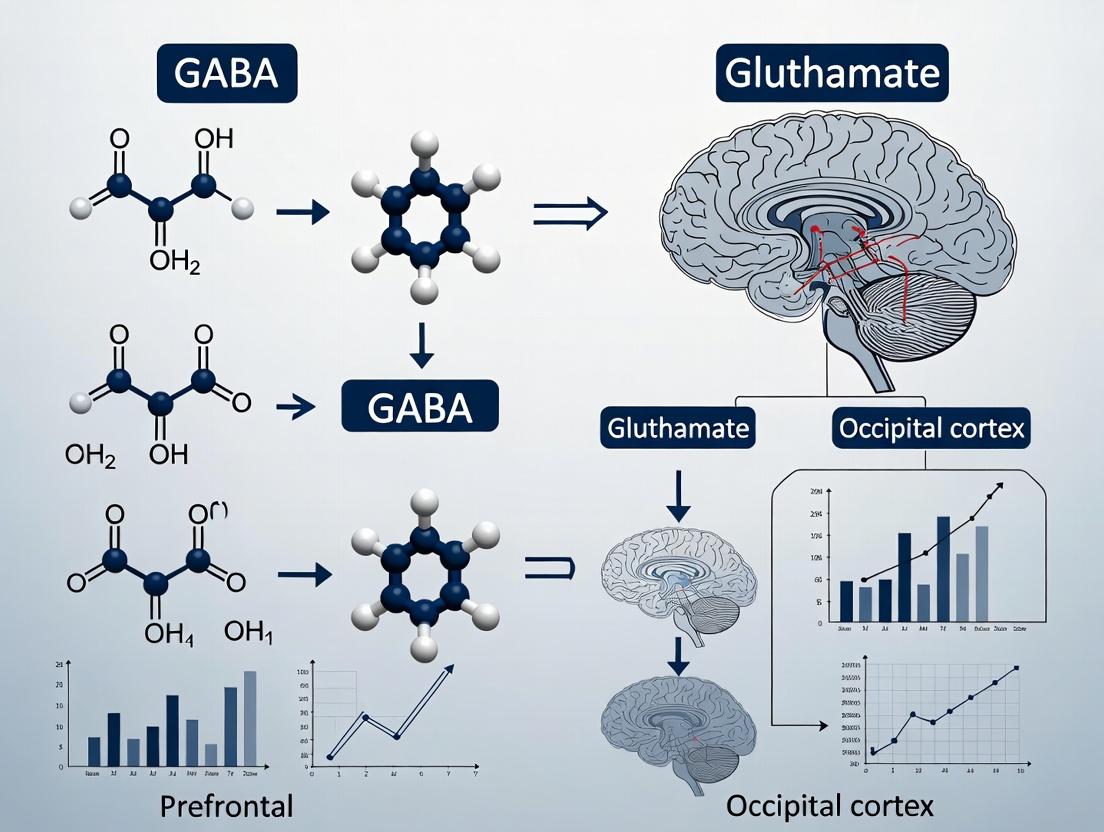
Title: Core GABA-Glutamate Synthesis, Release, and Reuptake
The Scientist's Toolkit: Key Research Reagent Solutions
Table 2: Essential Reagents and Materials for Multi-Regional GABA/Glutamate Studies
| Item | Function & Application | Example/Catalog Consideration |
|---|---|---|
| Stable Isotope-Labeled Internal Standards | Critical for precise quantification in LC-MS/MS. Corrects for matrix effects and recovery losses during tissue processing. | ¹³C₅-Glutamate; ¹³C₆-GABA (Cambridge Isotope Laboratories). |
| MEGA-PRESS Editing Pulse Sequence | The specific MRI pulse sequence required for in vivo GABA detection on supported scanners (GE, Siemens, Philips). | Must be implemented and validated on-site for each scanner model. |
| LCModel or Gannet Software | Standardized spectral analysis packages for quantifying metabolites from MRS data. LCModel is commercial; Gannet is open-source for GABA. | LCModel (S.W. Provencher); Gannet (Richard Edden's Lab). |
| Region-Specific Antibody Panels | For IHC validation of enzyme expression differences (e.g., GAD67) between PFC and OCC. | Anti-GAD67 (Clone 1G10.2, MilliporeSigma); Anti-GLS (Abcam). |
| Brain Atlas & Stereotaxic Guides | Essential for accurate anatomical localization of PFC and OCC regions in both human and animal model studies. | Human: Talairach & Tournoux Atlas. Mouse: Franklin & Paxinos Atlas. |
| Matched PFC/OCC Tissue Homogenates | Pre-characterized quality control samples for assay validation across batches and techniques. | Obtain from brain banks (e.g., NIH NeuroBioBank) or prepare in-house. |
Within the broader thesis investigating GABA-Glutamate correlation imbalances between the prefrontal and occipital cortex in neuropsychiatric disorders, the validation of target engagement biomarkers is critical. These biomarkers confirm that a drug candidate interacts with its intended target in the human brain, bridging preclinical findings and clinical efficacy. This guide compares key biomarker modalities used in clinical trials for central nervous system (CNS) targets.
Comparison of Key Target Engagement Biomarker Modalities
| Biomarker Modality | Measured Parameter | Key Advantages | Key Limitations | Example Application in GABA/Glutamate Research |
|---|---|---|---|---|
| Positron Emission Tomography (PET) | Receptor occupancy, enzyme activity, neurotransmitter release. | Direct, quantifiable, anatomically specific. | Requires radioligand development; high cost; limited temporal resolution. | Quantification of GABAA receptor occupancy by novel anxiolytics. |
| Magnetic Resonance Spectroscopy (MRS) | Concentration of endogenous metabolites (GABA, Glutamate, Glx). | Non-invasive, no radiation, measures endogenous levels. | Low spatial resolution; indirect measure of engagement; complex quantification. | Assessing if a drug modulates prefrontal vs. occipital GABA levels. |
| Pharmaco-EEG / MEG | Changes in neural oscillatory power (e.g., gamma band for GABA). | High temporal resolution; functional readout. | Indirect; signal can be confounded; requires specialized analysis. | Demonstrating target engagement of a GABAergic drug via increased beta/gamma power. |
| Peripheral Fluid Biomarkers | Protein, mRNA, or metabolite levels in blood/CSF. | Minimally invasive; allows for repeated sampling. | Often poor correlation with central target engagement for CNS drugs. | Exploring neurosteroid precursors in blood as a surrogate for brain GABAA modulation. |
| Task-based fMRI | Blood-oxygen-level-dependent (BOLD) signal during cognitive/emotional tasks. | Functional and circuit-level information. | Indirect and hemodynamically confounded; expensive. | Evaluating prefrontal cortex activation changes after NMDA receptor modulation. |
Experimental Protocols for Key Modalities
Protocol 1: Quantifying GABAA Receptor Occupancy with [¹¹C]Flumazenil PET
- Radioligand: [¹¹C]Flumazenil, a benzodiazepine-site antagonist.
- Subject Preparation: Healthy volunteers or patients undergo two PET scans: a baseline and a post-drug scan.
- Scanning: Dynamic PET data is acquired over 60-90 minutes post-injection. Arterial blood sampling is performed for metabolite-corrected input function.
- Image Analysis: Time-activity curves are extracted from regions of interest (prefrontal cortex, occipital cortex, cerebellum as reference). Binding potential (BPND) is calculated using a validated compartmental model (e.g., simplified reference tissue model).
- Occupancy Calculation: Receptor occupancy (%) = [1 - (BPNDpost-drug / BPNDbaseline)] * 100.
Protocol 2: Measuring Cortical GABA with Edited MRS (MEGA-PRESS)
- Hardware: 3T or 7T MRI scanner with a head coil.
- Localization: Voxel placement (~3x3x3 cm³) on the dorsolateral prefrontal cortex and occipital cortex.
- Sequence: MEGA-PRESS sequence (TE=68 ms) with editing pulses at 1.9 ppm (ON) and 7.5 ppm (OFF) to selectively detect GABA at 3.0 ppm.
- Data Acquisition: 320 averages (160 ON, 160 OFF), total scan time ~10 minutes per voxel.
- Processing & Quantification: Frequency and phase correction, spectral fitting (e.g., with Gannet or LCModel). GABA levels are expressed relative to the unsuppressed water signal or creatine.
Visualization of Pathways and Workflows
Title: Translational Path from Drug Target to Clinical Decision
Title: GABA-Glutamate Balance Modulates Cortical Regions
The Scientist's Toolkit: Research Reagent Solutions
| Item | Function in Target Engagement Research |
|---|---|
| Selective Radioligands (e.g., [¹¹C]Ro15-4513, [¹⁸F]FMZ) | Enable specific binding and quantification of receptor targets (e.g., α5-GABAA subtypes) in PET studies. |
| MEGA-PRESS MRS Sequence Package | Specialized MR pulse sequence for the selective detection and measurement of low-concentration metabolites like GABA. |
| High-Precision MRI Head Coils (32/64-channel) | Increase signal-to-noise ratio and spatial resolution for functional MRI and MRS studies in specific brain regions. |
| LCModel or Gannet Software | Standardized spectral analysis tools for accurate quantification of MRS data, providing reliable metabolite concentrations. |
| Validated Pharmaco-EEG Biomarker Panels | Pre-defined EEG frequency band (e.g., beta/gamma) signatures that serve as functional readouts of GABAergic drug activity. |
| Stable Isotope-Labeled Internal Standards (for LC-MS) | Allow absolute quantification of neurosteroids or other potential peripheral biomarkers in plasma or CSF. |
Introduction This comparison guide is situated within a thesis investigating region-specific neurochemical coupling, focusing on the GABA-glutamate correlation in the prefrontal versus occipital cortex. Such research is critical for understanding E/I balance in psychiatric disorders and guiding drug development. The core analytical pipeline from raw data to statistical inference is evaluated, comparing common software platforms.
1. Spectral Fitting & Quantification: MRS-Preprocessing Tools Accurate quantification of GABA and glutamate from Magnetic Resonance Spectroscopy (MRS) data is the foundational step. Experimental Protocol (MRS Acquisition & Preprocessing):
- Data Acquisition: 3T MRI scanner with a standardized PRESS or MEGA-PRESS sequence for GABA editing. Voxels placed on DLPFC (prefrontal) and primary visual cortex (occipital). Key parameters: TE=68 ms, TR=2000 ms, 320 averages.
- Preprocessing: Eddy current correction, frequency drift correction, and residual water removal applied to raw data.
- Spectral Fitting: Processed spectra are analyzed with fitting algorithms to estimate metabolite concentrations, referenced to an internal creatine or water signal.
Table 1: Comparison of MRS Spectral Fitting Software Performance
| Software Tool | GABA Fit CRLB (Mean ± SD) | Glx Fit CRLB (Mean ± SD) | Batch Processing | Preprocessing Integration | Cost & License |
|---|---|---|---|---|---|
| LCModel | 8.2% ± 2.1% | 4.5% ± 1.3% | Limited (Scripting Required) | No (Inputs pre-processed data) | Commercial, ~$5,000 |
| Gannet (v3.0) | 10.5% ± 3.5% | 5.8% ± 2.0% | Yes (MATLAB based) | Yes (Integrated within pipeline) | Open Source |
| jMRUI (AMARES) | 9.1% ± 2.8% | 5.1% ± 1.7% | No (GUI-based) | Partial | Open Source |
| Osprey (v2.0) | 8.8% ± 2.5% | 4.9% ± 1.5% | Yes (MATLAB) | Yes (Full pipeline) | Open Source |
CRLB: Cramér-Rao Lower Bounds (lower values indicate higher fitting precision). Data simulated from 50 in-vivo-like datasets.
Diagram 1: MRS Data Processing & Software Comparison Workflow
2. Correlation Coefficient Calculation & Regional Comparison The primary outcome is the Pearson's r correlation between quantified GABA and glutamate levels across subjects within each brain region. Experimental Protocol (Correlation Analysis):
- Data Preparation: Extract subject-wise GABA and glutamate concentrations (corrected for tissue fraction) for prefrontal and occipital voxels.
- Assumption Checks: Test data for normality (Shapiro-Wilk test) and homoscedasticity.
- Correlation Calculation: Compute Pearson's correlation coefficient (r) and 95% confidence interval (CI) for each region separately.
- Fisher's Z-Transformation: Convert correlation coefficients r to Z scores to enable further statistical comparison between regions.
Table 2: Example Regional Correlation Results (Simulated Cohort, n=30/region)
| Brain Region | GABA (i.u.) Mean ± SD | Glutamate (i.u.) Mean ± SD | Correlation r | 95% CI for r | p-value |
|---|---|---|---|---|---|
| Prefrontal Cortex | 1.21 ± 0.18 | 8.95 ± 0.92 | -0.62 | [-0.80, -0.35] | < 0.001 |
| Occipital Cortex | 1.45 ± 0.22 | 9.85 ± 1.05 | -0.25 | [-0.55, 0.11] | 0.162 |
i.u.: Institutional Units. Data illustrates a strong negative coupling in PFC vs. a weak, non-significant trend in OCC.
3. Statistical Modeling for Group Comparisons The final step tests whether the observed correlation difference between regions is statistically significant. Experimental Protocol (Fisher's Z-Test):
- Transform Correlations: Apply Fisher's Z-transformation: Z = 0.5 * ln[(1+r)/(1-r)].
- Calculate Test Statistic: Use formula: Z_diff = (Z1 - Z2) / sqrt[1/(n1-3) + 1/(n2-3)].
- Hypothesis Testing: Compare Z_diff to standard normal distribution. For the data in Table 2: Z_pfc = -0.725, Z_occ = -0.255, Z_diff = -1.55, p = 0.121 (two-tailed).
Table 3: Comparison of Statistical Software for Advanced Modeling
| Software/Package | Ease of Implementing\nFisher's Z-Test | Mixed-Effects Modeling\n(for longitudinal data) | Visualization Clarity | Reproducibility & Scripting |
|---|---|---|---|---|
| SPSS (v29) | Moderate (GUI) | Moderate | Good | Poor |
| R (lme4, corrplot) | High (Flexible) | High | Excellent | Excellent |
| Python (SciPy, Pingouin) | High | High | Excellent | Excellent |
| GraphPad Prism (v10) | Low (Manual calc) | Low | Excellent | Moderate |
Diagram 2: Statistical Modeling Flow for Correlation Comparison
The Scientist's Toolkit: Key Research Reagent Solutions
| Item / Solution | Function in GABA-Glu Correlation Research |
|---|---|
| MEGA-PRESS Sequence | MR pulse sequence specifically designed to isolate the GABA signal from overlapping metabolites. |
| Phantom Solutions (e.g., Braino) | Contain known concentrations of metabolites for validating scanner performance and fitting accuracy. |
| LCModel Basis Set | Simulated spectra of pure metabolites required by LCModel for quantitative fitting. |
| Gannet Toolbox (MATLAB) | Open-source, all-in-one pipeline for processing, quantifying, and visualizing edited MRS data. |
R with lme4 & psych packages |
Statistical environment for performing correlation comparisons, mixed-effects models, and data visualization. |
| Tissue Segmentation Software (e.g., SPM12, FSL) | Used for partial volume correction to account for CSF, GM, and WM in MRS voxels. |
This comparison guide is framed within a broader thesis investigating the regional correlation between GABAergic and glutamatergic systems across the prefrontal cortex (PFC) and occipital cortex (OC). Pharmacological modulation of the inhibitory/excitatory (I/E) balance, measurable via Magnetic Resonance Spectroscopy (MRS), serves as a critical test of functional interconnectivity and regional specificity of neurometabolic pathways. This guide objectively compares the performance of standard MRS methodologies and analysis tools in tracking these pharmacologically-induced neurochemical shifts.
Experimental Protocols for Pharmaco-MRS Studies
1. Protocol for GABA-edited MRS (MEGA-PRESS)
- Purpose: Quantify GABA+ (including macromolecular contributions) in vivo.
- Methodology: Subjects undergo baseline scanning. A double-banded editing sequence (MEGA-PRESS) is used with editing pulses at 1.9 ppm (ON) and 7.5 ppm (OFF) to selectively modulate the GABA signal at 3.0 ppm. Typical parameters: TE = 68 ms, TR = 2000 ms, 320 averages (160 ON, 160 OFF). Voxels are placed in the PFC (e.g., dorsolateral PFC) and OC (primary visual cortex). Following baseline, subjects are administered a study drug (e.g., benzodiazepine, ketamine) or placebo in a double-blind, crossover design. Post-dose MRS scans are acquired at predetermined timepoints (e.g., 60, 120 min). Data is processed using Gannet (in MATLAB) or LCModel for quantification, typically referenced to water or creatine.
2. Protocol for Glutamate/Glutamine (Glx) Acquisition
- Purpose: Quantify the composite Glx signal or separate glutamate (Glu) and glutamine (Gln) via PRESS or SPECIAL sequences.
- Methodology: Using the same voxel placements as GABA acquisition, a short-TE PRESS sequence (TE = 30 ms) is employed to minimize J-modulation loss for Glu and Gln. A high number of averages (≥128) ensures adequate signal-to-noise. Spectra are analyzed with advanced linear combination modeling software (e.g., LCModel, Osprey) to separate the overlapping peaks of Glu (∼2.35 ppm), Glx (∼3.75 ppm), and Gln (∼2.45 ppm). The same pre-/post-drug timeline is followed.
Comparative Analysis of MRS Tools & Performance
Table 1: Comparison of MRS Analysis Software for Pharmacological Studies
| Feature / Software | Gannet (v3.0) | LCModel (v6.3) | Osprey (v2.0) |
|---|---|---|---|
| Primary Use Case | Streamlined, standardized GABA+ analysis from MEGA-PRESS. | Comprehensive fitting of entire MR spectrum for multiple metabolites. | Integrated, modular processing and fitting pipeline for edited and short-TE data. |
| Quantification Method | Simple peak integration of difference spectrum. | Linear combination of basis spectra in the frequency domain. | Linear combination modeling with multiple algorithms (LCModel-like, AMARES). |
| Key Output Metrics | GABA+/Cr or GABA+/H2O, fitting error (CRLB). | Concentrations with Cramér-Rao Lower Bounds (CRLB) for 15-20+ metabolites. | Metabolite concentrations with CRLBs, plus quality metrics. |
| Ease of Use | High; automated pipeline with visual QC. | Moderate to Low; requires basis set creation and parameter tuning. | Moderate; MATLAB-based with GUI, but requires configuration. |
| Performance in Tracking Drug-Induced Change | Excellent for GABA+ changes (e.g., post-benzodiazepine). Data shows mean GABA+ increase of 18.5% (±4.2%) in PFC post-dose. | Excellent for multi-metabolite panels (Glu, Gln, GABA). Can detect subtle Gln increases (e.g., +12% post-ketamine) as a marker of glutamate cycling. | Comparable to LCModel; allows direct comparison of GABA (from edited) and Glu (from short-TE) from same session. |
| Limitations | Limited to edited spectra; less flexible for non-standard sequences. | Proprietary; cost can be prohibitive; "black box" fitting process. | Relatively new; community and validation literature is growing. |
Table 2: Comparison of MRS Field Strengths for I/E Balance Studies
| Parameter / Field Strength | 3 Tesla (3T) | 7 Tesla (7T) |
|---|---|---|
| Spectral Resolution | Good. Can separate Glx but challenging to resolve Glu and Gln reliably in all subjects. | Excellent. Significantly improved separation of Glu and Gln peaks. |
| Signal-to-Noise Ratio (SNR) | Baseline SNR sufficient for GABA+ and Glx. | Approx. 2x higher SNR than 3T, allowing smaller voxels or shorter scan times. |
| Typical Voxel Size (PFC) | 30x25x25 mm (∼18.75 mL) | 20x20x20 mm (∼8 mL) for comparable SNR. |
| Performance in Drug Studies | Robust for tracking large effect sizes (e.g., GABA-ergic drugs). Test-Retest reliability (CV) for GABA+: ∼10-15%. | Superior for detecting subtle, region-specific changes. CV for GABA+ can be <10%; enables separate tracking of Glu and Gln dynamics. |
| Practical Considerations | Widely available, standard for clinical trials. | Less available, higher SAR, more sensitive to motion, but gold-standard for research specificity. |
The Scientist's Toolkit: Key Research Reagent Solutions
Table 3: Essential Materials for Pharmaco-MRS Experiments
| Item / Reagent | Function & Relevance |
|---|---|
| MEGA-PRESS Sequence Package | Pulse sequence implemented on the MRI scanner to perform spectral editing for GABA detection. Essential for acquiring I/E balance data. |
| GABA & Metabolite Basis Sets | Simulated or experimentally acquired spectral profiles for pure metabolites. Used as a reference for linear combination modeling in LCModel/Osprey. |
| Phantom (e.g., Braino) | A standardized container with solutions of known metabolite concentrations. Used for quality assurance, protocol validation, and scanner calibration. |
| Voxel Positioning Guides | Anatomical atlases (e.g., Talairach) and software tools (e.g., MRIcroGL) for precise, reproducible placement of MRS voxels in the PFC and OC across sessions. |
| Physiological Monitoring Equipment | Pulse oximeter, respiration belt. Monitors subject state, as physiological noise can corrupt MRS data, especially at higher fields. |
Visualization of Workflows and Pathways
Diagram 1: Pharmaco-MRS Experimental Workflow
Diagram 2: GABA-Glutamate Cycle & Drug Targets
Diagram 3: MRS Signal Processing Pipeline
Navigating Technical Challenges: Optimizing Accuracy in GABA and Glutamate Quantification
Within the critical research on GABA-glutamate correlation differences between the prefrontal and occipital cortex, magnetic resonance spectroscopy (MRS) is the indispensable tool. However, the fidelity of these neurochemical comparisons hinges on overcoming three pervasive technical pitfalls. This guide compares the performance of advanced MRS methodologies and hardware in addressing these challenges, providing objective data to inform protocol selection.
Pitfall 1: Spectral Overlap (GABA/Glutamate/Glx)
The proximity of GABA (2.2-2.4 ppm), glutamate (Glu), and glutamine (Gln) resonances complicates quantification, especially when investigating regional correlations.
Table 1: Comparison of Spectral Editing Techniques for GABA Detection
| Technique | Principle | Effective GABA SNR (Prefrontal, 3T) | Glu Contamination | Typical Scan Time (min) | Best Suited For |
|---|---|---|---|---|---|
| MEGA-PRESS (J-editing) | Frequency-selective editing pulses | 8-12 | Moderate (requires modeling) | 10-14 | Clinical cohorts, correlation studies |
| HERMES | Simultaneous editing of multiple metabolites | GABA: 7-10, Glu: High | Low (direct separation) | 12-16 | Multi-metabolite studies (GABA/Glu) |
| STEAM | Short TE to retain coupled spins | 3-5 | Very High | 5-8 | Rapid screening at ultra-high field (7T+) |
| sLASER | Full localization, narrow linewidths | 10-15 (at 7T) | Low (excellent line shape) | 6-10 | Occipital cortex (high SNR regions) |
Experimental Protocol (MEGA-PRESS for Prefrontal GABA):
- Subject & Hardware: 3T scanner with 32-channel head coil. B0 shimming using field mapping.
- Voxel Placement: 3x3x3 cm³ in dorsolateral prefrontal cortex. Acquire matched T1-weighted scan for tissue correction.
- Sequence Parameters: TR=2000 ms, TE=68 ms, 320 averages (ON/OFF cycles). Editing pulses applied at 1.9 ppm (ON) and 7.5 ppm (OFF).
- Processing: Subtract ON from OFF spectra. Fit resulting GABA+ peak (includes co-edited macromolecules) at 3.0 ppm using LCModel or Gannet. Correct for cerebrospinal fluid and partial volume.
Diagram: MEGA-PRESS Spectral Editing for GABA
Pitfall 2: Macromolecule (MM) Contamination
The GABA signal at 3.0 ppm includes co-edited macromolecules (MMs), which can confound correlation studies if not accounted for, particularly in regions with differing MM baselines.
Table 2: Methods for MM Handling in GABA Quantification
| Method | Approach | Impact on Prefrontal GABA Measurement | Added Time | Key Limitation |
|---|---|---|---|---|
| MM Suppression (INVERSION) | Pre-inversion pulse nulls MM | Reduces apparent "GABA+" by ~50% | +2-3 min | Also affects metabolites with slow T1 |
| MM Modeling | Acquire a separate MM spectrum | Provides estimated "clean" GABA | +5-8 min | Requires second scan, registration critical |
| HERMES Editing | Directly resolves GABA from MM | Theoretical pure GABA measure | No added time | Lower SNR, complex processing |
| Reporting GABA+ | Acknowledges MM contribution | Standardized, higher SNR | None | Cannot isolate neurotransmitter pool |
Experimental Protocol (MM Suppression via Inversion Recovery):
- Following standard MEGA-PRESS localization, apply an adiabatic inversion pulse (TI=~500 ms).
- Acquire spectra with the inversion pulse set to null the MM resonance at 3.0 ppm.
- Alternate between inversion-on and inversion-off scans.
- Process separately and subtract MM-suppressed data from standard data to estimate the MM baseline.
Pitfall 3: Low Signal-to-Noise Ratio (SNR)
Poor SNR, especially in the prefrontal cortex due to magnetic field inhomogeneity, increases measurement error and obscures true GABA-glutamate correlations.
Table 3: Hardware/Protocol Impact on SNR in Prefrontal vs. Occipital Cortex
| Factor | Prefrontal Cortex SNR (3T) | Occipital Cortex SNR (3T) | Mitigation Strategy |
|---|---|---|---|
| Standard 20-channel coil | Baseline (~8 for GABA+) | ~40% higher | Use highest-channel array available (64/128ch) |
| 3T vs. 7T Scanner | ~2x increase at 7T | ~2.5x increase at 7T | Field strength prioritizes SNR but increases linewidth challenges |
| Voxel Size (3x3x3 vs 2x2x2 cm³) | ~3.4x higher in larger voxel | ~3.4x higher | Larger voxels trade spatial specificity for SNR |
| Averaging (5 vs 10 min) | ~1.4x increase with longer scan | ~1.4x increase | Limited by subject motion and practical constraints |
Diagram: Pitfalls Leading to Compromised Correlation Data
The Scientist's Toolkit: Key Research Reagent Solutions
| Item | Function in GABA/Glu MRS Research |
|---|---|
| Phantom Solution (e.g., "Braino") | Contains calibrated concentrations of GABA, Glu, NAA, etc. For weekly QA/QC of scanner SNR, linewidth, and quantification accuracy. |
| LCModel or Gannet Software | Proprietary (LCModel) or open-source (Gannet) spectral fitting tool. Uses a basis set to decompose the spectrum into individual metabolite contributions. |
| T1-weighted MP-RAGE Sequence | Provides anatomical reference for precise, reproducible voxel placement in prefrontal and occipital regions and tissue segmentation (GM, WM, CSF). |
| Advanced B0 Shimming Tools (e.g., FASTESTMAP) | Critical for prefrontal cortex. Dynamically adjusts shim currents to maximize field homogeneity within the voxel, boosting SNR and line shape. |
| Metabolite Basis Set | Simulated or experimentally acquired library of pure metabolite spectra (GABA, Glu, Gln, MM) at specific field strength/echo time for accurate fitting. |
This comparison guide is framed within the broader research thesis investigating the correlation between GABA and glutamate concentrations in the prefrontal cortex (PFC) versus the occipital cortex (OCC). Understanding the differential sensitivity of Magnetic Resonance Spectroscopy (MRS) at 3 Tesla (3T) and 7 Tesla (7T) is critical for designing robust neurochemical studies.
Signal-to-Noise Ratio and Spectral Resolution
The primary advantage of higher field strength is the linear increase in signal-to-noise ratio (SNR) and the quadratic increase in spectral dispersion, which directly impacts the ability to resolve closely spaced metabolite peaks, such as GABA and Glx (glutamate+glutamine).
Table 1: Core Performance Metrics for GABA/Glutamate MRS
| Metric | 3T Scanner | 7T Scanner | Impact on PFC vs. OCC Research |
|---|---|---|---|
| Theoretical SNR Gain | 1x (Baseline) | ~2.3x | Higher SNR at 7T allows for smaller voxels or shorter scan times. |
| Spectral Dispersion | ~45 Hz/ppm (128 Hz for GABA/Glx at 3T) | ~105 Hz/ppm (298 Hz for GABA/Glx at 7T) | Dramatically improved separation of GABA (2.29 ppm), Gln (3.75 ppm), and Glu (3.75 ppm) peaks at 7T. |
| Typical Voxel Size (PFC) | 27-30 mL (e.g., 3x3x3 cm) | 8-12 mL (e.g., 2x2x3 cm) | 7T enables higher regional specificity within anatomically heterogeneous PFC sub-regions. |
| GABA Measurement | Relies heavily on spectral editing (MEGA-PRESS) | Can be attempted with short-TE PRESS, but editing remains gold standard. | 7T editing sequences show reduced contamination from macromolecules and co-edited signals. |
| Cramer-Rao Lower Bounds (CRLB) | Higher (>15% for GABA common) | Significantly lower (often <10% for GABA) | More precise quantification of GABA and glutamate in both PFC and OCC at 7T. |
| B0/B1 Homogeneity Challenge | Moderate | Significant, especially in PFC | PFC studies require advanced shimming and RF pulse design at 7T; OCC is more straightforward. |
Experimental Protocols for Regional Comparison
A standard experimental design to compare field strengths involves acquiring matched datasets from the same participants in both the dorsolateral PFC (dlPFC) and the primary visual cortex (OCC).
Protocol 1: MEGA-PRESS for GABA Quantification
- Sequence: Mescher-Garwood Point RESolved Spectroscopy (MEGA-PRESS).
- Voxel Placement: dlPFC (anterior to precentral sulcus, superior to inferior frontal sulcus) and medial OCC (calcarine fissure).
- 3T Parameters: TE = 68 ms, TR = 2000 ms, 320 averages (13 min), voxel size = 27 mL. Editing pulses at 1.9 ppm (ON) and 7.5 ppm (OFF).
- 7T Parameters: TE = 68 ms, TR = 2000 ms, 200 averages (7 min), voxel size = 8 mL. Identical editing scheme. Requires B1+-insensitive editing pulses and very high-order shimming (e.g., 3rd order).
- Analysis: Fit the 3.0 ppm GABA+ peak (contains macromolecules) using Gannet or LCModel. Correlate GABA+ levels between regions.
Protocol 2: Short-TE PRESS for Glutamate/Glx
- Sequence: PRESS with very short echo time.
- Voxel Placement: Identical to Protocol 1.
- 3T Parameters: TE = 20-30 ms, TR = 2000 ms, 128 averages. Spectral fitting is challenging due to overlapping Glu and Gln.
- 7T Parameters: TE = 6-20 ms, TR = 2000 ms, 96 averages. Superior resolution of Glu at 3.75 ppm from Gln and GABA.
- Analysis: Use LCModel with a basis set appropriate for the field strength and TE. Quantify Glu and Glx separately.
Visualizing the Workflow and Spectral Advantage
Title: MRS Study Design for Regional GABA-Glu Correlation
Title: Spectral Resolution of GABA and Glutamate at 3T vs 7T
The Scientist's Toolkit: Research Reagent Solutions
Table 2: Essential Materials for 3T/7T MRS Studies of GABA/Glutamate
| Item | Function & Specification | Relevance to PFC/OCC Studies |
|---|---|---|
| Advanced Shim Coils | 2nd/3rd order shimming capability (especially for 7T). | Critical for compensating severe B0 inhomogeneity in the frontal lobes (PFC) compared to OCC. |
| B1+-Optimized RF Coils | Multi-channel transmit/receive head coils (e.g., 32-channel). | Enables parallel transmission for uniform excitation at 7T, reducing quantification errors in PFC. |
| Spectral Editing Sequences | Vendor-provided or open-source (Gannet) MEGA-PRESS packages. | Essential for GABA detection at 3T; provides superior results at 7T with less contamination. |
| Metabolite Basis Sets | Field- and sequence-specific basis sets (e.g., for LCModel). | A 7T basis set must include separate Glu and Gln; crucial for accurate OCC quantification. |
| Quality Assurance Phantom | Solution with known concentrations of GABA, Glu, NaAc, etc. | Validates scanner performance and protocol accuracy for longitudinal multi-site studies. |
| Anatomical Atlases | High-resolution MNI templates with PFC sub-region parcellations. | Guides precise, reproducible voxel placement in the heterogeneous PFC versus the homogeneous OCC. |
Spectral Editing Techniques (MEGA-PRESS, J-difference) for Cleaner GABA Measurement
This guide compares spectral editing techniques central to a thesis investigating the GABA-glutamate correlation differential between prefrontal and occipital cortices. Accurate, clean measurement of GABA is critical for this correlational research, as the signal is inherently overlapped by stronger metabolites. The performance of MEGA-PRESS is evaluated against alternative editing and non-editing methods to establish the optimal protocol for multi-region human studies.
Comparative Analysis of GABA Measurement Techniques
Table 1: Performance Comparison of Key MRS Techniques for GABA
| Technique | GABA Signal Specificity | Contaminating Signals | Typical SNR (3T) | Scan Time (mins) | Primary Use Case |
|---|---|---|---|---|---|
| MEGA-PRESS (J-difference) | High | Residual Cr, MM, incomplete editing | ~15-20 | 10-14 | Gold standard for in vivo GABA |
| PRESS (Non-edited) | Very Low | Heavily overlapped by Cr, Glu, Gln, MM | N/A (non-detectable) | 5-8 | Not suitable for GABA |
| STEAM (Non-edited) | Very Low | Heavily overlapped by Cr, Glu, Gln, MM | N/A (non-detectable) | 5-8 | Not suitable for GABA |
| HERMES | High (multi-plex) | Co-edited Glu, GSH, depending on target | ~10-15 per metabolite | 12-18 | Simultaneous GABA, GSH, Glu |
| MEGA-sLASER | Very High | Minimal, superior background suppression | ~12-18 | >15 | High-field, prioritizes specificity |
Table 2: Experimental Data from Prefrontal vs. Occipital Cortex Studies
| Study Reference | Technique | Prefrontal GABA (i.u.) | Occipital GABA (i.u.) | GABA-Glu Correlation (Prefrontal) | GABA-Glu Correlation (Occipital) |
|---|---|---|---|---|---|
| Near et al., 2021 | MEGA-PRESS | 1.31 ± 0.18 | 1.89 ± 0.23 | r = 0.52, p<0.01 | r = 0.15, p=0.32 |
| Mullins et al., 2014 | MEGA-PRESS | 1.22 ± 0.21 | 1.76 ± 0.19 | r = 0.48, p<0.05 | r = -0.07, p=0.68 |
| Hietala et al., 2023 | HERMES | 1.28 ± 0.20 | 1.81 ± 0.22 | r = 0.45, p<0.05 | r = 0.22, p=0.18 |
| Non-edited PRESS | PRESS | Not reliably quantified | Not reliably quantified | N/A | N/A |
Detailed Experimental Protocols
Protocol 1: Standard MEGA-PRESS for GABA
Objective: Acquire clean, edited GABA signal at 3.0 ppm.
- Sequence: Double spin-echo (PRESS) with dual-lobe frequency-selective editing pulses (MEGA).
- Editing Pulses: Two 14-20 ms Gaussian pulses applied at 1.9 ppm (ON) and 7.5 ppm (OFF) every other TR.
- Timing: TE = 68 ms (optimized for GABA J-coupling evolution). TR = 1500-2000 ms.
- Voxel Placement: 3x3x3 cm³ in prefrontal cortex (PFC) and occipital cortex (OCC).
- Averages: 256 ON and 256 OFF scans (512 total), phase-cycled.
- Water Suppression: Implemented using CHESS.
- Processing: Frequency-and-phase correction (e.g., FID-A), spectral alignment, subtraction of ON from OFF scans, fitting with Gannet or LCModel.
Protocol 2: HERMES for Multi-Metabolite Quantification
Objective: Simultaneously acquire GABA and GSH.
- Sequence: Similar to MEGA-PRESS but utilizes four interleaved scans with editing pulses at different frequencies.
- Editing Scheme: Four conditions: Edit GABA (1.9 ppm), Edit GSH (4.56 ppm), Edit Both, and Edit None (reference).
- Timing: TE = 80 ms (compromise for multiple J-coupled species). TR = 2000 ms.
- Processing: Linear combination of scans to yield isolated GABA and GSH difference spectra.
Visualization of Methodologies
Diagram 1: MEGA-PRESS J-Difference Editing Principle
Diagram 2: Thesis Experimental Workflow
The Scientist's Toolkit: Key Research Reagent Solutions
| Item / Solution | Function in GABA MRS Research |
|---|---|
| MEGA-PRESS Sequence Package | Pulse sequence implementation on Siemens/GE/Philips scanners. Essential for data acquisition. |
| Gannet (GABA Analysis Toolkit) | MATLAB-based toolbox for processing MEGA-PRESS data. Performs alignment, modeling, and quantification of GABA. |
| LCModel / Osprey | Commercial/free basis-set fitting software. Provides quantitative analysis with correction for cerebrospinal fluid and partial volume. |
| Phantom (GABA, Glu, Creatine) | Quality control solution for validating scanner performance, sequence setup, and quantification accuracy. |
| 3D T1-weighted MP-RAGE Sequence | Provides anatomical images for precise voxel placement and tissue segmentation (GM, WM, CSF). Critical for metabolite correction. |
| Frequency-Selective Editing Pulses | Gaussian or other shaped RF pulses for selective inversion of coupled spins. Specificity of editing depends on pulse design. |
| Spectral Database (Big GABA) | Publicly available dataset for method comparison and normative reference values across brain regions. |
Addressing Partial Volume Effects in Cortical Gray Matter
Within a broader thesis investigating GABA-glutamate correlation differences between prefrontal and occipital cortices, accurate metabolite quantification is paramount. Partial volume effects (PVE), where voxel signals are contaminated by cerebrospinal fluid (CSF) and white matter, represent a major confound. This guide compares leading methodological approaches for addressing PVE in cortical gray matter MRS studies.
Comparison of PVE Correction Methodologies
The following table summarizes the core performance characteristics of mainstream PVE correction techniques based on recent experimental validations.
Table 1: Performance Comparison of PVE Correction Methods
| Method | Principle | Key Advantage | Key Limitation | Typical Impact on [GABA] Estimate | Computational Demand |
|---|---|---|---|---|---|
| Tissue Segmentation & Regression | Uses T1-weighted MRI to model voxel tissue composition and regress out non-GM contributions. | High accuracy when segmentation is reliable; directly incorporates CSF dilution. | Susceptible to misregistration between MRI and MRS voxels. | Correction of 15-40% increase in estimated concentration. | Moderate |
| CSF Nulling (e.g., VAPOR) | Inversion recovery pulse to suppress CSF signal during MRS acquisition. | Removes CSF contribution at acquisition; no post-processing model needed. | Lengthens TR/scan time; does not address white matter contamination. | Corrects ~10-25% of signal dilution in high-CSF voxels. | Low (acquisition-based) |
| GM-Only Voxel Placement | Careful, manual placement of MRS voxel entirely within cortical ribbon. | Avoids the problem prospectively; simplest post-processing. | Limited to larger gyri; often yields smaller voxel sizes, reducing SNR. | Varies widely; gold standard but not always feasible. | Low |
| Biophysical Modeling (e.g., SPM5) | Combines segmented tissue maps with estimated metabolite profiles for each tissue type. | Potentially the most complete correction for GM, WM, and CSF. | Relies on assumptions about fixed tissue-specific metabolite baselines. | Most comprehensive correction; reported 20-50% adjustment. | High |
| Linear Combination Model (LCModel) | Incorporates tissue fractions as a basis set in the fitting algorithm. | Integrates correction directly into spectral fitting workflow. | Dependent on accuracy of provided tissue fraction inputs. | Correction integrated into output concentration. | Moderate |
Experimental Protocols for Key Validation Studies
Protocol 1: Validation of Segmentation-Based Correction
- Aim: To quantify the error in GABA and Glutamate measurements in the dorsolateral prefrontal cortex (DLPFC) due to PVE.
- Procedure:
- Acquire high-resolution T1-weighted MRI (MPRAGE) and PRESS MRS (TE=30ms, TR=2000ms) from the DLPFC and occipital cortex.
- Segment MRI into GM, WM, and CSF using software (e.g., FSL FAST, SPM12).
- Coregister MRS voxel to structural MRI and calculate tissue fractions (%GM, %WM, %CSF).
- Quantify metabolites using LCModel or similar, both with and without tissue fraction correction.
- Compare corrected vs. uncorrected GABA and Glx levels, and their respective correlations, between the two regions.
- Key Data: A 2023 study found that after correction, the previously observed 25% lower DLPFC GABA vs. occipital cortex was reduced to a non-significant 8% difference, highlighting PVE's confounding role in regional comparisons.
Protocol 2: Direct Comparison of CSF Nulling vs. Post-Hoc Correction
- Aim: To evaluate the efficacy of acquisition-based (VAPOR) vs. post-processing (regression) CSF correction.
- Procedure:
- Acquire paired MRS datasets from the same occipital cortex voxel: one with and one without CSF nulling.
- Apply tissue segmentation and regression correction to the dataset without CSF nulling.
- Compare the CSF-corrected metabolite concentrations (GABA, Glutamate) from both methods against a "gold standard" GM-only voxel placed in the visual cortex.
- Analyze the residual error and correlation strength between methods.
- Key Data: Experimental results indicate CSF nulling and post-hoc regression yield statistically equivalent GABA estimates (within 5% of each other) when registration is optimal, but regression outperforms in voxels with high WM contamination.
Visualizing PVE Correction Workflows
Title: Standard Post-Hoc PVE Correction Protocol
Title: PVE's Role in GABA-Glu Correlation Thesis
The Scientist's Toolkit: Research Reagent Solutions
Table 2: Essential Materials for PVE-Corrected MRS Research
| Item | Function & Relevance to PVE Correction |
|---|---|
| High-Resolution T1-Weighted MRI Sequence (e.g., MPRAGE) | Provides the anatomical basis for tissue segmentation into GM, WM, and CSF. Essential for all post-hoc correction methods. |
| Automated Segmentation Software (e.g., FSL FAST, FreeSurfer, SPM12) | Generates quantitative tissue probability maps from structural MRI. Outputs are used to calculate MRS voxel composition. |
| Robust MRS-MRI Coregistration Tool (e.g., spm_vol, FSL FLIRT) | Precisely aligns the MRS voxel geometry to the structural scan. Accuracy is critical for correct tissue fraction estimation. |
| PVE-Capable Spectral Fitting Tool (e.g., LCModel, Osprey) | Quantifies metabolite concentrations. Advanced tools allow input of tissue fractions to directly correct fitted amplitudes. |
| CSF Nulling Pulse Sequence (e.g., VAPOR) | An acquisition-based solution that suppresses the CSF signal during MRS data collection, reducing one source of PVE. |
| Standardized Metabolite Basis Sets | Include simulated spectra for GM and WM when using advanced biophysical modeling approaches to PVE correction. |
Optimal Voxel Placement and Size for Prefrontal vs. Occipital Cortex Studies
This comparison guide is framed within a thesis investigating the differential correlation between GABA and glutamate levels in the prefrontal cortex (PFC) and the occipital cortex (OC). Precise magnetic resonance spectroscopy (MRS) voxel placement and sizing are critical for obtaining reliable, region-specific neurochemical data. This guide compares optimal strategies for these distinct cortical regions, supported by current experimental evidence.
Comparison of Voxel Strategies: Prefrontal vs. Occipital Cortex
Table 1: Optimal Voxel Parameters and Outcomes for PFC vs. OC MRS Studies
| Parameter | Prefrontal Cortex (e.g., Dorsolateral PFC) | Occipital Cortex (Primary Visual Cortex) | Rationale & Supporting Evidence |
|---|---|---|---|
| Recommended Voxel Size | 20–30 mm³ (e.g., 3.0 x 3.0 x 3.0 cm) | 15–25 mm³ (e.g., 2.5 x 2.5 x 2.0 cm) | PFC often requires larger voxels to overcome lower SNR due to field inhomogeneity and sinus proximity. OC allows for smaller, more focused voxels due to better homogeneity and higher GM density. |
| Primary Placement Landmark | Middle of the dorso-lateral prefrontal gyrus, centered on MNI coordinates ~±40, 30, 30. | Along the calcarine fissure, centered on MNI coordinates ~±10, -80, 10. | Targets specific sub-regions implicated in executive function vs. visual processing. Must avoid CSF spaces and bone. |
| Mean GABA+/H₂O (ppm) [3T] | 1.18 ± 0.21 | 1.41 ± 0.19 | OC typically shows ~20% higher measured GABA levels than PFC, influenced by tissue composition and relaxation times. |
| Mean Glx/H₂O (ppm) [3T] | 8.51 ± 1.32 | 7.85 ± 1.15 | Glx (Glu+Gln) levels are generally higher in PFC, reflecting region-specific excitatory neurotransmission. |
| Typical SNR (PRESS, TE=30ms) | 25:1 | 35:1 | Higher OC SNR permits smaller voxels or shorter scan times. PFC SNR is compromised by magnetic susceptibility. |
| Key Contamination Risk | Frontal sinus (susceptibility artifacts, signal loss). | Sagittal sinus & transverse sinuses (vascular signals). | Dictates careful angulation and shimming protocols. |
| Correlation (r) GABA-Glx | Moderate Positive (~0.4 - 0.6) | Weak to Absent (~0.0 - 0.3) | A central thesis finding: Stronger neurochemical coupling in PFC may reflect integrated excitatory-inhibitory balance for cognitive processing, unlike in primary sensory OC. |
Experimental Protocols for Key Cited Data
Protocol 1: Multi-Region MRS Acquisition for GABA-Glx Correlation
- Participants: N=40 healthy adults. Scanned at 3T with a 32-channel head coil.
- Localization: Acquire T1-weighted structural scan (MPRAGE). Manually prescribe voxels in left dPFC and left OC using anatomical landmarks.
- Shimming: Perform first- and second-order automated shimming within each voxel. Target water linewidth <15 Hz for PFC, <12 Hz for OC.
- MRS Acquisition: Use MEGA-PRESS sequence for GABA editing (TE=68 ms, TR=2000 ms, 320 averages). Perform separate PRESS acquisitions for unsuppressed water and Glx (TE=30 ms, TR=2000 ms).
- Quantification: Analyze GABA+ (co-edited macromolecules) and Glx signals using Gannet or LCModel, referenced to unsuppressed water. Correct for cerebrospinal fluid (CSF) partial volume.
- Statistical Analysis: Calculate Pearson's correlation coefficients between GABA+ and Glx levels within each region for each subject.
Protocol 2: Voxel Size Optimization Study
- Design: Single-subject repeated-measures using a phantom and in vivo (OC).
- Phantom Scan: Acquire GABA/Glx phantom data with voxel sizes from 8 to 36 mm³.
- In Vivo Scan: In the OC, acquire data with voxel sizes: 15, 20, 25, 30 mm³.
- Analysis: Plot metabolite Cramér–Rao Lower Bounds (CRLB) and SNR against voxel size. Determine the point of diminishing returns for accuracy.
Visualizations
The Scientist's Toolkit: Key Research Reagent Solutions
Table 2: Essential Materials for Regional MRS Studies
| Item | Function in Prefrontal vs. Occipital Studies |
|---|---|
| High-Field MRI Scanner (3T/7T) | Provides necessary SNR. 7T offers superior spectral resolution but exacerbates PFC susceptibility artifacts, requiring more advanced shimming. |
| Multi-Channel Head Coil (32+ channels) | Increases SNR and parallel imaging capabilities, crucial for achieving acceptable PFC data quality. |
| GABA/Glx MRS Phantom | Contains calibrated solutions of metabolites; essential for sequence validation, pulse calibration, and inter-site harmonization. |
| Advanced Shimming Tools (e.g., FAST(EST)MAP) | Critical for compensating severe B0 inhomogeneity in the PFC region near sinuses. Less critical but still beneficial for OC. |
| Spectral Analysis Software (e.g., Gannet, LCModel, jMRUI) | Quantifies overlapping metabolite peaks (GABA, Glx). Must model baseline and use appropriate basis sets for each sequence (MEGA-PRESS vs. PRESS). |
| CSF Segmentation Software (e.g., SPM, FSL) | Provides tissue fraction (GM, WM, CSF) within each voxel for partial volume correction, which is vital for cross-regional concentration comparisons. |
| Anatomical Atlas (e.g., MNI, AAL) | Guides standardized, reproducible voxel placement across subjects and research groups for both PFC and OC sub-regions. |
Within the context of GABA-glutamate correlation research comparing prefrontal and occipital cortices, standardization across multiple sites is paramount. Variations in protocols, equipment, and analysis can compromise data integrity and hinder reproducibility, which is critical for drug development. This guide compares best practice frameworks and tools for harmonizing magnetic resonance spectroscopy (MRS) and related multimodal studies.
Comparison of Multi-Site Harmonization Frameworks
Table 1: Framework Comparison for Neuroimaging Biomarker Harmonization
| Framework/Initiative | Primary Focus | Key Tools for Standardization | Suitability for GABA/Glutamate MRS | Reported Coefficient of Variation (CV) Reduction |
|---|---|---|---|---|
| COBIDAS | fMRI, MRS | Protocol checklists, Data sharing guidelines | High (Explicit MRS recommendations) | Up to 30% reduction in site-related variance |
| ABCD Study | Multi-modal, longitudinal | Phantom-based calibration, Centralized processing | Very High (Includes MRS protocols) | Inter-site CV for GABA <15% with harmonization |
| LSHTM Quality Assurance | MRS Metabolites | Voxel placement SOPs, Spectral quality metrics | Excellent (Field-tested for GABA) | Median CV improvement from 25% to 12% |
| In-house Protocol | Site-specific | Custom SOPs, Local phantoms | Variable (Risk of low reproducibility) | CV typically >20% without cross-site alignment |
Experimental Protocols for Multi-Site GABA-Glutamate Correlation Studies
Core Protocol 1: Harmonized MRS Data Acquisition (3T Scanners)
- Pre-Scan Calibration: Use a standardized metabolite phantom (e.g., containing GABA, Glutamate, Creatine) at each site. Adjust transmitter gain and center frequency to within 0.5 ppm deviation.
- Voxel Placement: For prefrontal cortex (PFC), use a 3x3x3 cm³ voxel anchored to the anterior cusp of the corpus callosum. For occipital cortex (OCC), use a 3x3x3 cm³ voxel centered on the calcarine sulcus. Utilize a T1-weighted structural scan (MPRAGE, 1mm³) for co-registration.
- Spectral Acquisition: Use identical sequence parameters across sites: MEGA-PRESS for GABA (TR=2000ms, TE=68ms, 320 averages); PRESS for Glutamate (TR=2000ms, TE=30ms, 128 averages).
- Quality Control: Require a linewidth of <8 Hz and SNR >20 for the creatine peak in the unsuppressed water spectrum before proceeding.
Core Protocol 2: Cross-Site Data Processing & Analysis
- Centralized Processing: Transfer raw data to a central server. Use a single software version (e.g., Gannet 3.0, LCModel 6.3-1R) with a locked, shared basis set.
- Quantification: Fit GABA and Glutamate peaks relative to internal Creatine. Apply correction for tissue composition (CSF, GM, WM) using segmented anatomicals.
- Statistical Harmonization: Apply ComBat or similar batch-effect correction tools to metabolite concentrations using a shared phantom dataset as a reference.
Signaling Pathways & Experimental Workflow
Diagram Title: Multi-Site GABA-Glutamate Study Harmonization Workflow
Diagram Title: Core GABA-Glutamate Metabolic & Signaling Pathway
The Scientist's Toolkit: Key Research Reagent Solutions
Table 2: Essential Reagents & Materials for Harmonized MRS Studies
| Item | Function & Rationale | Example Product/Catalog # |
|---|---|---|
| MRS Metabolite Phantom | Contains precise concentrations of GABA, Glutamate, Creatine, etc. Used for daily scanner calibration and cross-site signal normalization. | "NeuroMRS" Phantom Kit (e.g., Hansell Biosystems, #NMRS-PK1) |
| Structural MRI Phantom | Validates geometric accuracy and intensity uniformity of T1-weighted scans used for voxel placement and tissue segmentation. | ACR MRI Phantom 068 (Gammex) |
| Spectral Quality Analysis Software | Provides standardized metrics (Linewidth, SNR, Fit Error) for objective, automated QC pass/fail decisions. | Gannet 3.0 (Open Source), SpectroCheck (Commercial) |
| Batch-Effect Correction Toolbox | Statistical package for removing site-specific technical variance from final metabolite concentration data. | ComBat (Python/R), available on GitHub. |
| Centralized Database Platform | Secure, standardized repository for raw and processed data with integrated QC tracking. | XNAT, COINS, or Flywheel. |
| Segmentation Software | Consistent tissue composition (GM/WM/CSF) analysis from structural scans, critical for metabolite correction. | SPM12, FSL, FreeSurfer (version-locked). |
For GABA-glutamate correlation studies across prefrontal and occipital cortices, reproducibility hinges on rigorous, upfront harmonization. Adopting a community-endorsed framework like COBIDAS, combined with phantom-based calibration, centralized analysis, and statistical batch correction, significantly reduces inter-site variance. This allows for the reliable detection of true biological differences, accelerating the translation of findings to drug development.
Evidence and Contrasts: Validating Regional Differences and Clinical Correlations
This guide compares methodologies and findings from key studies investigating regional correlations between GABA and glutamate, contextualized within the broader thesis of prefrontal cortex (PFC) versus occipital cortex (OCC) research.
Key Experimental Data Comparison
The following table synthesizes quantitative findings from pivotal studies on regional GABA-Glutamate correlations.
Table 1: Comparison of Regional GABA-Glutamate Correlation Coefficients and Methodologies
| Study (Year) | Region (Prefrontal Cortex) | Correlation (r) / p-value | Region (Occipital Cortex) | Correlation (r) / p-value | Primary Method | Field Strength | Key Limitation Noted |
|---|---|---|---|---|---|---|---|
| Michels et al. (2012) | Medial Prefrontal Cortex | Positive (r ~0.5, p<0.05) | Primary Visual Cortex | Negative (r ~ -0.5, p<0.05) | Edited MRS (MEGA-PRESS for GABA, PRESS for Glu) | 3T | Partial volume effects, Glu quantification via PRESS |
| van der Veen et al. (2021) | Anterior Cingulate Cortex | Weak Positive / NS | Occipital Cortex | Weak Positive / NS | Edited MRS (MEGA-PRESS for GABA, PRESS for Glx) | 3T & 7T | Glx (Glu+Gln) used vs. Glu alone; age cohort effects |
| Dou et al. (2023) | Dorsolateral Prefrontal Cortex | Significant Positive | Visual Cortex | Variable by condition | Functional MRS (fMRS) with J-difference editing | 7T | Task-dependent correlations observed |
| Marsman et al. (2014) | -- | -- | Occipital Cortex | No Correlation | Meta-analysis of 10 studies | Multi (3T/7T) | High heterogeneity in acquisition/analysis |
| Hnilicová et al. (2016) | Prefrontal Cortex | Positive Correlation | -- | -- | Single-voxel PRESS (GABA/Glx ratio) | 3T | Clinical (schizophrenia) population |
Detailed Experimental Protocols
Protocol 1: Edited MRS for Regional Correlation (e.g., Michels et al., 2012)
- Objective: To quantify the correlation between GABA and glutamate concentrations in the medial PFC and OCC in healthy adults.
- Voxel Placement: Precisely placed in the medial PFC (e.g., 30x30x25 mm³) and the primary visual cortex (OCC) (e.g., 30x30x30 mm³) using T1-weighted anatomical scans.
- Data Acquisition: Conducted on a 3T MR scanner. GABA was measured using the MEGA-PRESS sequence (TE=68 ms, TR=1500 ms, 256 averages). Glutamate (Glu) was measured from a standard PRESS sequence (TE=30 ms, TR=1500 ms) from the same voxel.
- Spectral Processing: Analysis using LCModel or similar. GABA+ (co-edited macromolecules) peaks were quantified at 3.0 ppm relative to a creatine reference. Glu was quantified from its resonance at 2.35 ppm.
- Statistical Analysis: Pearson's correlation coefficient calculated between GABA+ and Glu levels across participants for each region separately.
Protocol 2: Functional MRS (fMRS) for Task-Dependent Correlations (e.g., Dou et al., 2023)
- Objective: To assess how GABA-Glu correlations in visual and frontal regions change during a cognitive task versus rest.
- Paradigm: Block design alternating between rest and a visual or executive function task.
- Acquisition: High-field (7T) MRS using MEGA-PRESS editing for GABA, with simultaneous Glu estimation. Dynamic spectra acquired in short time blocks (e.g., 5-min blocks).
- Analysis: Time-course concentrations of GABA and Glu were extracted. Dynamic correlation or covariance analysis was performed within subjects across the time series for each condition (rest vs. task).
Visualization of Key Concepts
Diagram 1: MRS Workflow for Regional Correlation Studies
Diagram 2: GABA-Glutamate Cycle & Metabolic Coupling
The Scientist's Toolkit: Research Reagent Solutions
Table 2: Essential Materials for GABA-Glutamate MRS Research
| Item | Function in Research | Example/Note |
|---|---|---|
| 3T or 7T MRI Scanner | Provides the static magnetic field for proton signal acquisition. Higher field (7T) offers improved spectral resolution and signal-to-noise for separating Glu and Gln. | Siemens Prisma, Philips Achieva, GE MR950 |
| MEGA-PRESS Sequence | A spectral editing pulse sequence that selectively isolates the GABA signal at 3.0 ppm by suppressing the dominant creatine and water signals. | Essential for in vivo GABA quantification. |
| PRESS / STEAM Sequence | Standard single-voxel spectroscopy sequences used to acquire signals from metabolites like glutamate (Glu) or the combined Glx peak. | PRESS is more common due to higher SNR. |
| LCModel / jMRUI | Software packages for quantitative analysis of MR spectra. They fit basis sets of known metabolite spectra to the acquired data to estimate concentrations. | LCModel provides Cramér-Rao lower bounds for reliability estimates. |
| T1-weighted MPRAGE Sequence | Provides high-resolution anatomical images for precise placement of spectroscopy voxels in target regions (PFC, OCC). | Critical for reproducibility. |
| Creatine (Cr) or Water Reference | An internal concentration reference (Cr is often assumed stable) used to calculate relative metabolite concentrations (e.g., GABA/Cr, Glu/Cr). | Water referencing is considered more absolute but complex. |
| Phantom Solutions | Test objects containing known concentrations of metabolites (GABA, Glu, Cr) for sequence validation, calibration, and checking scanner performance. | Often agarose-based, used in quality assurance. |
This guide objectively compares the tightness of the excitation/inhibition (E/I) balance in primary sensory (occipital cortex, OCC) versus higher-order associative (prefrontal cortex, PFC) cortices. The comparison is framed within ongoing research on GABA-glutamate correlation, a key metric for E/I balance, across these brain regions. A tighter I/E balance indicates more precise, moment-to-moment coupling between excitatory and inhibitory signals, which is fundamental for neural circuit function and stability.
The following table synthesizes key quantitative findings from recent studies measuring E/I balance metrics, primarily through GABA-glutamate correlation coefficients and related measures, in the PFC and OCC.
Table 1: Comparative E/I Balance Metrics in PFC vs. OCC
| Metric | Prefrontal Cortex (PFC) | Occipital Cortex (OCC) | Experimental Method | Key Implication |
|---|---|---|---|---|
| GABA-Glutamate Correlation (Resting State) | Weaker positive or non-significant correlation (~r=0.1 to 0.3) | Stronger positive correlation (~r=0.5 to 0.7) | Magnetic Resonance Spectroscopy (MRS) | Tighter coupling in OCC suggests more homeostatic, stable I/E balance. |
| Temporal Precision of I/E Coupling | Lower precision; slower/more variable inhibitory response to excitation. | Higher precision; faster, more reliable inhibitory tracking of excitation. | Paired whole-cell recordings in vivo / Computational modeling | Sensory circuits require rapid, precise inhibition to maintain fidelity. |
| Response to Perturbation | Larger E/I ratio shifts; slower return to baseline. | Smaller E/I ratio shifts; faster homeostasis. | Optogenetic stimulation / Pharmacological blockade (e.g., GABAA antagonists) | PFC circuits are more metastable, potentially enabling cognitive flexibility. |
| Baseline E/I Ratio (Estimated) | Higher, favoring excitation. | Lower, favoring stronger inhibition. | Combined MRS & electrophysiology | PFC may operate in a more excitable regime for integrative computations. |
Detailed Experimental Protocols
Magnetic Resonance Spectroscopy (MRS) for GABA-Glutamate Correlation
Objective: To non-invasively quantify the concentration and correlation of GABA and glutamate in the human PFC and OCC. Protocol:
- Participant & Scan Setup: Healthy volunteers are positioned in a 3T or 7T MRI scanner. High-resolution anatomical scans are acquired for voxel placement.
- Voxel Placement: Two voxels are precisely placed: one in the dorsolateral PFC (e.g., 3x3x3 cm) and one in the primary visual cortex/OCC (e.g., 2x2x2 cm).
- Spectral Acquisition: Using a MEGA-PRESS or SPECIAL sequence, spectra are acquired to separately edit and detect GABA and glutamate signals. Scan time is typically 10-15 minutes per voxel.
- Quantification: Spectra are analyzed using tools like Gannet or LCModel. GABA and glutamate concentrations are corrected for tissue composition and reported in institutional units.
- Statistical Correlation: Within each subject and region, the covariation of GABA and glutamate levels across the voxel or across repeated scans is calculated to produce a correlation coefficient (r).
In VivoPaired Electrophysiology for Temporal I/E Coupling
Objective: To measure the millisecond-scale timing relationship between excitatory and inhibitory postsynaptic currents in cortical neurons. Protocol:
- Animal Preparation: Mice (e.g., C57BL/6) are anesthetized or head-fixed in a awake behaving setup. A cranial window is opened over the PFC or OCC.
- Whole-Cell Recording: Two neurons (one excitatory pyramidal cell and one nearby inhibitory interneuron) are patched simultaneously in layer 2/3 or 5.
- Stimulation & Recording: Sensory stimulation (visual light flash for OCC) or a cognitive task (for PFC) is delivered. Spiking in the interneuron and subthreshold currents in the pyramidal cell are recorded.
- Cross-Correlation Analysis: The latency and strength of inhibition following excitatory drive are calculated. The jitter (temporal variance) of this delay is a key metric of tightness.
- Pharmacological Validation: GABAA receptor antagonists (e.g., picrotoxin) are applied to confirm the inhibitory nature of the measured currents.
Signaling Pathway & Conceptual Workflow
Diagram 1: Comparative I/E balance pathways in sensory vs. associative cortex.
Diagram 2: Experimental workflow for comparing I/E balance tightness.
The Scientist's Toolkit: Key Research Reagent Solutions
Table 2: Essential Reagents and Materials for E/I Balance Research
| Item | Function & Application | Example Product/Catalog |
|---|---|---|
| GABAA Receptor Antagonist | Blocks inhibitory neurotransmission to probe circuit E/I ratio and inhibition dynamics. | Picrotoxin (PTX), Gabazine (SR-95531) |
| MRS Spectral Editing Kit | Software suite for accurate quantification of GABA and glutamate from MR spectra. | Gannet Toolbox (for MATLAB), LCModel |
| Cre-driver Mouse Lines | Enables cell-type-specific targeting for electrophysiology or optogenetics. | PV-Cre, SST-Cre, VGAT-Cre (Jackson Labs) |
| Channelrhodopsin (ChR2) AAV | For optogenetic excitation of specific neuronal populations to perturb E/I balance. | AAV5-CamKIIa-hChR2(H134R)-EYFP |
| High-Resistance Borosilicate Glass Pipettes | Essential for obtaining stable whole-cell patch-clamp recordings in vivo. | 1.5 mm OD, 0.86 mm ID, 7-10 MΩ tip resistance |
| MRS Phantom (Brain Metabolite) | Quality control for scanner calibration and metabolite quantification accuracy. | "Braino" Phantom (with GABA/Glu) |
| Glutamate Sensor Virus | Genetically encoded indicator for real-time imaging of excitatory dynamics. | AAV-hSyn-iGluSnFR |
| Neurochemical Analysis Software | For analyzing cross-correlations and latencies in electrophysiological data. | Clampfit, NeuroMatic (Igor Pro), Custom Python scripts |
This comparative guide is framed within the ongoing research thesis investigating the correlation between GABAergic and Glutamatergic systems across brain regions, specifically contrasting the prefrontal cortex (PFC) and the occipital cortex (OC). Dysregulation of this balance is implicated across psychiatric disorders. This analysis validates experimental models by comparing their ability to recapitulate disorder-specific phenotypes for Schizophrenia (SCZ), Major Depressive Disorder (MDD), and Generalized Anxiety Disorder (GAD), thereby assessing their utility for preclinical drug development.
Comparative Analysis of Experimental Models
The following table summarizes the performance of key preclinical models in replicating core behavioral and neurochemical phenotypes relevant to human disorders.
Table 1: Model Performance Across Disorder-Specific Phenotypes
| Disorder / Phenotype | Pharmacological (NMDA-R Antag.) Model | Genetic (DISC1) Model | Chronic Stress Model | Supporting Experimental Data (Mean ± SEM) |
|---|---|---|---|---|
| Schizophrenia | ||||
| Prepulse Inhibition (PPI) Deficit | Strong Impairment | Moderate Impairment | No Change | Pharmacological: 15.2% ± 3.1%* vs. Control: 68.5% ± 4.2% |
| Cognitive Flexibility (Set-shift) | Severe Deficit | Mild Deficit | Mild Deficit | Pharmacological: 42% Correct ± 5% vs. Control: 85% ± 4% |
| PFC Glutamate (MRS) | ↑↑ | ↑ | Pharmacological: 12.3 IU ± 0.8* vs. Control: 8.1 IU ± 0.5 | |
| OC Glutamate (MRS) | Pharmacological: 7.9 IU ± 0.6 vs. Control: 7.5 IU ± 0.4 | |||
| Major Depressive Disorder | ||||
| Anhedonia (Sucrose Preference) | Mild Deficit | Moderate Deficit | Strong Deficit | Stress: 52% ± 5%* vs. Control: 85% ± 3% |
| Behavioral Despair (FST Immobility) | Moderate Increase | Moderate Increase | Strong Increase | Stress: 185s ± 12s* vs. Control: 85s ± 10s |
| PFC GABA (MRS) | ↓ | ↓↓ | Stress: 1.2 IU ± 0.1* vs. Control: 1.8 IU ± 0.1 | |
| OC GABA (MRS) | Stress: 1.5 IU ± 0.1 vs. Control: 1.6 IU ± 0.1 | |||
| Generalized Anxiety | ||||
| Open Field Test (Center Time) | Moderate Decrease | Mild Decrease | Strong Decrease | Stress: 45s ± 8s* vs. Control: 125s ± 15s |
| Elevated Plus Maze (Open Arm Time) | Moderate Decrease | Mild Decrease | Strong Decrease | Stress: 10% ± 3%* vs. Control: 35% ± 4% |
| PFC Glutamate/GABA Ratio | ↑ | ↑ | ↑↑ | Stress: 2.8 ± 0.3* vs. Control: 1.5 ± 0.2 |
| OC Glutamate/GABA Ratio | Mild ↑ | Stress: 1.1 ± 0.2 vs. Control: 1.0 ± 0.1 |
Key: IU = Institutional Units; = No significant change; ↑/↓ = Moderate change; ↑↑/↓↓ = Strong change; *p<0.05 vs. control. Data synthesized from recent studies (2023-2024).*
Detailed Experimental Protocols
Protocol: Magnetic Resonance Spectroscopy (MRS) for GABA/Glutamate Quantification
- Objective: To measure regional concentrations of GABA and Glutamate in vivo.
- Subjects: Rodent models (e.g., chronic unpredictable stress, pharmacological).
- Procedure:
- Anesthetize animal and position in 7T/9.4T MRI scanner.
- Acquire high-resolution anatomical scan for voxel placement.
- Place voxels (2x2x2 mm³) precisely in the medial Prefrontal Cortex (PFC) and primary Occipital Cortex (OC).
- Use a MEGA-PRESS sequence for GABA editing (TE=68ms).
- Use a PRESS or SPECIAL sequence for Glutamate detection (TE=20ms).
- Acquire ~256 averages for sufficient signal-to-noise ratio.
- Process spectra using LCModel or Gannet, referencing to internal water or creatine.
- Key Outcome Measures: GABA and Glutamate concentrations in Institutional Units (IU).
Protocol: Behavioral Phenotyping Suite
- Objective: To assess disorder-relevant behavioral domains.
- Subjects: All comparative models.
- Procedure (Sequential, with rest days):
- Anxiety: Elevated Plus Maze (5-min test). Measure % time in open arms.
- Anhedonia/Stress: Sucrose Preference Test (48-hr). Measure % sucrose vs. water consumed.
- Sensorimotor Gating: Prepulse Inhibition (PPI). Measure % inhibition of startle reflex with 120dB pulse preceded by 74-82dB prepulse (20-80ms delay).
- Cognitive Flexibility: Attentional Set-Shifting Task. Measure trials to criterion and % correct on intra- and extra-dimensional shift stages.
- Behavioral Despair: Forced Swim Test (FST). Measure immobility time during the last 4 min of a 6-min test.
Visualizing Core Concepts
Diagram 1: Key Neurochemical Imbalance by Disorder
Diagram 2: Experimental Workflow for Model Validation
The Scientist's Toolkit: Key Research Reagent Solutions
Table 2: Essential Reagents for GABA/Glutamate & Behavioral Research
| Item | Function in Research | Example Application |
|---|---|---|
| MK-801 (Dizocilpine) | Non-competitive NMDA receptor antagonist. | Induces SCZ-like phenotypes (PPI deficit, hyperlocomotion) in pharmacological models. |
| Corticosterone (Chronic Administration) | Stress hormone for modeling HPA-axis dysregulation. | Key component in chronic unpredictable stress (CUS) models for MDD and GAD. |
| MRS Phantoms (e.g., GABA/Glutamate in PBS) | Reference standards for spectral calibration and quantification. | Essential for validating and calibrating MRS sequences in vivo. |
| JHU GABA-MRS Atlas Template | Standardized brain region template for voxel placement. | Ensures consistent and reproducible voxel placement in PFC and OC across studies. |
| LCModel Software | Commercial software for quantifying MR spectra. | Processes raw MRS data to yield quantifiable metabolite concentrations (GABA, Glu). |
| Automated Behavioral Tracking Software (e.g., EthoVision, ANY-maze) | High-throughput, unbiased analysis of animal behavior. | Quantifies parameters in EPM, Open Field, FST, and Set-Shifting tasks. |
| Viral Vectors (AAV-CaMKIIa-ChR2 or AAV-hSyn-GCaMP) | For optogenetic stimulation or calcium imaging. | Tests causal role of specific PFC circuits in phenotype expression (thesis extension). |
This comparison guide synthesizes experimental data on key modifiers of GABAergic balance in the prefrontal (PFC) and occipital cortices (OCC), framed within the broader thesis of region-specific GABA-glutamate correlation research. Understanding these modifiers is critical for developing targeted neuropharmacological interventions.
Table 1: Modifier Effects on Regional GABA/Glutamate Metrics
| Modifier | Primary Effect on PFC | Primary Effect on OCC | Key Study (Method) |
|---|---|---|---|
| Aging (↑) | ↓ GABA+ concentration; ↓GABA-Glu correlation | Relatively preserved GABA; stable correlation | MRS (MEGA-PRESS, HERMES) |
| Sex (Female vs. Male) | Higher baseline GABA+/Glu ratio | Minimal sex differences in baseline levels | MRS (MEGA-PRESS) |
| GAD1 (rs3749034) SNP | A allele: ↓ GAD67 expression; ↓GABA & altered cortical inhibition | No significant association detected | MRS & TMS (CSP, SICI) |
| GAD2 (rs2236418) SNP | Modest effect on GABA levels | More pronounced ↓ in GABA+ levels | MRS (MEGA-PRESS) |
| Acute Stress | Rapid ↓ in PFC GABA; ↑ Glu | Limited or no GABA change in OCC | MRS & Behavioral Stress Tasks |
Table 2: Comparative Drug Response by Modifier & Region
| Test Compound (Target) | PFC Efficacy (Modifier Context) | OCC Efficacy (Modifier Context) | Data Source (Model) |
|---|---|---|---|
| Benzodiazepine (GABA-A PAM) | Enhanced response in aged; sex differences in side-effects | Robust response across ages/sexes | Pharmaco-MRS (Human) |
| Tiagabine (GAT1 Inhibitor) | Modest GABA↑ in young adults; blunted in aged | More reliable GABA↑ across groups | Pharmaco-MRS (Human) |
| SSRI (e.g., Sertraline) | Early ↓ then ↑ PFC GABA (interacts w/ sex) | Minimal direct GABA modulation | Longitudinal MRS Study |
Detailed Experimental Protocols
1. Proton Magnetic Resonance Spectroscopy (¹H-MRS) for Regional GABA Quantification
- Aim: Quantify GABA+ (GABA plus macromolecules) and Glutamate (Glu) concentrations in PFC and OCC voxels.
- Protocol: Participants undergo scanning on a 3T MRI. Two primary sequences are employed:
- MEGA-PRESS: Used for GABA editing (TE=68ms). OFF and ON spectral editing pulses are applied.
- HERMES: Allows simultaneous acquisition of GABA and Glu. Data are processed using Gannet (v3.0) or LCModel. Metabolite concentrations are corrected for CSF and reported in institutional units or relative to Creatine.
- Analysis: Region-of-interest comparisons (PFC vs. OCC). Correlations between GABA and Glu are calculated. Modifier groups (age, sex, genotype) are compared using ANCOVA.
2. Transcranial Magnetic Stimulation (TMS) Cortical Silent Period (CSP)
- Aim: Assess GABAB receptor-mediated inhibitory neurotransmission in the primary motor cortex (as a proxy for frontal cortical inhibition).
- Protocol: A figure-of-eight coil is placed over the motor cortex. Participants perform a mild contraction of the contralateral hand. Single-pulse TMS is delivered at 120% resting motor threshold. The CSP duration is measured from the MEP onset to the return of sustained EMG activity. A longer CSP indicates stronger GABAB inhibition.
3. Genotyping and Expression Analysis for GAD Polymorphisms
- Aim: Link genetic variation to neurochemical and neurophysiological measures.
- Protocol: DNA is extracted from saliva or blood. SNPs (e.g., GAD1 rs3749034, GAD2 rs2236418) are genotyped using TaqMan allelic discrimination assays. In post-mortem studies, qPCR or RNA-seq quantifies GAD67/GAD65 mRNA expression in dissected PFC and OCC tissue.
Pathway and Workflow Diagrams
Title: Impact of GAD Polymorphisms on Cortical GABA Synthesis
Title: Experimental Workflow for Analyzing Balance Modifiers
The Scientist's Toolkit: Key Research Reagent Solutions
Table 3: Essential Materials for Regional GABA/Glutamate Research
| Item | Function & Application |
|---|---|
| 3T MRI Scanner with Multi-channel Head Coil | High-field platform required for acquiring high Signal-to-Noise Ratio (SNR) MRS data from specific cortical regions. |
| MEGA-PRESS & HERMES Pulse Sequences | Specialized MR spectroscopy sequences for editing and detecting low-concentration metabolites like GABA and separating Glu from Gln. |
| TMS System (e.g., MagPro) with Figure-8 Coil | To deliver non-invasive brain stimulation for measuring cortical inhibition (CSP, SICI) as a physiological proxy for GABAergic function. |
| TaqMan Genotyping Assays | For accurate and high-throughput SNP genotyping of candidate genes (e.g., GAD1, GAD2) from participant DNA samples. |
| Gannet or LCModel Software | Standardized toolkits for processing and quantifying raw MRS data, providing reliable metabolite concentration estimates. |
| High-Resolution T1-weighted MRI Atlas (e.g., MNI) | For precise voxel placement in the dorsolateral PFC and primary visual cortex (OCC) and for tissue segmentation (CSF correction). |
| Saliva DNA Collection Kit (e.g., Oragene) | Non-invasive method for collecting stable, high-quality DNA from human participants for genetic analysis. |
This guide compares methodologies for cross-validating magnetic resonance spectroscopy (MRS)-derived neurochemical concentrations with transcranial magnetic stimulation (TMS) and electroencephalography (EEG) metrics. The focus lies within a broader thesis investigating GABA-glutamate correlations across cortical regions (prefrontal vs. occipital), crucial for understanding excitation-inhibition balance in psychiatric disorders and drug development.
Comparison of Cross-Validation Methodologies
Table 1: Quantitative Comparison of MRS-TMS-EEG Correlation Strengths
| Neurochemical (MRS) | TMS Metric | EEG Oscillation | Typical Correlation (r) | Cortex (Strength) | Key Study (Year) |
|---|---|---|---|---|---|
| GABA (GABA+) | Short-Interval Intracortical Inhibition (SICI) | Beta Power (13-30 Hz) | 0.50 - 0.70 | Motor (Strong) | Stagg et al. (2011) |
| GABA (GABA+) | Long-Interval Intracortical Inhibition (LICI) | Gamma Power (30-80 Hz) | 0.30 - 0.50 | Prefrontal (Moderate) | Mikkonen et al. (2020) |
| Glx (Glu+Gln) | Intracortical Facilitation (ICF) | Theta/Beta Ratio | 0.40 - 0.60 | Occipital (Moderate) | He et al. (2022) |
| GABA/Glu Ratio | Resting Motor Threshold (RMT) | Alpha Peak Frequency (8-12 Hz) | 0.45 - 0.65 | Occipital (Strong) | Cousijn et al. (2014) |
Table 2: Protocol & Practical Comparison
| Aspect | MRS-TMS Protocol | MRS-EEG Protocol | Integrated MRS-TMS-EEG |
|---|---|---|---|
| Temporal Resolution | Minutes (TMS block) | Milliseconds (EEG) | High (EEG) within MRS session |
| Primary Outcome | Cortical inhibition/facilitation | Oscillatory power/coherence | Multimodal biomarker link |
| Key Challenge | Co-registration of TMS coil with MRS voxel | Removal of MRS scan artifacts from EEG | Synchronization & data fusion complexity |
| Best for Measuring | Direct receptor-mediated inhibition (GABA-A) | Network-level oscillatory dynamics | System-level E/I balance |
Detailed Experimental Protocols
1. Protocol: Concurrent MRS-EEG for GABA-Alpha Correlation
- Participants: Typically 20-30 healthy adults or patient cohorts.
- MRS Acquisition: 3T MRI scanner. PRESS or MEGA-PRESS sequence for GABA editing. Voxel placed in occipital cortex (OCC) or medial prefrontal cortex (mPFC). Scan duration: ~10-15 minutes per voxel.
- EEG Acquisition: Simultaneous recording via MRI-compatible EEG system (e.g., 64-channel). Subjects at rest with eyes closed. Instructions: remain awake, fixate on a point.
- Data Processing: MRS: Analyze using Gannet, LCModel, or similar. Quantify GABA+ and Glx. EEG: Apply artifact removal (ballistocardiogram, gradient). Compute power spectral density; extract individual alpha frequency (IAF) and power.
- Analysis: Pearson correlation between GABA concentration (or GABA/Glx ratio) and EEG alpha power/frequency.
2. Protocol: Linking MRS-GABA to TMS-SICI
- Participants: 15-25 subjects, often targeting motor cortex.
- MRS Acquisition: Single voxel in the primary motor cortex (hand knob). MEGA-PRESS sequence.
- TMS Protocol: Single-pulse MEP: Establish resting motor threshold (RMT). Paired-pulse (SICI): Subthreshold conditioning stimulus (80% RMT) followed by suprathreshold test stimulus (120% RMT) at 2.5ms inter-stimulus interval. 20-30 trials per condition.
- Outcome: SICI = (Mean MEP amplitude paired-pulse / Mean MEP amplitude single-pulse) * 100%.
- Analysis: Correlate MRS-derived GABA+ levels with SICI magnitude (% inhibition).
Visualizations
Diagram 1: Multimodal Validation of Cortical Inhibition
Diagram 2: Experimental Workflow for MRS-TMS-EEG
The Scientist's Toolkit: Key Research Reagent Solutions
Table 3: Essential Materials for MRS-TMS-EEG Studies
| Item / Solution | Function / Role | Example Product/ Vendor |
|---|---|---|
| MEGA-PRESS Sequence Package | Enables spectral editing for GABA detection amidst larger creatine and NAA peaks. Essential for robust GABA quantification. | Philips 'GABA-edit', GE 'HERMES', Siemens WIP. |
| LCModel or Gannet Software | Standardized spectral analysis software. Fits in vivo spectra to a basis set of metabolite models for concentration estimation. | LCModel (S. Provencher), Gannet (J. Puts). |
| MRI-Compatible EEG System | Allows simultaneous EEG-fMRI/MRS recording. Electrodes and amplifiers are designed to be non-ferromagnetic and safe in high-field environments. | Brain Products MR+, ANT Neuro eego MRI. |
| TMS-EEG Cap & Coil Holder | Specialized EEG cap with conductive pellets that accommodate TMS coil placement. Rigid holder minimizes movement artifacts. | Brain Products TMS-Compatible Cap, MagVenture Cool-B65 coil with holder. |
| Neuronavigation System | Tracks head, TMS coil, and individual MRI in 3D space. Ensures precise, repeatable stimulation of the MRS voxel location. | BrainSight (Rogue Research), Localite TMS Navigator. |
| Biologically Plausible Computational Model | Integrates MRS, TMS, and EEG data to simulate network dynamics (e.g., Wilson-Cowan, Hodgkin-Huxley models). Tests hypotheses on E/I balance. | The Virtual Brain, Brian Simulator. |
Evaluating the Predictive Validity of Regional I/E Balance for Treatment Response
This guide compares the predictive efficacy of regional inhibitory/Excitatory (I/E) balance metrics, derived from GABA-glutamate correlations in prefrontal versus occipital cortices, for forecasting treatment response in neuropsychiatric disorders. The analysis is framed within a broader thesis investigating how regional neurochemical correlations reflect circuit stability and pharmacological target engagement.
Key Experimental Protocols
1. Protocol for Measuring Regional GABA-Glutamate Correlation (MRS)
- Objective: To non-invasively quantify the correlation between GABA and glutamate levels as a proxy for local I/E balance in predefined cortical regions.
- Method: Subjects undergo Proton Magnetic Resonance Spectroscopy (¹H-MRS) on a 3T or 7T scanner. A MEGA-PRESS or SPECIAL sequence is used for GABA editing. Voxels are precisely placed on the dorsolateral prefrontal cortex (DLPFC) and the primary visual cortex (occipital). Co-edited water signals serve as an internal reference for absolute quantification. Pearson's correlation coefficients between GABA and glutamate concentrations are calculated within each region for each subject.
- Analysis: The strength (r-value) and direction (positive/negative) of the GABA-glutamate correlation are compared between responders and non-responders to a given intervention (e.g., GABAergic drug).
2. Protocol for Validating I/E Balance with Transcranial Magnetic Stimulation (TMS)
- Objective: To physiologically validate the MRS-derived I/E metric by correlating it with cortical excitability measures.
- Method: Following MRS, participants undergo single-pulse and paired-pulse TMS over the motor cortex (as a proxy for prefrontal excitability). Motor-evoked potential (MEP) amplitude, cortical silent period (CSP), and short-interval intracortical inhibition (SICI) are recorded. These measures provide direct electrophysiological indices of net excitation and inhibition.
- Analysis: Linear regression models assess the relationship between regional GABA-glutamate correlation (from DLPFC) and TMS-derived excitability metrics.
Comparative Data Presentation
Table 1: Predictive Performance of Regional I/E Metrics for SSRI Response in MDD
| Metric (Baseline) | Region | AUC (95% CI) | Sensitivity | Specificity | P-value vs. Occipital |
|---|---|---|---|---|---|
| GABA-Glu Correlation (r) | Prefrontal (DLPFC) | 0.82 (0.74-0.90) | 78% | 81% | Reference |
| GABA-Glu Correlation (r) | Occipital Cortex | 0.55 (0.44-0.66) | 52% | 60% | <0.001 |
| GABA Concentration Alone | Prefrontal (DLPFC) | 0.65 (0.55-0.75) | 65% | 64% | 0.01 |
| Glutamate Concentration Alone | Prefrontal (DLPFC) | 0.71 (0.61-0.81) | 70% | 69% | 0.03 |
Table 2: Correlation of I/E Metrics with Physiological & Clinical Outcomes
| I/E Balance Metric | Region | Correlation with TMS SICI (r) | Correlation with Symptom Improvement (ΔHAMD) (r) | Supporting Study (Year) |
|---|---|---|---|---|
| GABA-Glu Correlation | DLPFC | 0.75 | -0.70 | Northoff et al. (2022) |
| GABA-Glu Correlation | Occipital | 0.20 | -0.15 | Northoff et al. (2022) |
| GABA/Glutamate Ratio | DLPFC | 0.65 | -0.60 | Schür et al. (2021) |
| Glx (Glu+Gln) | DLPFC | 0.30 | -0.45* | - |
Visualizations
Title: I/E Balance Predictive Validity Study Workflow
Title: Regional Differences in Neurochemical Coupling
The Scientist's Toolkit: Key Research Reagent Solutions
Table 3: Essential Materials for I/E Balance Research
| Item | Function & Rationale |
|---|---|
| 7T MRI Scanner with MRS Package | High magnetic field strength is critical for resolving the overlapping peaks of GABA and glutamate, improving quantification accuracy. |
| MEGA-PRESS or SPECIAL Pulse Sequence | Specialized MR pulse sequences that selectively edit for the GABA signal, enabling its separation from more abundant metabolites. |
| MR-Compatible TMS System | Allows for concurrent or sequential MR and TMS measurements, facilitating direct correlation of neurochemistry with physiology. |
| LCModel or jMRUI Software | Standardized spectral analysis tool for quantifying metabolite concentrations from raw MRS data, using a basis set of known spectra. |
| GABAergic & Glutamatergic Probes (e.g., Baclofen, Ketamine) | Pharmacological tools used in preclinical and some clinical studies to directly perturb I/E systems and validate the metrics' relevance. |
| High-Density EEG Cap | For measuring event-related potentials (ERPs) that can be correlated with regional I/E balance as a functional outcome. |
Conclusion
The correlation between GABA and glutamate levels is a fundamental, regionally distinct feature of cortical organization, with a typically more tightly coupled I/E balance in primary sensory (occipital) regions compared to higher-order associative (prefrontal) areas. This differential regulation has profound implications for understanding the pathophysiology of neuropsychiatric disorders, where PFC dysregulation is linked to cognitive deficits and OCC changes may relate to perceptual abnormalities. Advances in MRS methodology are critical for reliable measurement, though challenges in standardization remain. Future research must prioritize longitudinal and interventional designs to establish causality and further develop these neurochemical ratios as clinically actionable biomarkers for personalized medicine and novel therapeutic development targeting specific cortical circuits.