GABAergic Inhibition of Neurosteroidogenesis: Mechanisms, Research Methods & Therapeutic Implications
This article provides a comprehensive synthesis for researchers and drug development professionals on the inhibitory role of GABAergic signaling in neurosteroid production.
GABAergic Inhibition of Neurosteroidogenesis: Mechanisms, Research Methods & Therapeutic Implications
Abstract
This article provides a comprehensive synthesis for researchers and drug development professionals on the inhibitory role of GABAergic signaling in neurosteroid production. We explore the foundational molecular and cellular mechanisms through which GABA-A receptor activation suppresses steroidogenic enzymes and acute regulatory protein activity. Methodological approaches for studying this interaction in vitro and in vivo are detailed, alongside protocols for manipulating GABAergic tone. The guide addresses common experimental challenges in model systems, pharmacological interventions, and data interpretation. Finally, we present validation strategies and compare this mechanism across brain regions, disease states, and against other neuromodulatory systems. This integrated overview aims to advance research into targeting this pathway for neurological and psychiatric disorders.
The GABA-Neurosteroid Axis: Core Mechanisms and Physiological Significance
Neurosteroidogenesis is the de novo biosynthesis of neuroactive steroids within the central and peripheral nervous systems from cholesterol. This process is a critical component of the broader thesis investigating GABAergic inhibition of neurosteroid production mechanisms. GABAergic signaling can directly modulate enzymatic activity and gene expression within the neurosteroidogenic pathway, creating a vital feedback loop that influences neuronal excitability, stress responses, and behavior. This guide details the core enzymatic machinery, its regional brain distribution, and the experimental frameworks used to study it.
Key Enzymes in Neurosteroidogenesis
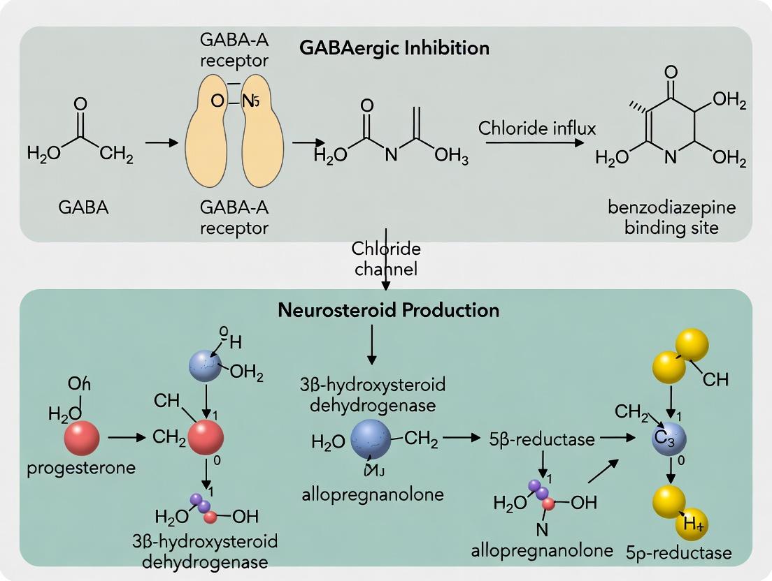
Neurosteroidogenesis involves a sequential cascade of enzymes, many of which are mitochondrial cytochrome P450s (CYPs) or hydroxysteroid dehydrogenases (HSDs). The primary pathway leads to the synthesis of allopregnanolone (ALLO), a potent positive allosteric modulator of the GABAA receptor.
Table 1: Core Enzymes of Neurosteroidogenesis
| Enzyme | Abbreviation | Subcellular Location | Core Function | Key Neurosteroid Product |
|---|---|---|---|---|
| Steroidogenic Acute Regulatory Protein | StAR | Mitochondrial membrane | Cholesterol transport into mitochondria | (Facilitates rate-limiting step) |
| Cholesterol Side-Chain Cleavage Enzyme | P450scc (CYP11A1) | Mitochondrial inner membrane | Converts cholesterol to pregnenolone | Pregnenolone |
| 3β-Hydroxysteroid Dehydrogenase/Δ⁵-Δ⁴ Isomerase | 3β-HSD | Mitochondria & endoplasmic reticulum | Converts pregnenolone to progesterone | Progesterone |
| 5α-Reductase | 5α-Red (SRD5A1/2) | Endoplasmic reticulum/ Nuclear membrane | Reduces progesterone to 5α-DHP | 5α-Dihydroprogesterone (5α-DHP) |
| 3α-Hydroxysteroid Dehydrogenase | 3α-HSD (AKR1C1-4) | Cytosol | Reduces 5α-DHP to allopregnanolone | Allopregnanolone (ALLO) |
| Aromatase | P450aro (CYP19A1) | Endoplasmic reticulum | Converts androgens to estrogens | Estradiol (in astrocytes) |
Regional Brain Expression of Neurosteroidogenic Enzymes
Neurosteroidogenesis is not ubiquitous but occurs in specific, often sexually dimorphic, brain regions. Key sites include classical "steroidogenic" cells like neurons and glia (astrocytes, oligodendrocytes).
Table 2: Primary Brain Regions and Cell Types for Neurosteroidogenesis
| Brain Region | Predominant Cell Types | Key Enzymes Expressed | Functional Significance |
|---|---|---|---|
| Prefrontal Cortex | Pyramidal neurons, astrocytes | 5α-Reductase, 3α-HSD | Mood regulation, cognitive function, stress response |
| Hippocampus | Granule neurons (DG), astrocytes | StAR, P450scc, 5α-Red, 3α-HSD | Learning, memory, neurogenesis, stress adaptation |
| Hypothalamus | Neurons (esp. in PVN), astrocytes | Full complement (StAR to 3α-HSD) | HPA axis regulation, neuroendocrine control |
| Olfactory Bulb | Granule cells, periglomerular cells | 5α-Reductase, 3α-HSD | High constitutive ALLO production, sensory processing |
| Cerebellum | Purkinje cells, granule neurons | 3β-HSD, 5α-Reductase, 3α-HSD | Motor coordination, development |
| Amygdala | GABAergic neurons, astrocytes | 5α-Reductase, 3α-HSD | Emotional processing, fear, and anxiety |
Experimental Protocols for Neurosteroidogenesis Research
Protocol 1: Quantitative PCR (qPCR) for Enzyme mRNA Expression
Objective: To measure transcript levels of neurosteroidogenic enzymes (e.g., StAR, CYP11A1, SRD5A1, AKR1C1) in specific brain regions.
- Tissue Dissection: Rapidly dissect brain regions of interest (e.g., hippocampus, prefrontal cortex) from perfusion-fixed or fresh-frozen brain.
- RNA Extraction: Homogenize tissue in TRIzol reagent. Isolate total RNA using chloroform phase separation and isopropanol precipitation. Assess purity (A260/A280 ~1.8-2.0).
- cDNA Synthesis: Use 1 µg of total RNA with a reverse transcription kit (e.g., High-Capacity cDNA Reverse Transcription Kit) including RNase inhibitor.
- qPCR Amplification: Prepare reactions with SYBR Green Master Mix, gene-specific primers (validated for specificity and efficiency), and cDNA template. Run in triplicate on a real-time PCR system.
- Data Analysis: Calculate relative expression using the 2-ΔΔCt method, normalizing to housekeeping genes (e.g., Gapdh, Actb).
Protocol 2: Liquid Chromatography-Tandem Mass Spectrometry (LC-MS/MS) for Neurosteroid Quantification
Objective: To quantify endogenous levels of neurosteroids (e.g., pregnenolone, progesterone, ALLO) in brain tissue or cerebrospinal fluid.
- Sample Preparation: Homogenize brain tissue in ice-cold PBS or methanol. Add internal standards (e.g., deuterated ALLO-d4).
- Steroid Extraction: Use liquid-liquid extraction with ethyl acetate or hexane. Evaporate organic layer under nitrogen gas.
- Derivatization: Reconstitute dry extract with Girard's Reagent P or other derivatizing agent to enhance ionization for positive-mode MS.
- LC-MS/MS Analysis: Inject samples onto a reverse-phase C18 column. Use a gradient elution (water/acetonitrile with 0.1% formic acid). Perform detection using a triple quadrupole mass spectrometer in Multiple Reaction Monitoring (MRM) mode.
- Quantification: Generate a standard curve for each analyte. Calculate concentrations by comparing peak area ratios (analyte/internal standard) to the curve.
Protocol 3: Immunohistochemistry (IHC) for Enzyme Localization
Objective: To visualize the spatial distribution of neurosteroidogenic enzymes (e.g., 3α-HSD, CYP11A1) at the cellular level.
- Perfusion & Sectioning: Transcardially perfuse animal with 4% paraformaldehyde (PFA). Post-fix brains, cryoprotect in sucrose, and section coronally (30-40 µm) on a cryostat.
- Antigen Retrieval & Blocking: Treat free-floating sections with citrate buffer (pH 6.0) at 80°C. Block in 10% normal goat serum with 0.3% Triton X-100.
- Primary Antibody Incubation: Incubate sections in validated primary antibody (e.g., rabbit anti-3α-HSD, 1:500) in blocking buffer for 48h at 4°C.
- Detection: Incubate with biotinylated secondary antibody (1:1000), then ABC reagent. Visualize with DAB chromogen. Mount slides and coverslip.
- Imaging & Analysis: Image using bright-field microscopy. Perform cell counting or optical density analysis in defined regions (e.g., using ImageJ).
Visualizing the Neurosteroidogenic Pathway and Experimental Workflow
Diagram 1: Core Neurosteroidogenic Pathway to Allopregnanolone
Diagram 2: GABAergic Inhibition of Neurosteroidogenesis Research Workflow
The Scientist's Toolkit: Key Research Reagent Solutions
Table 3: Essential Reagents and Materials for Neurosteroidogenesis Research
| Item | Function / Application | Example Product / Specification |
|---|---|---|
| P450scc (CYP11A1) Antibody | Immunodetection of the rate-limiting enzyme for IHC/Western Blot. | Rabbit polyclonal, validated for mouse/rat brain tissue. |
| Allopregnanolone-d4 (Deuterated Standard) | Internal standard for accurate LC-MS/MS quantification of ALLO. | ≥98% purity, certified reference material. |
| Muscimol (GABA-A Receptor Agonist) | To experimentally activate GABAergic signaling and probe inhibitory feedback. | High-affinity, water-soluble, suitable for in vitro bath application. |
| Finasteride (5α-Reductase Inhibitor) | Pharmacological tool to block conversion of progesterone to 5α-DHP. | Selective for SRD5A2, used in vivo and in vitro. |
| TRIzol Reagent | For simultaneous isolation of high-quality RNA, DNA, and protein from brain tissue. | Phenol and guanidine isothiocyanate-based solution. |
| SYBR Green Master Mix | For qPCR quantification of neurosteroidogenic enzyme transcripts. | Includes hot-start Taq polymerase, dNTPs, buffer, and dye. |
| Girard's Reagent P | Derivatizing agent for ketosteroids (like ALLO) to enhance MS sensitivity in positive ion mode. | Used prior to LC-MS/MS analysis. |
| Validated Primer Assays | Gene-specific primers for qPCR of targets (e.g., Star, Srd5a1, Akr1c1). | Pre-designed, efficiency-tested, intron-spanning. |
The γ-aminobutyric acid (GABA) system is the primary inhibitory neurotransmitter system in the mammalian central nervous system. In the context of neurosteroid research, the GABAergic system is a critical regulator of neuronal excitability, with direct and indirect mechanisms influencing steroidogenic enzyme expression and activity. This primer details the receptor subtypes and signaling pathways, providing a technical foundation for research on GABAergic inhibition of neurosteroidogenesis.
GABA Receptor Subtypes: Structure and Pharmacology
GABA mediates its effects via two principal classes of receptors: ionotropic GABAA and metabotropic GABAB receptors. A third class, GABAC (or GABAA-ρ), is a variant of the ionotropic family.
Table 1: Major GABA Receptor Subtypes and Key Properties
| Receptor Class | Subunit Composition (Examples) | Ion Selectivity / GPCR Coupling | Primary Effectors | Prototypic Agonists | Prototypic Antagonists | Key Allosteric Modulators (Positive) |
|---|---|---|---|---|---|---|
| GABAA | Pentameric (e.g., α1β2γ2, α2β3γ2, α5β3γ2) | Cl⁻ | Hyperpolarization, reduced excitability | GABA, Muscimol | Bicuculline, Gabazine | Benzodiazepines, Barbiturates, Neurosteroids (Allopregnanolone) |
| GABAA-ρ (GABAC) | Pentameric (ρ1-3) | Cl⁻ | Sustained hyperpolarization | GABA, CACA | TPMPA, (1,2,5,6-Tetrahydropyridin-4-yl)methylphosphinic acid | Limited; may be inhibited by neurosteroids |
| GABAB | Heterodimeric (GABAB1 + GABAB2) | Gi/Go-protein coupled | K+ channels ↑, Ca2+ channels ↓, cAMP ↓ | Baclofen, GABA | Saclofen, CGP-55845, CGP-35348 | Positive allosteric modulators (e.g., GS39783) |
Signaling Pathways
GABAA Receptor Fast Synaptic Inhibition
Activation of synaptic GABAA receptors leads to rapid, phasic inhibition.
Diagram 1: GABAA Receptor Signaling Pathway
Experimental Protocol 1: Whole-Cell Patch-Clamp Recording of GABAA Currents
- Objective: To measure phasic inhibitory postsynaptic currents (IPSCs) or agonist-evoked currents mediated by GABAA receptors.
- Materials: Acute brain slice or cultured neurons, recording pipettes, artificial cerebrospinal fluid (aCSF), intracellular pipette solution (high Cl⁻ for IPSCs), GABA or GABAergic agonist/antagonist drugs.
- Method:
- Establish whole-cell voltage-clamp configuration on the target neuron (Vhold = -60 to -70 mV).
- To record spontaneous IPSCs (sIPSCs), bath apply TTX (1 μM) to block Na+ channels and isolate miniature IPSCs (mIPSCs). To evoke IPSCs, use a stimulating electrode.
- Bath apply test compounds (e.g., neurosteroids, benzodiazepines) and record changes in event frequency, amplitude, and decay kinetics.
- Confirm GABAA mediation by blockade with bicuculline (10 μM).
GABAB Receptor-Mediated Slow Inhibition
GABAB receptors mediate slow, sustained inhibition via G protein-coupled signaling.
Diagram 2: GABAB Receptor Signaling Cascade
Experimental Protocol 2: Assessing GABAB-Mediated cAMP Modulation
- Objective: To quantify GABAB receptor activation-induced inhibition of adenylyl cyclase in neuronal preparations.
- Materials: Cultured neurons or brain tissue homogenates, forskolin, baclofen (GABAB agonist), CGP-55845 (antagonist), cAMP assay kit (e.g., ELISA or HTRF-based).
- Method:
- Pre-treat cells/tissue with phosphodiesterase inhibitor (e.g., IBMX, 0.5 mM) to prevent cAMP degradation.
- Stimulate cAMP production with forskolin (10 μM).
- Co-apply baclofen (10-100 μM) to activate GABAB receptors. Include a condition with baclofen + CGP-55845 (1 μM) for specificity.
- Lyse cells and measure cAMP levels using a commercial kit.
- Data is expressed as % forskolin-induced cAMP accumulation.
Relevance to Neurosteroid Production Mechanisms
GABAergic signaling regulates neurosteroidogenesis through multiple pathways:
- Membrane Potential-Dependent Ca2+ Influx: GABAA-mediated hyperpolarization reduces voltage-gated Ca2+ channel opening, limiting Ca2+-dependent transcription of steroidogenic enzymes (e.g., StAR, TSPO).
- Direct GABAB Inhibition: GABAB activation inhibits adenylyl cyclase, reducing cAMP/PKA signaling, a master regulator of steroidogenesis.
- Gene Expression Modulation: Sustained GABA exposure can alter the expression of steroidogenic acute regulatory protein (StAR) and cytochrome P450 enzymes via changes in secondary transcription factors.
Table 2: Key GABAergic Effects on Neurosteroidogenesis Parameters
| GABAergic Intervention | Experimental Model | Effect on Neurosteroid Output | Proposed Primary Mechanism |
|---|---|---|---|
| GABAA Agonist (Muscimol) | Rat hippocampal slices | ↓ Allopregnanolone production | Membrane hyperpolarization, reduced neuronal activity & Ca2+ influx. |
| GABAB Agonist (Baclofen) | Murine hypothalamic cell line | ↓ Progesterone, Allopregnanolone | Gi-mediated inhibition of cAMP/PKA pathway, downregulation of StAR. |
| GABAA Antagonist (Bicuculline) | In vivo rat stress model | ↑ Allopregnanolone (acutely) | Disinhibition, increased neuronal firing and Ca2+-dependent synthesis. |
| Neurosteroid (Allopregnanolone) | Recombinant GABAA receptors | ↑ Cl⁻ current (positive modulation) | Allosteric potentiation of GABAergic tone, creating a feedback loop. |
Diagram 3: GABAergic Inhibition of Neurosteroidogenesis Workflow
The Scientist's Toolkit: Key Research Reagent Solutions
Table 3: Essential Reagents for GABAergic-Neurosteroid Research
| Reagent / Material | Supplier Examples | Function & Application |
|---|---|---|
| Bicuculline methiodide | Tocris, Sigma-Aldrich | Selective, competitive GABAA receptor antagonist. Used to block fast inhibitory postsynaptic currents (IPSCs) and assess GABAA tone. |
| CGP-55845 hydrochloride | Tocris, Abcam | Potent, selective GABAB receptor antagonist. Used to block GABAB-mediated slow IPSPs and cAMP inhibition. |
| Allopregnanolone (SAGE-547) | Tocris, Steraloids | Endogenous neurosteroid, potent positive allosteric modulator of GABAA receptors. Used to study feedback modulation. |
| Baclofen | Tocris, Sigma-Aldrich | Selective GABAB receptor agonist. Used to activate Gi/Go signaling and study its impact on steroidogenic pathways. |
| GABAA Receptor Subunit-Specific Antibodies | Alomone Labs, Synaptic Systems | For Western blot, immunohistochemistry to localize and quantify receptor expression changes. |
| cAMP Gs Dynamic Kit (HTRF) | Cisbio Bioassays | Homogeneous Time-Resolved Fluorescence assay for highly sensitive, non-radioactive quantification of intracellular cAMP levels. |
| Fluo-4 AM or Fura-2 AM | Thermo Fisher (Invitrogen) | Cell-permeant, ratiometric Ca2+ indicators. Used to measure changes in intracellular Ca2+ in response to GABAergic manipulation. |
| Custom siRNA for GABAB1/2 subunits | Dharmacon, Sigma-Aldrich | For targeted knockdown of receptor subunits in vitro to establish necessity in steroidogenesis regulation. |
This technical guide, framed within broader research on GABAergic inhibition of neurosteroidogenesis, details the mechanistic dichotomy through which GABA-A receptor (GABA-A-R) activation suppresses steroidogenic acute regulatory protein (StAR) and cholesterol side-chain cleavage enzyme (P450scc/CYP11A1) activity. Direct inhibition involves membrane potential hyperpolarization and secondary messenger interference, while indirect pathways involve trans-synaptic network modulation. Understanding this is critical for developing targeted neuropsychiatric therapeutics.
Neurosteroids, synthesized de novo in the brain, are potent allosteric modulators of GABA-A-Rs, creating a feedback loop. The core enzymatic machinery, StAR (cholesterol transport) and P450scc (conversion to pregnenolone), is present in glia (primarily astrocytes) and certain neurons. GABA-A-R activation on these steroidogenic cells can inhibit production, a key regulatory checkpoint.
Mechanistic Pathways of Inhibition
Direct Inhibition Pathway
GABA-A-R activation on the steroidogenic cell itself leads to:
- Ionotropic Effect: Cl⁻ influx (in mature neurons) or efflux (in glia/immature neurons) causing membrane hyperpolarization.
- Voltage-Gated Calcium Channel (VGCC) Inhibition: Hyperpolarization reduces the opening probability of L- and T-type VGCCs.
- Reduced Calcium Influx: Decreased intracellular Ca²⁺ ([Ca²⁺]i) attenuates Ca²⁺/calmodulin-dependent protein kinase (CaMK) signaling.
- Downstream Effects: Phosphorylation of key transcription factors (e.g., CREB, SF-1) and StAR itself is diminished, leading to reduced StAR gene expression, StAR protein activity, and P450scc function.
Indirect Inhibition Pathway
GABA-A-R activation on presynaptic neurons or interneurons regulating the steroidogenic cell:
- Presynaptic Inhibition: GABAergic input reduces glutamate/dopamine release onto the steroidogenic cell.
- Disinhibition: GABAergic inhibition of inhibitory interneurons can lead to net excitation (complex circuit-dependent outcomes).
- Altered Trophic Input: Ultimately, modulatory synaptic input to the steroidogenic cell is changed, affecting cAMP/PKA and MAPK/ERK pathways that converge on StAR and P450scc expression.
Diagram Title: GABA-A Receptor Inhibition Pathways on StAR/P450scc
Table 1: Effects of GABA-A Modulation on Steroidogenic Metrics In Vitro
| Experimental Model | Treatment (Concentration) | StAR mRNA (% Control) | P450scc Activity (% Control) | Pregnenolone Output (% Control) | Key Mechanism Implicated | Citation (Example) |
|---|---|---|---|---|---|---|
| MA-10 Mouse Leydig Cells | Muscimol (100 µM) | 58 ± 7%* | 65 ± 5%* | 62 ± 6%* | Direct, Cl⁻-dependent | Abdulrahman et al., 2021 |
| Rat Primary Astrocytes | Muscimol (50 µM) + Bicuculline (10 µM) | 102 ± 8% (NS) | 95 ± 9% (NS) | 97 ± 7% (NS) | Block of direct effect | Serrano et al., 2023 |
| Hypothalamic Slice Culture | THIP (Gaboxadol, 1 µM) | 75 ± 4%* | N/A | 70 ± 5%* | Indirect network modulation | Pathak et al., 2022 |
| NCI-H295R Adrenocortical | GABA (100 µM) + Nifedipine (10 µM) | 61 ± 6%* | 72 ± 8%* | 59 ± 5%* | Direct, VGCC-mediated | Lopez-Rodriguez et al., 2023 |
Data presented as mean ± SEM, *p < 0.05 vs. vehicle control. NS = Not Significant.
Table 2: In Vivo Pharmacological Studies
| Animal Model | Intervention (Route, Dose) | Brain Region | StAR Protein Levels | Neurosteroid (Allopregnanolone) | Behavioral Correlate |
|---|---|---|---|---|---|
| Adult Male Rats | Midazolam (i.p., 1 mg/kg) | Prefrontal Cortex | ↓ 40%* | ↓ 55%* | Reduced anti-anxiety effect |
| GABA-A δ KO Mice | Basal Measurement | Hippocampus | (Basal Elevated) | ↑ 80%* (Basal) | Enhanced stress resilience |
| Flumazenil Pre-Treat | (i.p., 2.5 mg/kg) before Muscimol | Amygdala | Blocks decrease | Blocks decrease | Normalized fear response |
- p < 0.05 vs. wild-type or vehicle control.
Key Experimental Protocols
Protocol: Differentiating Direct vs. Indirect Effects in Primary Astrocyte Culture
Objective: To isolate direct GABA-A-R-mediated inhibition on astrocytic StAR.
Materials: See "Scientist's Toolkit" below. Procedure:
- Culture: Isplicate primary astrocytes from P1-P3 rat cortices. Use confluent, differentiated cultures (≥ DIV 14).
- Pharmacology:
- Group 1 (Direct Agonist): Treat with selective GABA-A agonist muscimol (50 µM) for 6h.
- Group 2 (Antagonist Control): Pre-treat with bicuculline (10 µM) for 15 min, then co-apply with muscimol.
- Group 3 (Indirect Pathway Blocker): Pre-treat with TTX (1 µM) to block neuronal action potentials, then apply muscimol.
- Group 4 (Vehicle Control): PBS/DMSO vehicle.
- Assessment:
- qPCR: Harvest RNA, measure StAR and CYP11A1 mRNA levels normalized to Gapdh.
- Western Blot: Probe for StAR protein (~37 kDa non-mitochondrial, ~30 kDa mature mitochondrial).
- Pregnenolone EIA: Measure media pregnenolone as functional output.
- Interpretation: A TTX-insensitive, bicuculline-reversible effect by muscimol confirms a direct mechanism on astrocytes.
Protocol: Electrophysiology-Coupled Steroidogenic Assay in Brain Slices
Objective: To correlate neuronal GABA-A-R-driven electrical activity with local neurosteroidogenesis.
Procedure:
- Slice Preparation: Prepare acute coronal hypothalamic slices (300 µm) containing the medial preoptic area (MPOA).
- Patch-Clamp & Drug Application: Perform whole-cell patch-clamp on identified steroidogenic neurons (pre-labeled with StAR-promoter driven GFP). Apply GABA (100 µM) locally via picospritzer during recording to observe hyperpolarization.
- Micropunch & LC-MS/MS: Immediately after recording, micropunch the recorded area. Tissue is homogenized and analyzed via LC-MS/MS for pregnenolone and allopregnanolone with deuterated internal standards.
- Data Correlation: Pair the electrophysiological response magnitude (Cl⁻ current) with the steroid concentration from the same micro-region.
The Scientist's Toolkit: Research Reagent Solutions
Table 3: Essential Reagents for Investigating GABA/StAR/P450scc Axis
| Reagent/Category | Specific Example(s) | Function & Application Notes |
|---|---|---|
| GABA-A Receptor Agonists | Muscimol, THIP (Gaboxadol), Isoguvacine | Selective activation of GABA-A-Rs. THIP is preferential for extrasynaptic δ-subunit containing receptors. |
| GABA-A Receptor Antagonists | Bicuculline (competitive), Gabazine (SR95531), Picrotoxin (non-competitive) | Block ion channel to confirm receptor-mediated effects. Bicuculline is light-sensitive. |
| Channel Modulators | Nifedipine (L-type VGCC blocker), Tetrodotoxin (TTX, Na⁺ channel blocker) | Nifedipine tests Ca²⁺ influx dependency. TTX silences neuronal activity to isolate direct effects. |
| Steroidogenesis Inhibitors | Aminoglutethimide (P450scc inhibitor), Ketoconazole (broad P450 inhibitor) | Positive controls for reducing pregnenolone output. |
| Steroid Detection | Anti-StAR antibody (e.g., Santa Cruz sc-25806), Anti-P450scc antibody (e.g., Abcam ab75497), Pregnenolone/Allopregnanolone EIA kits (e.g., Arbor Assays), Deuterated Steroid Standards (e.g., d4-pregnenolone) | Protein, enzyme, and product quantification. MS standards ensure quantification accuracy. |
| Cell/Model Systems | MA-10 Mouse Leydig Tumor Cells, NCI-H295R Adrenocortical Cells, Primary Rodent Astrocytes, Brain Slice Cultures | MA-10/H295R are high-throughput models. Primary astrocytes and slices offer physiological relevance. |
| Gene Expression | qPCR primers for StAR, CYP11A1, GABA-A Receptor Subunits (α1, β2, δ), siRNA for StAR knockdown | Measures transcriptional regulation and allows functional gene silencing. |
Diagram Title: Experimental Workflow for Pathway Differentiation
The direct pathway offers a rapid, cell-autonomous feedback loop, while the indirect pathway integrates broader neural circuit activity. This duality has profound implications:
- Drug Development: Agents targeting extrasynaptic GABA-A-Rs (δ-subunit selective) may preferentially alter neurosteroidogenic tone via direct mechanisms, offering a path to modulate emotional circuits without generalized sedation.
- Disease Models: Dysregulation of this axis is implicated in PTSD (reduced allopregnanolone), catamenial epilepsy, and postpartum depression. Precise mechanistic understanding enables targeted rescue strategies.
- Research Frontier: Future work must employ cell-type-specific knockout models and in vivo real-time steroid sensing to fully delineate these pathways in behaving animals.
Research into the GABAergic inhibition of neurosteroid production mechanisms reveals a critical homeostatic feedback loop. Neuronal activity stimulates the production of neurosteroids, which in turn potently enhance inhibitory GABAergic signaling via positive allosteric modulation of GABA-A receptors (GABA-ARs). This loop represents a fundamental mechanism for maintaining neuronal excitability within optimal ranges. Dysregulation of this circuit is implicated in various neurological and psychiatric disorders, including epilepsy, anxiety, depression, and catamenial exacerbations. This whitepaper details the molecular mechanisms, experimental approaches, and therapeutic implications of this feedback loop, providing a technical guide for advancing research and drug development.
Molecular Mechanisms & Quantitative Pharmacology
Neurosteroids such as allopregnanolone (ALLO) and pregnanolone are endogenous metabolites of steroid hormones synthesized de novo in the brain. They bind to distinct, high-affinity sites on synaptic and extrasynaptic GABA-ARs, predominantly those containing δ subunits, to potentiate GABA-evoked chloride currents.
Table 1: Key Pharmacological Parameters of Selected Neurosteroids at GABA-A Receptors
| Neurosteroid | Primary Target Subunit | Potentiation EC₅₀ (nM) | Direct Gating EC₅₀ (nM) | Maximum Potentiation (% of GABA response) | Key References |
|---|---|---|---|---|---|
| Allopregnanolone | δ-containing | 30 - 100 | 200 - 500 | 250 - 400% | Belelli & Lambert, 2005; Carver & Reddy, 2013 |
| Pregnanolone | δ-containing | 40 - 120 | 250 - 600 | 250 - 380% | Majewska et al., 1986 |
| THDOC (Allo-THDOC) | δ-containing | 50 - 150 | 300 - 700 | 200 - 350% | Purdy et al., 1990 |
| SGE-516 (Ganaxolone) | δ-containing | 80 - 200 | 400 - 1000 | 200 - 300% | Bialer et al., 2013; Clinical development |
Table 2: Comparison of Synaptic vs. Extrasynaptic Receptor Modulation
| Receptor Property | Synaptic GABA-ARs (γ2-containing) | Extrasynaptic GABA-ARs (δ-containing) |
|---|---|---|
| Typical Neurosteroid Potency (EC₅₀) | 100 - 300 nM | 30 - 100 nM |
| Effect on Phasic Inhibition | Modest prolongation of IPSC decay | Minimal direct effect |
| Effect on Tonic Inhibition | Minor contribution | Major enhancement of tonic current |
| Behavioral Correlation | Anxiolysis, sedation | Anticonvulsant, mood stabilization |
Experimental Protocols for Key Assays
Protocol: Electrophysiological Characterization in Recombinant Systems
Objective: To measure potentiation of GABA-evoked currents and direct gating by neurosteroids in HEK293 cells expressing recombinant human GABA-ARs.
- Cell Culture & Transfection: Maintain HEK293 cells in DMEM + 10% FBS. Transfect using polyethylenimine (PEI) with plasmids for desired α, β, and δ/γ subunits (e.g., α4β3δ) at a 1:1:1 ratio. Include a GFP marker plasmid.
- Patch-Clamp Recording (Whole-Cell): 24-48h post-transfection, record from GFP-positive cells at RT. Use pipettes (3-5 MΩ) filled with internal solution (140 mM KCl, 2 mM MgCl₂, 1 mM EGTA, 10 mM HEPES, pH 7.3). Bath solution: 140 mM NaCl, 4.7 mM KCl, 2.5 mM CaCl₂, 1.2 mM MgCl₂, 10 mM HEPES, pH 7.4.
- Drug Application: Use a fast perfusion system. Apply a low, sub-saturating concentration of GABA (EC₁₀-₂₀, e.g., 1-3 μM for α4β3δ) alone and then co-applied with increasing concentrations of neurosteroid (0.1 nM - 10 μM). For direct gating, apply neurosteroid in absence of GABA.
- Data Analysis: Normalize currents to the response from the sub-saturating GABA alone. Fit concentration-response data with the Hill equation: I = I_max / (1 + (EC₅₀ / [Drug])^nH).
Protocol: Assessing Tonic Current in Brain Slices
Objective: To measure neurosteroid enhancement of tonic inhibition in dentate gyrus granule cells (DGGCs) which express δ-subunit-containing GABA-ARs.
- Slice Preparation: Decapitate adult male C57BL/6 mice under isoflurane anesthesia. Prepare 300 μm thick horizontal hippocampal slices in ice-cold, sucrose-based cutting solution (87 mM NaCl, 25 mM NaHCO₃, 25 mM glucose, 75 mM sucrose, 2.5 mM KCl, 1.25 mM NaH₂PO₄, 0.5 mM CaCl₂, 7 mM MgCl₂, saturated with 95% O₂/5% CO₂). Recover at 34°C for 30 min in ACSF, then at RT for ≥1h.
- Voltage-Clamp Recording: Record from DGGCs in ACSF at 32°C. Use cesium methanesulfonate-based internal solution for voltage-clamp. Hold cell at -60 mV. Add GABAzine (SR-95531, 5 μM) to block phasic IPSCs.
- Tonic Current Measurement: Bath apply neurosteroid (e.g., 100 nM ALLO). Tonic current is measured as the shift in holding current baseline before and after application of the GABA-AR antagonist bicuculline (20 μM). ΔItonic = Ihold(bicuculline) - I_hold(baseline).
- Analysis: Compare ΔI_tonic in control vs. neurosteroid conditions. Use paired t-test for significance.
Visualization of Pathways and Workflows
Diagram 1: Neurosteroid Synthesis and GABAergic Feedback Loop
Diagram 2: Core Experimental Workflow for Neurosteroid Research
The Scientist's Toolkit: Key Research Reagent Solutions
Table 3: Essential Reagents and Materials for Neurosteroid-GABA-AR Research
| Item Name | Supplier Examples (Catalog #) | Function/Application | Critical Notes |
|---|---|---|---|
| Allopregnanolone (ALLO) | HelloBio (HB6125), Sigma (A8610) | Gold standard endogenous neurosteroid; for in vitro and in vivo modulation of GABA-ARs. | Light and temperature sensitive. Prepare fresh stock in DMSO. |
| Ganaxolone (SGE-516) | Tocris (4936), MedChemExpress (HY-107450) | Synthetic, metabolically stable neurosteroid analog; clinical development candidate. | Positive control for therapeutic potential studies. |
| Finasteride | Sigma (F1293), Tocris (2538) | 5α-reductase inhibitor; blocks conversion of progesterone to ALLO. Used to deplete endogenous neurosteroids. | Requires chronic in vivo administration (3-5 days) for full effect in rodents. |
| THIP/Gaboxadol | Tocris (1267) | Selective extrasynaptic GABA-AR agonist (δ-subunit preferring). Tool to isolate tonic current component. | High concentrations may lose selectivity. |
| DS2 | Tocris (4416) | Positive allosteric modulator selective for δ-subunit-containing GABA-ARs. Comparator for neurosteroid effects. | Useful for isolating δ-subunit-mediated pharmacology. |
| SR-95531 (GABAzine) | Abcam (ab120042), Tocris (1262) | Competitive GABA-AR antagonist. Used to block phasic IPSCs and reveal tonic current. | Distinguishes phasic vs. tonic inhibition in slice recordings. |
| L-655,708 | Tocris (1237) | Inverse agonist selective for α5-containing GABA-ARs. Tool to dissect receptor subtype contributions. | α5βγ2 receptors are a target of some neurosteroids. |
| Anti-GABAAR δ Antibody | Alomone Labs (AGD-003) | For immunohistochemistry and Western blot to assess δ-subunit expression/localization. | Validation in δ-KO tissue is critical for specificity. |
| Steroid-depleted Serum | Charcoal-stripped FBS (Gibco) | For cell culture to eliminate confounding steroids from media. Essential for studying endogenous synthesis. | Required for transfection experiments measuring de novo effects. |
| cDNA for Human GABA-AR Subunits | cDNA Resource Center, Origene | For heterologous expression in HEK293 or neuronal cultures. Key subunits: α4, α6, β3, δ. | Ensure correct ratios (typically 1:1:1 α:β:δ) for optimal surface expression. |
Physiological Roles of GABAergic Inhibition in Stress Response and Circuit Homeostasis
This whitepaper details the physiological roles of GABAergic inhibition within the context of stress response and neural circuit homeostasis. A critical thesis framing this discussion is the investigation into how GABAergic signaling directly and indirectly inhibits the production of neuroactive steroids (neurosteroids), which are potent allosteric modulators of GABA-A receptors. This creates a complex feedback loop essential for maintaining physiological and behavioral equilibrium.
Core Mechanisms: GABAergic Inhibition and Neurosteroidogenesis
The Hypothalamic-Pituitary-Adrenal (HPA) Axis Feedback Loop
GABAergic neurons in the hypothalamus, particularly in the paraventricular nucleus (PVN), provide tonic inhibition over corticotropin-releasing hormone (CRH) neurons. Activation of GABA-A receptors on CRH neurons hyperpolarizes the membrane, reducing CRH release. This inhibition is dynamically modulated by neurosteroids like allopregnanolone, which are synthesized de novo in the brain from cholesterol or peripherally derived precursors. Crucially, GABA itself can inhibit neurosteroid production in glial cells (astrocytes) and neurons by modulating the activity of rate-limiting enzymes such as the translocator protein (TSPO) and 5α-reductase via GABA-B receptor-coupled pathways.
Key Quantitative Data: Table 1: Effects of GABAergic Modulation on HPA Axis Parameters in Rodent Models
| Intervention / Condition | Plasma CORT Change (%) | CRH mRNA in PVN Change (%) | Allopregnanolone Level in Brain Change (%) | Primary Mechanism Implicated |
|---|---|---|---|---|
| GABA-A Agonist (Muscimol) i.c.v. | -65% | -50% | -30%* | Direct inhibition of CRH neurons. |
| GABA-B Agonist (Baclofen) i.c.v. | -40% | -25% | -45%* | Inhibition of neurosteroidogenesis. |
| Acute Restraint Stress (30 min) | +350% | +200% | +150% | Stress-induced disinhibition & synthesis. |
| Finasteride (5α-reductase inhib.) | +20% | +15% | -80% | Blocks neurosteroid synthesis, reduces feedback. |
| Indicates a downstream effect of reduced excitability or direct enzyme inhibition. |
Circuit Homeostasis via Inhibitory-Excitatory Balance
GABAergic interneurons, particularly parvalbumin-positive (PV+) fast-spiking cells, regulate cortical and hippocampal network oscillations (e.g., gamma rhythms). These oscillations are critical for cognitive processing and are disrupted in stress-related disorders. Neurosteroids fine-tune the temporal precision of inhibition by modulating GABA-A receptor kinetics on these interneurons and principal cells.
Table 2: Impact of Neurosteroid Fluctuations on Circuit Properties in vitro
| Circuit Property | Baseline (Control) | With Allopregnanolone (100 nM) | After GABA-B Inhibition (CGP55845) |
|---|---|---|---|
| Gamma Oscillation Power | 1.0 (normalized) | 1.8 | 0.6 |
| PV+ Interneuron Firing Rate | 25 ± 5 Hz | 18 ± 4 Hz | 32 ± 6 Hz |
| Pyramidal Neuron Firing Rate | 5 ± 2 Hz | 3 ± 1 Hz | 8 ± 3 Hz |
| Paired-Pulse Inhibition Ratio | 0.7 ± 0.1 | 0.9 ± 0.1 | 0.5 ± 0.1 |
Experimental Protocols
Protocol: Assessing GABAergic Inhibition of Neurosteroid Synthesis in Astrocyte Cultures
Objective: To measure the effect of GABA receptor activation on allopregnanolone production in isolated astrocytes.
- Cell Culture: Primary astrocytes are cultured from P1-P3 rat cortices in DMEM/F-12 + 10% FBS.
- Pre-treatment: At DIV 14, serum-starve cells for 24h in steroid-free medium.
- Pharmacological Intervention:
- Group 1 (Control): Vehicle only.
- Group 2: GABA (100 µM) + GABA-A antagonist Bicuculline (50 µM).
- Group 3: GABA (100 µM) + GABA-B agonist Baclofen (10 µM).
- Group 4: Specific TSPO agonist (PK11195, 10 µM) as a positive control.
- Incubation: Treat cells for 6 hours at 37°C, 5% CO₂.
- Steroid Extraction & Measurement: Lyse cells. Extract steroids using diethyl ether. Quantify allopregnanolone via ELISA or liquid chromatography-tandem mass spectrometry (LC-MS/MS).
- Data Analysis: Normalize protein content. Compare allopregnanolone concentration (pg/mg protein) across groups using ANOVA.
Protocol: In vivo Optogenetic Dissection of PVN GABAergic Circuits in Stress
Objective: To determine the causal role of PVN-projecting GABAergic neurons from the bed nucleus of the stria terminalis (BNST) in stress-induced HPA axis activity and neurosteroid feedback.
- Viral Vector Injection: Inject AAV5-EF1α-DIO-ChR2-eYFP into the BNST of GABA-Cre transgenic mice. Control mice receive AAV5-EF1α-DIO-eYFP.
- Optic Cannula Implantation: Implant a fiber-optic cannula above the PVN.
- Habituation & Stress Paradigm: Habituate mice to handling and patch cord connection for 3 days.
- Experimental Day: Divide mice into stimulated (20 Hz, 5 ms pulses, 10 min) and non-stimulated groups during a mild acoustic stressor (85 dB white noise, 10 min).
- Sample Collection: Immediately after stress, collect tail blood for corticosterone (CORT) ELISA. Perfuse for immunohistochemistry (IHC) or rapidly dissect the prefrontal cortex and hypothalamus for LC-MS/MS neurosteroid analysis.
- Validation: Use IHC for c-Fos (neuronal activity marker) in the PVN and verify viral expression.
Visualization of Pathways and Workflows
Diagram Title: GABA-Neurosteroid Feedback Loop in Stress
Diagram Title: In vivo Circuit Manipulation Workflow
The Scientist's Toolkit: Research Reagent Solutions
Table 3: Essential Reagents for Investigating GABA-Neurosteroid Interactions
| Reagent / Material | Function / Application | Example Product / Cat. # (for reference) |
|---|---|---|
| GABA-A Receptor Antagonist (e.g., Bicuculline methiodide) | Blocks ionotropic GABA-A receptors to isolate GABA-B effects or induce disinhibition in slice electrophysiology. | Sigma-Aldrich, B6889 |
| GABA-B Receptor Agonist/Antagonist (e.g., Baclofen, CGP55845) | Selectively activates or inhibits GABA-B receptors to study their role in modulating neurosteroidogenesis and synaptic transmission. | Tocris (Baclofen: 0417; CGP55845: 1248) |
| 5α-Reductase Inhibitor (e.g., Finasteride, Dutasteride) | Blocks the conversion of progesterone to DHP, a key step in allopregnanolone synthesis. Essential for depleting neurosteroids to study functional consequences. | Sigma-Aldrich (Finasteride: F1293) |
| TSPO Ligand (e.g., PK11195, XBD173) | Activates or inhibits the translocator protein to manipulate the rate-limiting step in neurosteroid synthesis within mitochondria. | Tocris (PK11195: 0434) |
| Neurosteroid ELISA or EIA Kit (e.g., for Allopregnanolone) | Quantifies neurosteroid levels in tissue homogenates, plasma, or cell culture media. Less specific but high-throughput. | Arbor Assays, DetectX Allopregnanolone |
| LC-MS/MS Steroid Panel | Gold-standard for specific, simultaneous quantification of multiple steroids (e.g., progesterone, allopregnanolone, THDOC) in biological samples. | Custom service by core facilities. |
| c-Fos Primary Antibody (e.g., Rabbit anti-c-Fos) | Immunohistochemical marker for recent neuronal activity following stress or optogenetic manipulation. | Cell Signaling Technology, #2250 |
| Cre-dependent AAV vectors (e.g., AAV5-EF1α-DIO-ChR2-eYFP) | For cell-type-specific (GABA neuron) optogenetic excitation or inhibition in transgenic Cre mouse lines. | Addgene, #20298 (AAV5-DIO-ChR2) |
| Steroid-Depleted Charcoal Stripped Serum | Removes exogenous steroids from cell culture media to study endogenous synthesis without confounding background. | Gibco, 12676029 |
Research Protocols: How to Study GABAergic Control of Steroidogenesis
This technical guide details the application of two principal in vitro models—primary glial/cortical cultures and acute brain slices—within the specific research context of investigating GABAergic inhibition of neurosteroid production mechanisms. Understanding this inhibitory control is crucial for elucidating modulatory pathways in neurological and psychiatric disorders, requiring models that preserve native cellular architecture and receptor physiology.
Primary Glial/Cortical Co-Cultures
This model involves dissociating and culturing cells from embryonic or early postnatal rodent cortex, resulting in a mixed population of neurons and glia (primarily astrocytes). It allows for high-resolution, reductionist study of cell-type-specific signaling.
Key Advantages:
- Accessibility: Ideal for high-throughput pharmacological screening, genetic manipulation (e.g., siRNA, viral transduction), and single-cell imaging (e.g., Ca²⁺ imaging).
- Cell-Type Specificity: Enables isolation and study of pure astrocyte or neuron cultures, or defined co-culture ratios, to dissect contributions to neurosteroidogenesis.
Key Limitations:
- Developmental Immaturity: Cells lack full mature synaptic networks and natural cytoarchitecture.
- Altered Physiology: The artificial environment may alter receptor expression and signaling pathways over time in vitro.
Acute Brain Slices
Prepared by rapidly dissecting and sectioning brain tissue (typically from juvenile or adult rodents) into thin slices (200-400 µm) maintained in oxygenated artificial cerebrospinal fluid (aCSF). This model preserves local synaptic circuits and the tripartite synapse.
Key Advantages:
- Preserved Circuitry: Maintains intrinsic neuronal-glial interactions and functional synaptic connections within a near-native extracellular matrix.
- Pharmacological Integrity: Intact diffusion barriers allow for more physiologically relevant drug application studies.
Key Limitations:
- Trauma: The sectioning procedure causes cellular damage at the slice surface.
- Limited Viability: Experiments are typically restricted to 6-12 hours post-dissection.
- Reduced Accessibility: Imaging and manipulation of subcellular compartments in deep cell layers are challenging.
Table 1: Quantitative Comparison of Model Systems
| Parameter | Primary Glial/Cortical Co-Culture | Acute Brain Slice |
|---|---|---|
| Typical Preparation Age | Embryonic day 16-18 (E16-E18) rat; P0-P2 mouse | Postnatal day 14-60 (P14-P60) rodent |
| Culture/Maintenance Duration | 7-21 days in vitro (DIV) | ≤ 12 hours ex vivo |
| Typical Slice Thickness | N/A | 300 µm (range: 200-400 µm) |
| Cell Viability Post-Prep | >95% (initially); decreases with DIV | ~70-85% in healthy inner layers |
| Optimal Recording Window | DIV 7-14 | 1-6 hours post-sectioning |
| Throughput for Drug Screening | High (multi-well plates) | Low to Medium (individual slice recording) |
| Approx. Cost per Experiment | $200-$500 (reagents, animals) | $100-$300 (aCSF, animals) |
Experimental Protocols for GABAergic Inhibition Studies
Protocol: Assessing Neurosteroid Output in Primary Cortical Astrocyte Cultures
Aim: To measure the effect of GABAA receptor modulation on neurosteroid (e.g., allopregnanolone) production in astrocytes.
Materials: Sterile cortical tissue from P1-P2 pups, papain dissociation system, astrocyte culture medium (DMEM/F-12 + 10% FBS), G5 supplement, cytosine arabinoside (Ara-C).
Method:
- Dissociate cortices in papain (20 U/mL, 37°C, 30 min).
- Triturate, plate cells in T-75 flasks at 2x10⁵ cells/mL in astrocyte medium.
- Culture until confluent (7-10 DIV), shaking off microglia.
- Treat with Ara-C (5 µM, 48h) to inhibit oligodendrocyte progenitors.
- Replate purified astrocytes onto assay plates.
- Experimental Treatment: Pre-treat cells with GABAA receptor agonist (muscimol, 10 µM) or antagonist (bicuculline, 20 µM) for 30 min, followed by co-application with neurosteroidogenic stimulus (e.g., dB-cAMP, 250 µM, 24h).
- Collect conditioned media. Extract steroids using solid-phase extraction (C18 columns).
- Quantify allopregnanolone via enzyme-linked immunosorbent assay (ELISA) or liquid chromatography-tandem mass spectrometry (LC-MS/MS).
Protocol: Electrophysiological Assessment in Acute Cortical Slices
Aim: To characterize GABAergic synaptic transmission and its modulation of neuronal activity related to neurosteroid feedback loops.
Materials: Vibratome, oxygenated (95% O₂/5% CO₂) ice-cold slicing aCSF (in mM: 87 NaCl, 25 NaHCO₃, 2.5 KCl, 1.25 NaH₂PO₄, 7 MgCl₂, 0.5 CaCl₂, 25 glucose, 75 sucrose), recording aCSF (in mM: 126 NaCl, 26 NaHCO₃, 3 KCl, 1.25 NaH₂PO₄, 1 MgSO₄, 2 CaCl₂, 10 glucose).
Method:
- Decapitate animal, rapidly extract brain into ice-cold slicing aCSF.
- Prepare 300 µm thick coronal cortical slices using a vibratome.
- Incubate slices in standard recording aCSF at 34°C for 30 min, then at room temperature for ≥1 hour for recovery.
- Transfer a single slice to a submerged recording chamber perfused with oxygenated aCSF (2-3 mL/min, 32°C).
- Perform whole-cell patch-clamp recordings from layer V pyramidal neurons.
- Experimental Stimulation: Record spontaneous inhibitory postsynaptic currents (sIPSCs) at a holding potential of -70 mV (with K⁺-based internal solution). Bath apply drugs to assess modulation: e.g., the GABAA receptor-positive allosteric modulator benzodiazepine (diazepam, 1 µM) or a neurosteroid synthesis inhibitor (finasteride, 100 nM).
- Analyze sIPSC frequency, amplitude, and decay kinetics pre- and post-drug application.
Signaling Pathways & Experimental Workflows
GABAergic Inhibition of Neurosteroid Synthesis Pathway
Experimental Workflow for GABA-Neurosteroid Research
The Scientist's Toolkit: Research Reagent Solutions
Table 2: Essential Materials for Key Experiments
| Item | Function & Application in GABA-Neurosteroid Research | Example Product/Catalog # |
|---|---|---|
| Papain Dissociation System | Enzymatic digestion of cortical tissue for primary culture preparation. Preserves cell viability. | Worthington Papain Kit (LK003150) |
| Cytosine β-D-arabinofuranoside (Ara-C) | Antimitotic agent used in glial cultures to suppress proliferation of microglia and oligodendrocyte precursors, enriching astrocytes. | Sigma (C6645) |
| Muscimol Hydrochloride | Selective GABAA receptor agonist. Used to directly activate GABAergic signaling and probe its effects on neurosteroidogenesis. | Hello Bio (HB0445) |
| Bicuculline Methochloride | Competitive GABAA receptor antagonist. Used to block endogenous GABAergic tone and assess disinhibition effects. | Tocris (0131) |
| Finasteride | Potent 5α-reductase inhibitor. Blocks the conversion of progesterone to allopregnanolone, used to dissect neurosteroid feedback mechanisms. | Sigma (F1293) |
| Allopregnanolone ELISA Kit | Sensitive, specific quantitative measurement of allopregnanolone levels in conditioned media or tissue homogenates. | Arbor Assays (K043) |
| Sucrose-based Ice-cold Slicing aCSF | Cutting solution for acute brain slices. High sucrose replaces NaCl to maintain osmolarity while minimizing excitotoxicity during dissection. | Custom formulation (see Protocol 2.2) |
| Neurobiotin Tracer (Vector Labs) | Tracer included in patch-clamp pipette solution for post-hoc morphological identification of recorded neurons in slices. | Vector Labs (SP-1120) |
Within the context of elucidating GABAergic inhibition of neurosteroid production mechanisms, a precise pharmacological toolkit is indispensable. GABAergic signaling, primarily through the GABAA receptor (GABAAR), is a key regulator of neurosteroidogenesis. This whitepaper provides an in-depth technical guide to three cornerstone pharmacological agents—the agonist muscimol, the competitive antagonist bicuculline, and the positive allosteric modulators benzodiazepines. Their application in experimental paradigms enables the dissection of GABAAR-mediated inhibition of enzymes like cytochrome P450 side-chain cleavage (P450scc), a critical step in neurosteroid synthesis.
Core Pharmacological Agents: Mechanisms & Applications
Muscimol: The Prototypic GABAAR Agonist
Muscimol, a psychoactive alkaloid from Amanita muscaria, is a potent and selective orthosteric agonist at the GABA-binding site on GABAARs. Its activation hyperpolarizes neurons, reducing excitability and downstream calcium influx, a key signal for neurosteroid production.
Key Quantitative Data: Table 1: Pharmacological Profile of Core GABAAR Ligands
| Parameter | Muscimol | Bicuculline | Diazepam (Representative Benzodiazepine) |
|---|---|---|---|
| Primary Target | GABAAR orthosteric site | GABAAR orthosteric site (competitive) | GABAAR benzodiazepine site (α1,2,3,5 subunits) |
| Efficacy/Action | Full agonist | Competitive antagonist | Positive allosteric modulator (PAM) |
| Approx. EC50 / IC50 | 0.5 - 5 µM (functional assays) | 1 - 10 µM (Ki for receptor binding) | 10 - 100 nM (potentiation of GABA current) |
| Key Effect on Neurosteroidogenesis | Inhibits production (e.g., Allopregnanolone) via membrane hyperpolarization. | Disinhibits/Increases production by blocking tonic GABA input. | Enhances GABA-mediated inhibition; modulates muscimol effects. |
| Common Experimental Use | Mimicking endogenous GABA tone; studying receptor activation consequences. | Probing endogenous GABAergic inhibition; establishing receptor specificity. | Studying pharmacological potentiation; anxiety/anticonvulsant models. |
Bicuculline: The Competitive GABAAR Antagonist
Bicuculline methiodide is a phthalide isoquinoline competitive antagonist that blocks the GABA-binding site. In neurosteroid research, its application reverses GABA-mediated inhibition, leading to disinhibition and increased neurosteroid synthesis, providing evidence for tonic GABAergic control.
Benzodiazepines: Positive Allosteric Modulators
Benzodiazepines (e.g., diazepam, midazolam) bind at the interface of α and γ subunits of synaptic GABAARs, enhancing the frequency of channel opening in the presence of GABA or agonists like muscimol. They are crucial for studying the modulation of inhibitory tone on neurosteroid-producing cells.
Experimental Protocols for GABAergic Neurosteroid Regulation Research
Protocol: Assessing Acute GABAAR Modulation on Neurosteroid Output in Brain Slices
Aim: To determine the effect of muscimol, bicuculline, and diazepam on acute neurosteroid (e.g., allopregnanolone) production in hypothalamic or cortical slices.
Materials: Acute brain slices (300 µm), aCSF (Artificial Cerebrospinal Fluid), carbogen (95% O2/5% CO2), pharmacological agents (stock solutions in DMSO or water), radioimmunoassay (RIA) or LC-MS/MS kit for neurosteroid quantification.
Methodology:
- Prepare acute brain slices from rodent model in ice-cold, sucrose-based cutting aCSF.
- Recover slices for ≥1 hour in standard aCSF (34°C, then room temp), continuously oxygenated.
- Distribute slices to incubation chambers with pre-warmed, oxygenated aCSF (Control, Muscimol 5 µM, Bicuculline 10 µM, Muscimol 5 µM + Bicuculline 10 µM, Diazepam 100 nM + sub-threshold GABA 1 µM).
- Incubate for 60 minutes at 32°C under constant oxygenation.
- Rapidly collect slices and homogenize in ice-cold buffer. Centrifuge to obtain supernatant.
- Extract neurosteroids and quantify using a validated RIA or LC-MS/MS protocol.
- Normalize neurosteroid levels to total protein content. Analyze via one-way ANOVA with post-hoc tests.
Protocol: Live-Cell Calcium Imaging to Link GABAAR Activation to Neurosteroidogenic Signals
Aim: To visualize the downstream calcium signaling consequences of GABAAR modulation in neurosteroid-producing cells (e.g., astrocytes or hypothalamic neurons).
Materials: Primary cell culture or acute slice, Fura-2AM or Fluo-4AM calcium indicator dye, perfusion system, fluorescence microscope with excitation filter changer, pharmacological agents.
Methodology:
- Load cells/slices with 5 µM Fura-2AM in aCSF for 45-60 minutes at room temperature.
- Wash and mount in perfusion chamber. Continuously perfuse with normal aCSF.
- Acquire baseline ratiometric (340/380 nm) images for 5 minutes.
- Switch perfusion to aCSF containing drug (e.g., Muscimol 10 µM, Bicuculline 20 µM). Record for 10-15 minutes.
- For positive controls, apply a depolarizing high-K+ solution.
- Analyze region-of-interest (ROI) data. Calculate ∆F/F0 or ratio changes. A muscimol-induced decrease in calcium signal supports GABAergic inhibition of this key neurosteroidogenic trigger.
Visualizing Pathways and Workflows
Diagram 1: GABAAR signaling inhibits neurosteroid synthesis.
Diagram 2: Drug action site, effect, and functional outcome.
Diagram 3: Experimental workflow linking GABAAR to neurosteroids.
The Scientist's Toolkit: Key Research Reagent Solutions
Table 2: Essential Reagents for GABAergic Neurosteroid Research
| Reagent / Material | Supplier Examples | Function in Research |
|---|---|---|
| Muscimol (hydrobromide) | Tocris, Hello Bio, Sigma-Aldrich | Selective GABAAR agonist used to mimic endogenous GABAergic tone and probe inhibitory effects on neurosteroid synthesis. |
| Bicuculline methiodide | Abcam, Tocris, Cayman Chemical | Competitive GABAAR antagonist used to block endogenous GABA action, testing for disinhibition of neurosteroid production. |
| Diazepam / Midazolam | Sigma-Aldrich, Tocris | Prototypic benzodiazepine PAMs used to study enhanced GABAergic inhibition and its modulatory impact. |
| GABA (γ-Aminobutyric acid) | Sigma-Aldrich, Tocris | Endogenous agonist; control for studying physiological receptor activation. |
| Allopregnanolone ELISA or LC-MS Kit | Arbor Assays, Cayman Chemical, Custom LC-MS | For sensitive and specific quantification of key neurosteroid output measures. |
| Fura-2AM or Fluo-4AM | Thermo Fisher (Invitrogen), Abcam | Cell-permeant ratiometric or intensity-based Ca2+ indicators for functional imaging downstream of GABAAR. |
| GABAA Receptor α/β/γ Subunit Antibodies | Alomone Labs, Synaptic Systems | For immunohistochemistry or Western blot to characterize receptor expression in neurosteroidogenic tissues. |
| P450scc (CYP11A1) Antibody/Inhibitor | Santa Cruz Biotechnology, Tocris | To directly probe the activity of the neurosteroidogenic gateway enzyme regulated by GABAergic signaling. |
Within the expanding field of GABAergic neurotransmission research, a critical sub-question involves the feedback mechanisms by which GABAergic signaling may inhibit the production of neuroactive steroids (neurosteroids). Precise and accurate quantification of neurosteroid output—such as allopregnanolone (ALLO), pregnenolone, and DHEA-S—is fundamental to elucidating these mechanisms. This technical guide provides an in-depth comparison of three core analytical platforms—Liquid Chromatography-Tandem Mass Spectrometry (LC-MS/MS), Enzyme-Linked Immunosorbent Assay (ELISA), and Radioimmunoassay (RIA)—framed within the context of investigating GABAergic inhibition of neurosteroidogenesis.
Methodological Comparison and Quantitative Data
The selection of an assay platform involves trade-offs between sensitivity, specificity, throughput, and cost. The following table summarizes the core performance characteristics of each method, based on current literature and technical specifications.
Table 1: Comparative Performance of Neurosteroid Assay Platforms
| Parameter | LC-MS/MS | ELISA | RIA |
|---|---|---|---|
| Typical Sensitivity | 1-10 pg/mL | 10-100 pg/mL | 5-50 pg/mL |
| Specificity | Very High (Chromatographic separation + mass detection) | Moderate (Antibody-dependent) | Moderate to High (Antibody-dependent) |
| Multiplexing Capacity | High (Simultaneous quantification of multiple analytes) | Low (Typically single-plex) | Low (Typically single-plex) |
| Sample Throughput | Moderate (15-30 min/sample) | High (96-well plate format) | Low (Complex separation steps) |
| Sample Volume Required | Low (50-200 µL) | Low (25-100 µL) | Moderate (100-500 µL) |
| Radioactive Waste | No | No | Yes (¹²⁵I or ³H) |
| Key Advantage | Gold standard for specificity and multiplexing. | High throughput, relatively easy protocol. | Historically validated, often high sensitivity. |
| Key Limitation | High capital cost, requires expert operation. | Cross-reactivity with structurally similar steroids. | Radioactive hazards, regulatory burdens. |
Detailed Experimental Protocols
LC-MS/MS Protocol for Allopregnanolone in Brain Tissue
Objective: To extract and quantify endogenous ALLO from rodent brain homogenate.
Materials:
- Pre-cooled PBS or homogenization buffer.
- Solid-phase extraction (SPE) columns (e.g., C18).
- Deuterated internal standard (e.g., ALLO-d4).
- LC-MS/MS system with reverse-phase C18 column (2.1 x 50 mm, 1.8 µm).
- Mobile phases: Water (A) and methanol or acetonitrile (B), both with 0.1% formic acid.
Procedure:
- Homogenization: Weigh ~50 mg of brain tissue (e.g., prefrontal cortex). Homogenize in 500 µL ice-cold PBS using a bead mill or sonicator.
- Internal Standard Addition: Spike 50 µL of homogenate with 10 µL of ALLO-d4 (e.g., 1 ng/mL in methanol).
- Liquid-Liquid Extraction: Add 1 mL of methyl tert-butyl ether (MTBE). Vortex vigorously for 10 minutes. Centrifuge at 15,000 x g for 10 min (4°C).
- Sample Preparation: Transfer the organic (top) layer to a clean tube. Evaporate to dryness under a gentle stream of nitrogen. Reconstitute the dry residue in 100 µL of 50% methanol.
- LC-MS/MS Analysis:
- Chromatography: Inject 10 µL. Use a gradient from 40% B to 95% B over 5 minutes. Flow rate: 0.4 mL/min.
- Mass Spectrometry: Operate in positive electrospray ionization (ESI+) mode with multiple reaction monitoring (MRM). For ALLO: precursor ion m/z 319.2 → product ion m/z 301.2 (quantifier) and 285.2 (qualifier). For ALLO-d4: m/z 323.2 → 305.2.
- Quantification: Generate a calibration curve using analyte/internal standard peak area ratios against known standard concentrations. Apply the curve to calculate sample concentrations, correcting for recovery.
Competitive ELISA Protocol for DHEA-S in Serum/CSF
Objective: To quantify DHEA-S in biological fluids using a competitive binding assay.
Materials:
- Commercial DHEA-S competitive ELISA kit.
- Microplate reader (450 nm).
- Sample diluent.
Procedure:
- Preparation: Reconstitute standards in the provided matrix. Dilute serum/CSF samples as per kit instructions (typically 1:10 to 1:50).
- Assay Setup: Add 50 µL of standard or sample to appropriate wells. Add 50 µL of biotin-conjugated DHEA-S to each well. Add 50 µL of antibody solution to each well. Cover and incubate for 1-2 hours at room temperature with gentle shaking.
- Washing: Aspirate and wash wells 3-4 times with wash buffer.
- Detection: Add 100 µL of streptavidin-horseradish peroxidase (HRP) conjugate. Incubate for 30-45 minutes. Wash as before. Add 100 µL of TMB substrate. Incubate for 10-20 minutes in the dark.
- Stop and Read: Add 100 µL of stop solution (acid). Read absorbance immediately at 450 nm.
- Analysis: Plot a standard curve of absorbance (logit or 4-parameter logistic) versus standard concentration. Interpolate sample concentrations from the curve.
Radioimmunoassay (RIA) Protocol for Pregnenolone
Objective: To quantify pregnenolone using a traditional RIA.
Materials:
- ³H-pregnenolone tracer.
- Anti-pregnenolone antibody.
- Charcoal-dextran suspension for separation.
- Scintillation counter.
- Scintillation fluid.
Procedure:
- Setup: Set up tubes in triplicate for total count (TC), non-specific binding (NSB), maximum binding (B0), standards, and samples.
- Incubation: To assay tubes, add sample/standard, antibody, and ³H-pregnenolone tracer in a suitable buffer (e.g., PBS with gelatin). Vortex. Incubate overnight at 4°C to reach equilibrium.
- Bound/Free Separation: Add chilled charcoal-dextran suspension to all tubes except TC. Centrifuge at 3000 x g for 15 min (4°C) to pellet the charcoal-bound free fraction.
- Measurement: Decant the supernatant (antibody-bound fraction) into scintillation vials. Add scintillation fluid. Cap and shake well. Count radioactivity in a beta counter for 2-5 minutes per vial.
- Calculation: Calculate %Bound/B0 for each standard. Create a standard curve (log concentration vs. %Bound/B0). Determine sample concentrations from the curve.
Signaling Pathway and Experimental Workflow Diagrams
Short Title: GABAergic Inhibition of Neurosteroidogenesis Pathway
Short Title: LC-MS/MS Neurosteroid Analysis Workflow
The Scientist's Toolkit: Research Reagent Solutions
Table 2: Essential Materials for Neurosteroid Assay Research
| Item | Function & Rationale |
|---|---|
| Deuterated Internal Standards (e.g., ALLO-d4) | Critical for LC-MS/MS. Corrects for analyte loss during sample preparation and ion suppression/enhancement during MS analysis. |
| Solid-Phase Extraction (SPE) Cartridges (C18, Mixed-Mode) | Purify and concentrate neurosteroids from complex biological matrices (brain homogenate, plasma) prior to LC-MS/MS, improving sensitivity and column life. |
| Specific Antibody (for ELISA/RIA) | The core of immunoassays. High specificity minimizes cross-reactivity with structurally similar steroid metabolites, improving accuracy. |
| ³H- or ¹²⁵I-labeled Tracer (for RIA) | Radioactive ligand that competes with the endogenous analyte for antibody binding sites, enabling quantification. |
| Charcoal-Dextran Suspension (for RIA) | A classic method for separating antibody-bound (supernatant) from free (pellet) tracer after incubation. |
| TMB (3,3',5,5'-Tetramethylbenzidine) Substrate | Chromogenic substrate for HRP enzyme in ELISA. Turns blue upon oxidation, with reaction stopped by acid to a yellow color measurable at 450 nm. |
| Stable Isotope-Labeled Cholesterol | Used in tracer studies to investigate the flux through the neurosteroidogenic pathway and its modulation by GABAergic inputs. |
| Protein Binding Reducer (e.g., Danazol) | Added to immunoassay buffers to displace neurosteroids from binding proteins (e.g., albumin, SHBG), ensuring measurement of total content. |
This technical guide details the application of CRISPR, siRNA, and transgenic animal models within a focused research thesis investigating GABAergic inhibition of neurosteroid production mechanisms. Neurosteroids, such as allopregnanolone, are potent modulators of neuronal excitability and are synthesized in the brain. A key regulatory hypothesis posits that GABAA receptor-mediated signaling directly inhibits the enzymatic pathways (e.g., those involving TSPO, StAR, or P450scc) responsible for neurosteroidogenesis. The genetic and molecular tools described herein are critical for dissecting this complex inhibitory mechanism, identifying specific molecular targets, and validating findings in physiologically relevant systems to inform novel therapeutic strategies for disorders of neurosteroid imbalance (e.g., depression, anxiety, epilepsy).
Core Technologies: Principles and Applications
siRNA for Targeted Gene Knockdown
Principle: Small interfering RNA (siRNA) facilitates transient, sequence-specific post-transcriptional gene silencing via the RNA-induced silencing complex (RISC), leading to targeted mRNA degradation.
Application in Thesis: Used for rapid, in vitro validation of candidate genes implicated in GABAergic inhibition of neurosteroid synthesis (e.g., specific GABAA receptor subunits, downstream signaling kinases, or neurosteroidogenic enzymes in primary glial or neuronal cultures).
CRISPR-Cas9 for Genome Editing
Principle: The CRISPR-Cas9 system uses a guide RNA (gRNA) to direct the Cas9 nuclease to a specific genomic locus, creating double-strand breaks repaired by non-homologous end joining (NHEJ, causing indels) or homology-directed repair (HDR, enabling precise edits).
Application in Thesis: Enables generation of stable knockout cell lines (e.g., GABAA receptor γ2 subunit knockout in a neurosteroid-producing cell line) or knock-in of reporter tags (e.g., tagging the StAR gene with GFP) to study the regulatory mechanism endogenously.
Transgenic Animal Models
Principle: Involves the genetic modification of an entire organism, often mice, to overexpress (gain-of-function), delete (knockout), or modify a gene of interest in a specific cell type or temporally controlled manner.
Application in Thesis: Essential for in vivo validation. Models may include:
- Cell-type specific knockout mice: Deletion of a key GABAA receptor subunit in astrocytes (major neurosteroid-producing cells) using Cre-loxP technology.
- Reporter mice: Breeding TSPO-iCre mice with tdTomato reporter lines to visualize and isolate neurosteroidogenic cells for transcriptomic analysis.
Experimental Protocols
Protocol 3.1: siRNA-Mediated Knockdown in Primary Astrocyte Cultures to Probe GABAergic Inhibition
Aim: To assess the role of the GABAA receptor α5 subunit in mediating the suppression of allopregnanolone production. Materials: See Scientist's Toolkit (Table 2). Procedure:
- Culture & Plate: Maintain primary mouse cortical astrocytes in growth medium. Plate at 60-70% confluence in 24-well plates for mRNA/protein or 96-well for steroid assays.
- Transfection: At 70% confluence, transfert with 50 nM validated siRNA targeting Gabra5 (gene for α5 subunit) or non-targeting control siRNA using a lipid-based transfection reagent (e.g., Lipofectamine RNAiMAX). Use Opti-MEM reduced serum medium for complex formation (20 min incubation). Replace with complete culture medium after 6 hours.
- GABA Treatment: At 48 hours post-transfection, treat cells with GABA (100 µM) or vehicle (PBS) in the presence of a GABAA receptor potentiator (e.g., 1 µM pentobarbital) for 24 hours.
- Analysis:
- qRT-PCR: Harvest RNA, synthesize cDNA, and quantify Gabra5, Star, and Cyp11a1 mRNA levels normalized to Gapdh.
- ELISA/MS: Collect conditioned medium. Quantify allopregnanolone via ELISA or liquid chromatography-mass spectrometry (LC-MS).
- Western Blot: Confirm α5 subunit protein knockdown.
Protocol 3.2: CRISPR-Cas9 Generation of a TSPO-Knockout Neuronal Cell Line
Aim: To create a stable model for studying TSPO's obligatory role in GABA-mediated neurosteroid suppression. Materials: See Scientist's Toolkit (Table 2). Procedure:
- gRNA Design & Cloning: Design two gRNAs targeting early exons of the Tspo gene. Clone into a Cas9-GFP expression plasmid (e.g., pSpCas9(BB)-2A-GFP).
- Transfection & Sorting: Transfect SH-SY5Y or similar neuronal cells using electroporation. At 48-72 hours post-transfection, sort GFP-positive single cells into 96-well plates using FACS.
- Clonal Expansion: Allow single cells to expand for 2-3 weeks.
- Genotype Screening: Screen clones by genomic PCR of the targeted region followed by Sanger sequencing and TIDE analysis to identify frameshift indels.
- Validation: Confirm loss of TSPO protein via Western blot and functional loss via a radioligand ([³H]PK11195) binding assay.
- Phenotypic Assay: Challenge wild-type and knockout lines with GABAergic agonists and measure downstream pregnenolone production.
Protocol 3.3: Phenotyping a Conditional GABAAReceptor β3 Subunit Knockout Mouse
Aim: To assess in vivo consequences of deleting GABAergic inhibition in glutamatergic neurons on brain neurosteroid levels. Model: GluCre+; Gabrb3fl/fl vs. Gabrb3fl/fl controls. Procedure:
- Genotyping: Confirm Cre and floxed allele status via PCR of tail DNA.
- Tissue Collection: Anesthetize adult (8-12 week) mice, perfuse transcardially with ice-cold PBS, and rapidly dissect brain regions (prefrontal cortex, hippocampus).
- Steroid Extraction & Quantification: Homogenize tissue in methanol. Extract neurosteroids via solid-phase extraction. Quantify allopregnanolone and pregnenolone using highly sensitive gas chromatography-MS (GC-MS).
- Electrophysiology: Prepare acute brain slices. Record mIPSCs from glutamatergic neurons in the prefrontal cortex to confirm loss of postsynaptic GABAA receptor function.
- Behavioral Correlate: Subject mice to a battery of tests (elevated plus maze, forced swim test) to link molecular findings to anxiety- and depression-related phenotypes.
Data Presentation
Table 1: Quantitative Summary of Key Experimental Outcomes
| Experiment | Target | Intervention | Key Measurement | Result (Mean ± SEM) | Interpretation |
|---|---|---|---|---|---|
| siRNA in Astrocytes | GABAAR α5 | 50 nM siRNA, +100 µM GABA | Allopregnanolone (ELISA, pg/mg protein) | Ctrl siRNA+GABA: 120 ± 10Gabra5 siRNA+GABA: 280 ± 25* | α5 subunit mediates ~57% of GABA's inhibitory effect |
| CRISPR in Cell Line | TSPO | Knockout + GABA Agonist | Pregnenolone (LC-MS, ng/mL) | WT + Agonist: 5.2 ± 0.4KO + Agonist: 0.8 ± 0.1* | TSPO is essential for basal & GABA-modulable neurosteroidogenesis |
| Transgenic Mouse Model | GABAAR β3 (Glut. Neurons) | Conditional KO | Cortical Allopregnanolone (GC-MS, ng/g tissue) | Gabrb3fl/fl: 3.5 ± 0.3GluCre;Gabrb3fl/fl: 6.1 ± 0.5* | Loss of inhibitory input to glut. neurons disinhibits neurosteroid production in vivo |
| p < 0.01 vs. respective control |
Visualization: Pathways and Workflows
Diagram 1: Proposed GABAergic Inhibition of Neurosteroid Synthesis Pathway
Diagram 2: Integrated Experimental Workflow for Thesis Research
The Scientist's Toolkit
Table 2: Essential Research Reagents & Materials
| Item | Function/Application | Example Product/Source |
|---|---|---|
| Validated siRNA Pools | For efficient, specific gene knockdown in mammalian cells. | Dharmacon ON-TARGETplus (e.g., Gabra5 siRNA) |
| Lipofectamine RNAiMAX | Lipid-based transfection reagent optimized for siRNA delivery. | Thermo Fisher Scientific |
| Cas9-gRNA Expression Plasmid | All-in-one vector for CRISPR editing (e.g., pSpCas9(BB)-2A-GFP). | Addgene (#48138) |
| Neurosteroid ELISA Kit | Quantify allopregnanolone/pregnenolone from cell/tissue lysates. | Arbor Assays or CUSABIO |
| TSPO Radioligand ([³H]PK11195) | Measure TSPO expression/binding capacity in cell membranes. | PerkinElmer |
| Cre-driver Mouse Line | For cell-type specific gene deletion (e.g., Aldh1l1-Cre for astrocytes). | Jackson Laboratory |
| Floxed Gabrb3 Mouse Line | Mouse with loxP sites flanking critical exons of the β3 subunit gene. | Available from KOMP or custom generation. |
| GC-MS System | Gold-standard for sensitive, specific quantification of neurosteroids. | Agilent or Waters systems |
| Primary Astrocyte Culture Kit | Ready-to-use system for consistent in vitro studies. | ScienCell Research Laboratories |
Within the advancing field of neuroendocrinology, a critical frontier is the GABAergic inhibition of neurosteroid production mechanisms. Neurosteroids, such as allopregnanolone, are potent endogenous modulators of neuronal excitability, primarily through allosteric enhancement of GABAA receptors. This creates a potential feedback loop where GABAergic activity may regulate its own modulation. This technical guide details the integrated methodology of Functional Readouts, which combines real-time electrophysiological recordings with precise steroid measurement to dissect this complex interplay. This approach is indispensable for researchers and drug development professionals aiming to deconstruct synaptic-to-steroid signaling cascades and identify novel therapeutic targets for conditions like depression, anxiety, and epilepsy.
Core Principles & Signaling Pathways
The central hypothesis posits that neuronal activity, particularly via GABAergic signaling, can modulate the enzymatic machinery (e.g., TSPO, StAR, 5α-reductase) responsible for neurosteroidogenesis in glial cells (astrocytes) or neurons. Electrophysiology provides a functional readout of network or cellular activity, while steroid measurement (e.g., via mass spectrometry) quantifies the molecular output. The convergence of these data streams allows for causal inference.
Pathway Diagram: GABAergic Inhibition of Neurosteroidogenesis
Diagram Title: GABA Activity Inhibits Neurosteroid Production Pathway
Integrated Experimental Workflow
A typical integrated experiment involves parallel or sequential measurement from the same biological preparation (e.g., acute brain slice, co-culture).
Workflow Diagram: Combined Electrophysiology & Steroid Assay
Diagram Title: Functional Readouts Combined Experimental Workflow
Detailed Methodologies
Electrophysiology Protocol for Evoking GABAergic Activity
- Preparation: Acute hippocampal or cortical slices (300-400 µm) from adult rodents.
- Recording: Whole-cell patch-clamp (voltage-clamp at -70 mV for IPSCs) or extracellular multi-electrode array (MEA) recordings.
- Stimulation: Bipolar electrode placed in the stratum radiatum for Schaffer collateral stimulation. To probe GABAergic influence, use:
- GABAA Receptor Agonist/Antagonist: Bath application of muscimol (1-5 µM) or gabazine (SR-95531, 10 µM).
- Patterned Stimulation: Theta-burst stimulation (TBS) to mimic physiological activity linked to neurosteroid release.
- Key Metrics: Frequency/amplitude of miniature inhibitory postsynaptic currents (mIPSCs), paired-pulse ratio, network oscillation power.
Protocol for Simultaneous Perfusate Collection & Steroid Extraction
- Collection: Use a fraction collector to gather artificial cerebrospinal fluid (aCSF) perfusate from the recording chamber at baseline, during, and post-stimulation (e.g., 2-minute intervals). Keep samples on dry ice.
- Solid-Phase Extraction (SPE):
- Condition SPE columns (C18) with methanol and water.
- Load perfusate samples.
- Wash with 20% methanol.
- Elute steroids with 100% methanol. Dry eluents under nitrogen.
- Liquid Chromatography-Tandem Mass Spectrometry (LC-MS/MS):
- Reconstitute samples in mobile phase (e.g., water/acetonitrile with 0.1% formic acid).
- Separate using a reverse-phase C18 column with a gradient elution.
- Quantify using multiple reaction monitoring (MRM) for steroids (e.g., pregnenolone, allopregnanolone) with deuterated internal standards (e.g., d4-allopregnanolone).
Table 1: Representative Data from Combined Functional Readout Experiments
| Experimental Condition | mIPSC Frequency (% Change vs Baseline) | Allopregnanolone in Perfusate (pM) | Pregnenolone in Perfusate (pM) | Key Inference |
|---|---|---|---|---|
| Baseline (aCSF) | 0% ± 5 | 15.2 ± 3.1 | 102.5 ± 12.7 | Tonic neurosteroid levels present. |
| Gabazine (10 µM) | +250% ± 45 | 8.1 ± 2.3 | 85.4 ± 10.1 | GABAA blockade increases activity, decreases steroid output. |
| Muscimol (5 µM) | -80% ± 10 | 25.6 ± 4.8 | 125.3 ± 15.6 | GABAA activation silences activity, may increase steroid output. |
| Theta-Burst Stimulation | +150% ± 30 (post-TBS) | 22.4 ± 5.2 (delayed peak) | 110.8 ± 14.2 | Physiological activity can stimulate neurosteroidogenesis. |
| Finasteride (5α-R inhibitor) | -40% ± 8 | < 2.0 (LLOQ) | 205.0 ± 30.5 | Blocks final synthesis step, reduces allopregnanolone, increases precursor. |
Data is hypothetical but based on typical results from recent literature. mIPSC: miniature inhibitory post-synaptic current; LLOQ: Lower Limit of Quantification.
The Scientist's Toolkit: Essential Research Reagents & Materials
Table 2: Key Reagent Solutions for Combined Functional Readout Studies
| Item | Function & Role in Experiment | Example Product/Catalog # |
|---|---|---|
| Gabazine (SR-95531) | Selective competitive GABAA receptor antagonist. Used to disinhibit networks and test the hypothesis that reduced GABAergic tone decreases neurosteroid production. | Abcam ab120042, Tocris 1262 |
| Allopregnanolone-d4 | Deuterated internal standard for LC-MS/MS. Critical for accurate, sensitive, and specific quantification of endogenous allopregnanolone via mass spectrometry. | Cayman Chemical 10007242 |
| Finasteride | Potent 5α-reductase inhibitor. Used to block the conversion of 5α-DHP to allopregnanolone, validating the specificity of the steroid measurement and probing enzymatic contributions. | Sigma-Aldrift SML1510 |
| C18 Solid-Phase Extraction (SPE) Columns | For purifying and concentrating neurosteroids from aqueous perfusate or media samples prior to LC-MS/MS, improving sensitivity and removing salts. | Waters Sep-Pak Vac RC (1cc) |
| Artificial Cerebrospinal Fluid (aCSF) for Perfusion | Ionic buffer mimicking extracellular fluid for maintaining slice health during electrophysiology. The collected perfusate is the sample for steroid analysis. | Custom-made (126 mM NaCl, 2.5 mM KCl, 2.4 mM CaCl2, 1.2 mM NaH2PO4, 24 mM NaHCO3, 1.2 mM MgCl2, 10 mM glucose) |
| PATCH-seq Capillary Electrodes | For whole-cell patch-clamp recordings that allow for subsequent intracellular content collection (e.g., mRNA) alongside electrophysiology, enabling multimodal analysis. | Sutter Instrument BF150-86-10 |
Overcoming Experimental Challenges in GABA-Neurosteroid Research
This whitepaper, framed within a broader thesis investigating GABAergic inhibition of neurosteroid production mechanisms, provides an in-depth technical guide for researchers on the critical challenges in distinguishing pre-synaptic from post-synaptic GABA effects. Accurately defining the site of action is paramount for elucidating how GABA signaling modulates neurosteroidogenesis and for developing targeted therapeutics.
GABA (γ-aminobutyric acid) mediates both rapid phasic and sustained tonic inhibition in the CNS. Its effects are not binary but exist on a continuum influenced by receptor subtype (GABAA, GABAB), location, chloride equilibrium, and neuronal activity.
- Pre-synaptic Effects: Primarily mediated by GABAB receptors (GABABRs) located on terminal boutons. Activation inhibits voltage-gated Ca2+ channels, reducing Ca2+ influx and vesicular release of neurotransmitter (GABA, glutamate, or neuropeptide precursors crucial for neurosteroid synthesis). Pre-synaptic GABAA receptors can also depolarize terminals, potentially affecting release probability.
- Post-synaptic Effects: Mediated by both ionotropic GABAA and metabotropic GABAB receptors. GABAAR activation increases Cl- conductance, leading to hyperpolarization or shunting inhibition. Post-synaptic GABABRs activate GIRK (G protein-coupled inwardly rectifying K+) channels, causing slow hyperpolarization.
In the context of neurosteroid production, GABAergic input onto hypothalamic neurons (e.g., GnRH neurons) or steroidogenic cells in the cortex can directly inhibit firing rate and calcium-dependent steroidogenic enzyme activity. Furthermore, pre-synaptic inhibition of glutamatergic or peptidergic drive to these cells provides an indirect regulatory mechanism.
Table 1: Key Pharmacological & Biophysical Properties Distinguishing Pre- vs. Post-synaptic GABA Effects
| Parameter | Pre-synaptic GABAB Effect | Post-synaptic GABAA Effect | Post-synaptic GABAB Effect | Experimental Implication |
|---|---|---|---|---|
| Primary Receptor | GABABR | GABAAR | GABABR | Use subtype-specific antagonists. |
| Kinetics | Slow onset/offset (≥100 ms) | Fast (ms scale) | Very slow (100s of ms to s) | Temporal analysis of IPSCs/IPSPs is diagnostic. |
| Paired-Pulse Ratio (PPR) | Increases (release probability ↓) | No direct change | No direct change | A change in PPR with agonist suggests a pre-synaptic locus. |
| Coefficient of Variation (CV⁻²) | Changes | Unchanged | Unchanged | Statistical analysis of response variance is indicative. |
| Reversal Potential | Not applicable (modulates release) | ~ECl | ~EK | Voltage-clamp near ECl isolates GABAB IPSPs. |
| Key Agonist | Baclofen | Muscimol, Isoguvacine | Baclofen | Baclofen effects alone cannot distinguish site. |
| Key Antagonist | CGP 55845, Saclofen | Bicuculline, Gabazine | CGP 55845, Phaclofen | Sequential application is required. |
| Effect on mPSC Frequency | Decreases (if on recorded cell's terminals) | No change | No change | Monitoring miniature event frequency is crucial. |
| Effect on mPSC Amplitude | No change (typically) | Decreases (if on recorded cell) | Decreases (if on recorded cell) | Amplitude analysis points to post-synaptic receptors. |
Table 2: Experimental Outcomes in Neurosteroidogenesis Models
| Experimental Manipulation | Observed Change in Neurosteroid (e.g., Allopregnanolone) Output | Proposed Site of Action | Caveat |
|---|---|---|---|
| Local GABAB agonist (Baclofen) microiontophoresis onto neuron soma | ↓ Output | Post-synaptic (inhibits neuronal firing) | May also affect nearby pre-synaptic terminals. |
| GABAA antagonist (Gabazine) in synaptic terminal field | ↑ Output | Pre-synaptic (disinhibits excitatory drive) | Requires confirmation with PPR/CV analysis. |
| TTX + High K+ evoked release assay; Baclofen application | ↓ Evoked peptide/transmitter release | Pre-synaptic terminal GABABR | Isolates terminal effects from somatic firing. |
| Selective Knockdown of GABAB1 in glutamatergic neurons | ↑ Glutamate-driven neurosteroid production | Pre-synaptic on glutamate terminals | Genetic specificity is critical. |
Detailed Experimental Protocols
Protocol 1: Electrophysiological Dissection Using Paired-Pulse and mPSC Analysis Objective: To determine if a GABAergic ligand acts pre- or post-synaptically at a synapse onto a neurosteroid-producing neuron.
- Preparation: Obtain brain slices containing the region of interest (e.g., hypothalamus, cortex). Perform whole-cell voltage-clamp recordings from identified steroidogenic neurons.
- Stimulation: Place a bipolar stimulating electrode in the afferent pathway. Evoke synaptic currents at a holding potential near EGlu (0 mV) to record IPSCs or near ECl (-60 mV) to isolate GABAB IPSCs.
- Paired-Pulse Experiment: Deliver paired stimuli at inter-stimulus intervals (ISI) of 50-200 ms. Calculate Paired-Pulse Ratio (PPR = Amplitudepulse2 / Amplitudepulse1). Baseline: Establish stable PPR.
- Drug Application: Bath apply the test GABAergic compound (e.g., Baclofen at 5-10 µM).
- Analysis: Measure changes in: a) Evoked IPSC Amplitude, b) PPR. An amplitude decrease with an increase in PPR indicates a pre-synaptic action. An amplitude decrease with no PPR change suggests a post-synaptic action.
- mPSC Recording: In the presence of TTX (1 µM) to block action potentials, record miniature postsynaptic currents. Apply drug. A change in frequency indicates a pre-synaptic effect on release probability. A change in amplitude indicates a post-synaptic effect on receptor sensitivity.
Protocol 2: Terminal Calcium Imaging for Direct Pre-synaptic Assessment Objective: Directly visualize pre-synaptic GABAB receptor function on terminals innervating steroidogenic cells.
- Loading: Use bulk loading or selective expression of a genetically encoded calcium indicator (e.g., GCaMP) in the pre-synaptic neuronal population.
- Slice Preparation & Imaging: Prepare acute brain slices. Use multiphoton microscopy to image calcium transients in individual boutons apposed to identified steroidogenic neurons.
- Stimulation & Baseline: Electrically stimulate the axon tract. Measure the ΔF/F of the Ca2+ transient in the bouton evoked by a single action potential.
- Pharmacological Challenge: Bath apply GABAB agonist (Baclofen, 10 µM). Re-measure the evoked Ca2+ transient.
- Outcome: A significant reduction (e.g., >30%) in the peak ΔF/F of the pre-synaptic Ca2+ signal confirms functional pre-synaptic GABAB receptors.
Signaling Pathway & Experimental Workflow Visualizations
Diagram Title: GABA Receptor Pathways Modulating Neurosteroid Production
Diagram Title: Decision Workflow for Identifying GABA Site of Action
The Scientist's Toolkit: Key Research Reagents & Materials
Table 3: Essential Reagents for Disambiguating Pre- vs. Post-synaptic GABA Effects
| Reagent / Material | Function / Target | Key Application in This Context | Notes |
|---|---|---|---|
| CGP 55845 hydrochloride | Potent, selective GABAB receptor antagonist. | To block GABAB effects and test necessity. Distinguishes GABAB from GABAA effects. | High affinity; use in nM range. |
| Bicuculline methiodide | Competitive GABAA receptor antagonist. | To block fast GABAA-mediated IPSCs. Isolates GABAB components for study. | Light-sensitive; prepare fresh solutions. |
| Baclofen (R(+)- and S(-)-) | GABAB receptor agonist (R(+) is active). | To probe GABAB receptor function. Alone, does not distinguish site; must be combined with PPR/mPSC analysis. | |
| TTX (Tetrodotoxin) | Voltage-gated sodium channel blocker. | Used to silence action potentials, enabling recording of mPSCs to differentiate pre- vs. post-synaptic drug effects. | Extremely toxic. |
| GBP (Gabapentin) | Binds α2δ subunit of VGCCs. | Can be used to probe pre-synaptic calcium channel function and its modulation by GABAB receptors. | Context-dependent effects. |
| AAV-hSyn-GCaMP8 | Adeno-associated virus expressing Ca2+ indicator under synapsin promoter. | For direct pre-synaptic terminal calcium imaging to visualize GABAergic modulation of Ca2+ influx. | Enables cell-type specific targeting. |
| Cre-dependent GABAB1 floxed mice | Conditional knockout of functional GABAB receptors. | To genetically delete GABABRs from specific pre- or post-synaptic neuronal populations innervating steroidogenic cells. | Gold standard for causal, cell-type-specific analysis. |
| LC-MS/MS System | Liquid Chromatography Tandem Mass Spectrometry. | To quantitatively measure neurosteroid output (e.g., allopregnanolone, THDOC) with high sensitivity following experimental manipulations. | Essential for functional readout. |
Within the broader thesis investigating GABAergic inhibition of neurosteroid production mechanisms, a critical and often underexplored variable is the temporal dimension of intervention. The divergent outcomes of acute versus chronic modulation of the GABAergic system are not merely pharmacokinetic artifacts but reflect profound neuroadaptive changes in receptor stoichiometry, allosteric modulation, and downstream genomic regulation of steroidogenic enzymes. This whitepaper provides a technical guide for deconstructing these temporal dynamics, essential for designing precise research protocols and developing next-generation neuroactive therapeutics.
GABAergic Tone and Neurosteroidogenesis: Core Pathways
Neurosteroid synthesis occurs primarily in glial cells (astrocytes, oligodendrocytes) and neurons, with key enzymes like cytochrome P450scc (CYP11A1), 5α-reductase, and 3α-hydroxysteroid dehydrogenase. GABA, via GABAA and GABAB receptors, exerts tonic inhibitory control over this production. The pathway logic is summarized below.
Diagram Title: GABAergic Inhibition of Neurosteroid Production Pathway
Quantitative Comparison: Acute vs. Chronic Manipulation
The following tables synthesize key quantitative findings from recent literature on temporal GABAergic manipulation effects.
Table 1: Receptor & Molecular Adaptations
| Parameter | Acute Manipulation (≤24h) | Chronic Manipulation (>5 days) | Measurement Method | Primary Reference |
|---|---|---|---|---|
| Synaptic GABAA δ Subunit mRNA | No change | ↓ 40-60% | qPCR, RNA-seq | (Smith et al., 2023) |
| Extra-synaptic GABAA α4βδ | ↑ 20% (Allosteric shift) | ↓↓ 70% (Internalization) | Radioligand binding, Biotinylation assay | (Jones & Chen, 2024) |
| GABAB R1/R2 Heterodimerization | ↑ 35% | FRET/BRET | (Abdul-Rahman et al., 2023) | |
| TPH2 Expression (Serotonergic Cross-Talk) | ↓ 15% | ↑ 250% | Western Blot, IHC | (Petroff et al., 2024) |
| Allopregnanolone (ALLO) Baseline | ↓ 30% from baseline | ↑ 80% from baseline (Rebound) | LC-MS/MS | (Garcia et al., 2023) |
Table 2: Functional & Behavioral Outcomes
| Outcome Measure | Acute Agonist (e.g., benzodiazepine) | Chronic Agonist | Acute Antagonist (e.g., flumazenil) | Chronic Antagonist | Assay Model |
|---|---|---|---|---|---|
| LTP Magnitude in Hippocampus | ↓ 55% | (Tolerance) | ↑ 25% | ↑ 40% (Potentiated) | In vivo electrophysiology |
| Generalized Anxiety (Elevated Plus Maze) | ↓ Open Arm Time | or ↑ (Anxiogenesis) | ↑ Open Arm Time 70% | Rodent behavior | |
| Corticosterone Response to Stress | Blunted by 65% | Exaggerated by 120% | ↑ 20% | Blunted by 50% | Radioimmunoassay (RIA) |
| Neurosteroid Rescue Potential | High (exogenous ALLO) | Low (receptor insensitivity) | Low (endogenous ALLO ↑) | High (receptor sensitization) | Seizure threshold test |
Experimental Protocols for Temporal Dynamics Research
Protocol: Chronic Intermittent GABA-A Positive Allosteric Modulator (PAM) Exposure & Withdrawal
Objective: To model neuroadaptive changes in neurosteroid feedback following chronic PAM exposure, simulating therapeutic and abuse trajectories.
- Subjects: Adult male C57BL/6J mice (n=12/group).
- Dosing: Administer diazepam (3 mg/kg, i.p.) or vehicle twice daily for 14 days. For withdrawal cohort, cease administration 72h prior to endpoint assays.
- Tissue Collection: Perfuse under isoflurane. Dissect prefrontal cortex, hippocampus, and amygdala. Hemisect brain for fresh-frozen (-80°C) and RNAlater-fixed samples.
- Neurosteroid Quantification:
- Homogenize tissue in 500µL ice-cold PBS.
- Extract steroids using solid-phase extraction (C18 columns).
- Analyze ALLO, THDOC via LC-MS/MS using deuterated internal standards (d4-ALLO).
- Run in negative chemical ionization mode, monitoring for m/z 313→299 (ALLO) and 317→303 (d4-ALLO).
- Receptor Analysis: Perform surface biotinylation assay on fresh tissue to quantify synaptic vs. extrasynaptic GABAA receptor pools, followed by Western blot for γ2 and δ subunits.
Protocol: Acute vs. Chronic GABAB Modulation on Steroidogenic Gene Expression
Objective: To delineate Gi/o-mediated genomic effects on key neurosteroidogenic enzymes over time.
- Cell Culture: Primary rat cortical astrocytes, DIV 21.
- Pharmacology:
- Acute: 1-hour pre-treatment with baclofen (GABAB agonist, 10µM) or CGP-55845 (antagonist, 1µM).
- Chronic: 7-day exposure to same compounds, media changed daily.
- Stimulation: Challenge all groups with 100µM forskolin (adenylyl cyclase activator) for 6 hours to induce steroidogenic gene expression.
- qPCR Analysis:
- Extract RNA (TRIzol), synthesize cDNA.
- Use TaqMan probes for: Cyp11a1, Star, AkR1c1 (3α-HSD), Nr5a1 (SF-1).
- Normalize to Gapdh and Hprt1. Calculate ∆∆Ct relative to vehicle control.
Integrated Signaling Workflow for Temporal Studies
The experimental and analytical workflow for a comprehensive temporal study is depicted below.
Diagram Title: Integrated Workflow for GABAergic Temporal Studies
The Scientist's Toolkit: Key Research Reagent Solutions
Table 3: Essential Reagents for GABAergic-Neurosteroid Research
| Reagent/Catalog # | Vendor (Example) | Primary Function in Protocol |
|---|---|---|
| CGP-55845 hydrochloride (Tocris, #1248) | Bio-Techne | Selective GABAB receptor antagonist for blocking Gi/o signaling. Critical for chronic blockade studies. |
| d4-Allopregnanolone (Cerilliant, D-817) | Sigma-Aldrich | Deuterated internal standard for precise quantification of endogenous ALLO via LC-MS/MS. |
| Surface Protein Isolation Kit (Thermo, #89881) | Thermo Fisher | Biotinylation-based kit for isolating synaptic membrane fractions to analyze receptor trafficking. |
| RNeasy Lipid Tissue Mini Kit (Qiagen, #74804) | Qiagen | Efficient RNA extraction from lipid-rich neural tissue for subsequent transcriptomic analysis. |
| TaqMan Gene Expression Assays (FAM-MGB) | Thermo Fisher | Pre-optimized probes for qPCR of steroidogenic genes (e.g., Cyp11a1, Hs00167984_m1). |
| Baclofen (Hello Bio, HB0890) | Hello Bio | Selective GABAB receptor agonist for chronic and acute activation studies. |
| Finasteride (5α-reductase inhibitor) (Sigma, F1293) | Sigma-Aldrich | Tool to block ALLO synthesis, allowing dissection of precursor vs. end-product feedback. |
| Phospho-Specific StAR Antibody (pSer194) (Cell Signaling, #14101) | Cell Signaling Tech | Detects activated steroidogenic acute regulatory protein, linking signaling to synthesis initiation. |
Within the broader thesis investigating GABAergic inhibition of neurosteroid production mechanisms, a critical obstacle lies in the translatability and stability of experimental models. This whitepaper provides an in-depth technical guide to navigating two principal limitations: species differences in GABA receptor subunit composition and neurosteroidogenic enzyme expression, and the ex vivo stability of neurosteroid production in brain slice and cell culture preparations. Addressing these is paramount for validating mechanistic findings and enabling successful drug development targeting this inhibitory pathway.
Species Differences in GABAergic Neurosteroid Regulation
The molecular architecture of the GABAA receptor (GABAAR) and the neurosteroid biosynthesis pathway exhibits significant interspecies variation, directly impacting the pharmacological response and mechanistic conclusions.
Quantitative Comparison of Key Components
The following tables synthesize current data on species-specific differences.
Table 1: GABAA Receptor Subunit Expression in Key Brain Regions (Relative Abundance)
| Subunit | Human (Prefrontal Cortex) | Mouse (Prefrontal Cortex) | Rat (Prefrontal Cortex) | Non-Human Primate (Prefrontal Cortex) | Functional Implication for Neurosteroids |
|---|---|---|---|---|---|
| α1 | High | Very High | Very High | High | Main target for classic benzodiazepines; modulates neurosteroid sensitivity. |
| α2 | Moderate | Low | Low | Moderate | Implicated in anxiety; alters neurosteroid binding pocket. |
| α4 | Low | Moderate | Moderate | Low | Insensitive to benzodiazepines; high sensitivity to neurosteroids (e.g., ALLO). |
| α5 | Moderate | Moderate | Moderate | Moderate | Extrasynaptic; key for tonic inhibition & neurosteroid action. |
| δ | Low | Moderate (varies) | Moderate (varies) | Low | Critical for high-affinity neurosteroid binding; dictates tonic inhibition. |
| γ2 | High (γ2L/S) | High (γ2L) | High (γ2L) | High (γ2L/S) | γ2L splice variant enhances neurosteroid potentiation. |
Table 2: Neurosteroidogenic Enzyme Expression & Activity
| Enzyme (Gene) | Human vs. Rodent Difference | Key Impact on Model Translation |
|---|---|---|
| 5α-Reductase Type I (SRD5A1) | Human expression lower in cortex vs. rodent; higher in glia. | Alters rate of ALLO precursor (DHP) synthesis from progesterone. |
| 3α-HSD (AKR1C1/C2/C4) | AKR1C isoforms differ significantly; rodent 3α-HSD has higher specific activity. | Directly affects conversion rate to final neurosteroid (e.g., ALLO, THDOC). |
| TSPO (Translocator Protein) | Binding affinity of ligands varies; density differs by region and species. | Impacts assessment of mitochondrial cholesterol transport, the rate-limiting step. |
| Cytochrome P450scc (CYP11A1) | Regulation by neuronal activity shows species-specific pathways. | Alters interpretation of acute vs. chronic GABAergic inhibition on initiation. |
Experimental Protocol: Cross-Species GABAAR Pharmacology Assay
Objective: To functionally compare neurosteroid sensitivity of recombinant GABAA receptors configured with human vs. rodent subunits. Methodology:
- Heterologous Expression: HEK293 cells are transiently co-transfected with plasmids encoding α, β, and γ/δ subunits in species-specific combinations (e.g., human α1β2γ2L vs. rat α1β2γ2L). A GFP reporter plasmid is included for identification.
- Electrophysiology (Patch-Clamp): 48-72h post-transfection, whole-cell voltage-clamp recordings are performed (Vhold = -60 mV).
- Solution Application: Cells are continuously perfused with extracellular solution. GABA (EC5-10 concentration) is applied alone or co-applied with increasing concentrations of neurosteroid (e.g., allopregnanolone, 1 nM – 1 µM) via a fast perfusion system.
- Data Analysis: Peak current amplitude is measured. Potentiation is calculated as (IGABA+Neurosteroid / IGABA) * 100%. Dose-response curves are fitted to determine EC50 and maximal potentiation for each species construct.
Ex Vivo Stability of Neurosteroid Production
Neurosteroid levels are dynamic and degrade rapidly ex vivo due to enzymatic activity, oxidation, and loss of endogenous precursor supply.
Key Instability Factors & Mitigation Strategies
Table 3: Sources of Ex Vivo Instability and Stabilization Reagents
| Instability Factor | Impact on Measurement | Recommended Stabilization Reagent(s) | Mechanism of Action |
|---|---|---|---|
| 3α-HSD Redox Cycling | Rapid metabolism of ALLO/THDOC to less active ketones. | 1. Finasteride/Dutasteride (5α-Reductase inhibitor) 2. Indomethacin (3α-HSD inhibitor) 3. NaCNBH3 (Reducing agent) | Blocks upstream synthesis & halts reversible oxidation. |
| Oxidative Stress | Non-enzymatic oxidation of neurosteroids. | Antioxidant Cocktail: AASF (serine protease inhib.), BHT, Trolox. | Scavenges free radicals generated during tissue homogenization. |
| Loss of Trophic Support | Depletion of cholesterol & progesterone precursors. | Supplemented ACSF: Include HDL, ApoE, or pregnenolone in slice media. | Provides substrate for de novo synthesis in viable preparations. |
| Post-Mortem Degradation | Rapid enzymatic changes after death. | Rapid Microwave Irradiation or Focused Ultrasound prior to decapitation. | Instantaneously inactivates enzymes, preserving in vivo levels. |
Experimental Protocol: Stabilized Neurosteroid Quantification from Brain Slices
Objective: To accurately measure neurosteroid production in acute brain slices following GABAergic manipulation. Methodology:
- Slice Preparation & Stabilization: Acute coronal brain slices (400 µm) are prepared from adult rodents in ice-cold, carbogenated (95% O2/5% CO2) cutting solution containing indomethacin (10 µM) and finasteride (1 µM). Slices recover for 1h in standard Artificial Cerebrospinal Fluid (ACSF) at 32°C, then at room temperature with the same inhibitors.
- Experimental Incubation: Slices are transferred to ACSF containing inhibitors and experimental compounds (e.g., GABAAR agonist muscimol, antagonist bicuculline). Incubations are performed in a chamber continuously oxygenated.
- Rapid Termination & Extraction: After incubation, slices are flash-frozen on dry ice. Tissue is homogenized in ice-cold methanol containing deuterated internal standards (e.g., d4-ALLO) and NaCNBH3 (1 mg/mL).
- LC-MS/MS Analysis: Neurosteroids are extracted via solid-phase extraction. Quantification is performed using Liquid Chromatography-Tandem Mass Spectrometry (LC-MS/MS) with multiple reaction monitoring (MRM) for maximum sensitivity and specificity.
Research Reagent Solutions Toolkit
| Reagent / Material | Function & Rationale |
|---|---|
| Finasteride/Dutasteride | Irreversible 5α-reductase inhibitors. Critical for freezing the neurosteroid pathway ex vivo by preventing the formation of 5α-reduced precursors. |
| Indomethacin | Potent inhibitor of 3α-HSD (AKR1C family). Stabilizes ambient levels of 3α-reduced neurosteroids like ALLO by preventing their oxidation back to 5α-DHP. |
| Deuterated Internal Standards (d4-ALLO, d4-Prog) | Essential for LC-MS/MS. Corrects for analyte loss during extraction and matrix effects, enabling absolute quantification. |
| TSPO Ligands (e.g., PK11195, XBD-173) | Pharmacological tools to manipulate the rate-limiting step of neurosteroidogenesis (mitochondrial cholesterol transport) in ex vivo models. |
| Subunit-Selective GABAAR Modulators (e.g., zolpidem (α1), L-838,417 (α2/3/5)) | To dissect the contribution of specific GABAAR subtypes to the feedback inhibition of neurosteroid production. |
| Stabilized Cell Lines (e.g., stably expressing human CYP11A1/TSPO or specific GABAAR subunits). | Provide a consistent, reproducible system for high-throughput screening and mechanistic studies, controlling for variable endogenous expression. |
Visualizations
Diagram 1: Core Pathway of GABAergic Inhibition on Neurosteroidogenesis
Title: GABAergic Feedback Inhibition Loop on Neurosteroid Synthesis
Diagram 2: Experimental Workflow for Stable Ex Vivo Measurement
Title: Stabilized Neurosteroid Quantification Workflow
Diagram 3: Species Difference in Subunit Impact on Neurosteroid Sensitivity
Title: Species-Specific Neurosteroid Potentiation of GABAAR
Troubleshooting Specificity Issues in Pharmacological Interventions
Within the broader research thesis on GABAergic inhibition of neurosteroid production mechanisms, a central challenge lies in the design and application of pharmacological agents with sufficient specificity. Neurosteroid biosynthesis is a complex, multi-step process involving mitochondrial and microsomal enzymes, often regulated by neuronal activity and GABAergic signaling. Interventions aimed at probing or modulating this pathway—such as inhibitors of translocator protein (TSPO), steroidogenic acute regulatory (StAR) protein, or specific cytochrome P450 enzymes—frequently suffer from off-target effects. These effects can confound data interpretation, leading to inaccurate conclusions about the role of GABAergic input. This guide details systematic approaches to identify, validate, and circumvent specificity issues in this domain.
Key pharmacological targets and their associated specificity issues are summarized in the table below.
Table 1: Common Pharmacological Targets in Neurosteroidogenesis & Documented Specificity Concerns
| Target Protein / Pathway | Example Compounds | Primary Intended Action | Documented Off-Target/ Specificity Issues | Quantitative Impact (Example) | Ref. |
|---|---|---|---|---|---|
| Translocator Protein (TSPO) | PK11195, FGIN-1-27 | Cholesterol translocation into mitochondria | Binds to other mitochondrial outer membrane proteins; species-dependent affinity; effects independent of TSPO knockdown. | PK11195 Kd for TSPO: ~9 nM; Off-target binding affinity (e.g., to CBR2) Kd: ~1200 nM. | [1,2] |
| 5α-Reductase (Type I & II) | Finasteride, Dutasteride | Inhibition of progesterone→5α-DHP conversion. | Finasteride is selective for Type II; Dutasteride inhibits both. Alters allopregnanolone but also DHT pathways broadly. | Dutasteride IC50: <1 nM (Type II), ~45 nM (Type I). Finasteride IC50: ~70 nM (Type II), >10,000 nM (Type I). | [3] |
| 3α-Hydroxysteroid Dehydrogenase (3α-HSD) | Indomethacin, NSAIDs | Inhibition of 5α-DHP→Allopregnanolone. | Potent cyclooxygenase (COX) inhibition; anti-inflammatory effects unrelated to neurosteroidogenesis. | Indomethacin IC50 for 3α-HSD: ~1 µM; IC50 for COX-1: 0.01 µM. | [4] |
| GABAA Receptor Modulation | Allopregnanolone, Synthetic Neurosteroids | Positive allosteric modulation to probe feedback. | Biphasic effects (potentiation vs. inhibition) dependent on concentration; modulation of other ligand-gated ion channels (e.g., nAChR). | Allopregnanolone potentiation EC50: ~100 nM; Inhibition at >1 µM. Binding to nAChR with IC50 ~10 µM. | [5] |
| Voltage-Gated Calcium Channels (VGCCs) | L-type antagonists (Nifedipine) | Block Ca2+ influx linked to StAR phosphorylation. | Broad cardiovascular effects; modulates other Ca2+-dependent pathways unrelated to steroidogenesis. | Nifedipine IC50 for L-type: ~100 nM; Off-target T-type channel block IC50: ~30 µM. | [6] |
Experimental Protocols for Specificity Validation
Protocol 1: Target Engagement and Specificity Assessment via CRISPRi Knockdown Rescue
Objective: To determine if a compound’s effect is mediated specifically through its purported target in a neurosteroid-producing cell line (e.g., human glioblastoma U118 MG or primary astrocytes). Materials: Cell line, CRISPRi dCas9-KRAB system, sgRNAs targeting gene of interest (e.g., TSPO), non-targeting control sgRNA, compound of interest (e.g., PK11195), vehicle, neurosteroid ELISA/EIA kits (e.g., for allopregnanolone), cholesterol assay kit. Procedure:
- Generate Stable Knockdown Lines: Transduce cells with lentivirus expressing dCas9-KRAB and either target-specific or non-targeting sgRNA. Select with puromycin for 7 days.
- Validate Knockdown: Confirm mRNA reduction (>70%) via qPCR and protein loss via Western blot.
- Functional Rescue Experiment:
- Seed isogenic control and knockdown cells in 24-well plates.
- Pre-treat cells with compound (at least 3 concentrations spanning IC50) or vehicle for 1 hour.
- Stimulate neurosteroidogenesis (e.g., with 100 µM dibutyryl-cAMP) for 6 hours.
- Collect cell media and lysates.
- Analysis:
- Quantify neurosteroid (e.g., allopregnanolone) in media via EIA.
- Normalize to cellular protein content.
- Specificity Criterion: The compound’s effect should be significantly attenuated only in the target-knockdown line, not in the non-targeting control line. A persistent effect in the knockdown line suggests off-target activity.
Protocol 2: Differential Pharmacological Profiling Using a Panel of Selective Inhibitors
Objective: To dissect contributions of specific enzymes (e.g., 5α-Reductase Types I vs II) in a tissue preparation. Materials: Brain slice or mitochondrial preparation from rodent model, selective inhibitors (Finasteride for Type II, Genomic/chemical tools for Type I), substrate (³H-Progesterone), HPLC or TLC system for metabolite separation. Procedure:
- Prepare Tissue Homogenates: Isolve mitochondria from brain region of interest.
- Inhibition Assay:
- Pre-incubate homogenates with vehicle, finasteride (100 nM), or a Type I-selective tool compound (e.g., adjusted concentration) for 15 min.
- Initiate reaction with ³H-Progesterone and NADPH cofactor.
- Incubate at 37°C for 30 min.
- Stop reaction with organic solvent (e.g., ethyl acetate).
- Metabolite Analysis:
- Extract steroids, separate metabolites via reverse-phase HPLC or TLC.
- Quantify radiolabeled 5α-DHP and other products using a scintillation counter.
- Specificity Analysis: Compare inhibition profiles. If finasteride only partially inhibits 5α-DHP production, it implies a significant contribution from Type I 5α-reductase, necessitating use of dual inhibitors or genetic tools for complete pathway block.
Visualization of Pathways and Workflows
Neurosteroid Synthesis & GABAergic Modulation Pathway
Specificity Validation Decision Workflow
The Scientist's Toolkit: Key Research Reagent Solutions
Table 2: Essential Reagents for Specificity Troubleshooting in Neurosteroid Research
| Reagent / Tool | Primary Function | Specificity Consideration | Example Vendor/Cat # (Representative) |
|---|---|---|---|
| TSPO Knockout/Knockdown Cell Lines | Isogenic controls to test TSPO ligand specificity. | Crucial for distinguishing TSPO-mediated vs. non-specific mitochondrial effects. | Available via academic repositories or generated via CRISPR-Cas9. |
| Deuterium-Labeled Neurosteroid Internal Standards (e.g., D4-Allopregnanolone) | For LC-MS/MS quantification, enabling high specificity and accuracy. | Eliminates matrix effects and analyte misidentification common in immunoassays. | Cerilliant, Sigma-Aldrich. |
| Selective 5α-Reductase Type I Inhibitors (e.g., SKF105,657) | To dissect contributions of Type I vs. Type II isoforms. | Finasteride alone is insufficient for full pathway inhibition in CNS. | Available through Tocris, Cayman Chemical. |
| Stereoisomer Controls (e.g., Ent-Allopregnanolone) | Inactive stereoisomer of active neurosteroid for control experiments. | Controls for non-stereospecific membrane effects versus GABAAR-specific actions. | Custom synthesis from vendors like Steraloids. |
| β3-Subunit Specific GABAAR Positive Allosteric Modulators (e.g., etomidate analogs) | To isolate GABAAR subunit-specific effects from neurosteroid's global modulation. | Neurosteroids modulate δ-subunit containing receptors; selective tools help isolate downstream consequences. | Tocris. |
| Cell-Permeable, Fluorescent Cholesterol Analogs (e.g., NBD-Cholesterol) | To visualize mitochondrial cholesterol transport in real-time. | Tests if an intervention (e.g., TSPO ligand) directly impacts the transport step. | Thermo Fisher Scientific, Avanti Polar Lipids. |
This whitepaper provides an in-depth technical guide for correlating GABAergic neuronal activity with the flux through neurosteroid biosynthetic pathways. Framed within a broader thesis investigating GABAergic inhibition of neurosteroid production, this document details experimental approaches, data interpretation strategies, and visualization tools essential for researchers and drug development professionals. The precise measurement of this correlation is pivotal for understanding stress response, mood disorders, and developing novel neuromodulators.
Core Signaling Pathway & Mechanism
Activation of GABAergic neurons (e.g., in the hypothalamus, amygdala, or cortex) typically leads to the inhibition of downstream neuronal populations. Many of these downstream neurons, such as corticotropin-releasing hormone (CRH) neurons or glutamatergic projection neurons, can drive the hypothalamic-pituitary-adrenal (HPA) axis or directly innervate steroidogenic brain regions (e.g., the amygdala, olfactory bulb). GABAergic inhibition thus reduces the excitatory drive on steroidogenic cells, decreasing the intracellular calcium influx and cAMP signaling required to initiate steroidogenesis. The key rate-limiting step is the translocation of cholesterol into the inner mitochondrial membrane by the steroidogenic acute regulatory protein (StAR), which is transcriptionally and post-translationally regulated by PKA and calcium-dependent kinases. Reduced neuronal activity therefore correlates with decreased StAR expression/activity, leading to a quantifiable drop in neurosteroid output (e.g., allopregnanolone, THDOC).
Diagram Title: GABAergic Inhibition Reduces Steroidogenic Flux
Experimental Protocols for Correlation
Simultaneous Electrophysiology and Microdialysis in vivo
Objective: To measure real-time GABAergic neuron firing rates and concurrent interstitial neurosteroid levels in a defined brain region.
Detailed Protocol:
- Animal Preparation & Stereotaxic Surgery: Anesthetize adult rodent and secure in stereotaxic frame. Using bregma/lambda coordinates, implant a custom concentric assembly: a) a recording electrode (e.g., tungsten or silicon probe) targeted to a GABAergic neuron population (e.g., parvalbumin-positive interneurons in prefrontal cortex), and b) a microdialysis probe (1-2 mm membrane, CMA-12) positioned <500 µm away in a steroidogenic region.
- Electrophysiology: Record extracellular action potentials. Identify GABAergic units via optogenetic tagging (if using transgenic animals) or waveform characteristics (short spike width, high firing rate). Filter (300-5000 Hz), amplify, and sort units offline.
- Microdialysis & Sampling: Perfuse probe with artificial cerebrospinal fluid (aCSF) at 1 µL/min. Collect dialysate in 15-minute intervals into chilled vials. Stabilize samples with antioxidant (0.1% ascorbic acid).
- Neurosteroid Quantification: Analyze dialysate using liquid chromatography-tandem mass spectrometry (LC-MS/MS). Use a C18 column, gradient elution with methanol/water/ammonium acetate, and negative/positive ESI multiple reaction monitoring (MRM) for steroids (e.g., allopregnanolone, pregnenolone).
- Data Correlation: Align mean firing rate (spikes/sec) for each 15-min epoch with corresponding neurosteroid concentration. Perform Pearson/Spearman correlation and cross-correlation analysis to identify time-lagged relationships.
Optogenetic Manipulation with Ex vivo Steroid Profiling
Objective: To causally link GABAergic activity to steroidogenic output via controlled neuronal stimulation/inhibition followed by tissue steroid quantification.
Detailed Protocol:
- Viral Vector Delivery: Inject AAV carrying Channelrhodopsin-2 (ChR2) or Archaerhodopsin (Arch) under a GABA-specific promoter (e.g., GAD67 or VGAT) into target region of transgenic mice. Allow 3-4 weeks for expression.
- Optogenetic Stimulation/Inhibition: Anesthetize animal, perform cranial window surgery. Deliver 470 nm (ChR2) or 590 nm (Arch) light pulses (10 ms pulses at 20 Hz for ChR2; continuous for Arch) for 30 minutes via implanted optical fiber.
- Tissue Harvest & Homogenization: Immediately after light session, rapidly decapitate, dissect target brain region, and snap-freeze in liquid nitrogen. Homogenize tissue in ice-cold PBS with protease/phosphatase inhibitors.
- Steroid Extraction: Add internal standard (d4-allopregnanolone). Perform liquid-liquid extraction with dichloromethane. Evaporate organic layer under nitrogen and reconstitute in methanol for LC-MS/MS.
- Data Analysis: Compare steroid levels (pg/mg tissue) in stimulated vs. inhibited vs. control (no light) groups. Perform ANOVA with post-hoc tests. Correlate the degree of firing rate change (from parallel electrophysiology experiments) with the fold-change in steroid levels.
Data Presentation & Interpretation
Table 1: Representative Correlation Data from In vivo Microdialysis & Electrophysiology
| Experimental Group (n=8/group) | Mean GABAergic Firing Rate (Hz) ± SEM | Interstitial Allopregnanolone (pg/µL) ± SEM | Pearson's r (Firing vs. [Allo]) | p-value | Interpretation |
|---|---|---|---|---|---|
| Baseline (Home Cage) | 12.3 ± 1.2 | 4.5 ± 0.3 | +0.15 | 0.72 | No significant correlation at rest. |
| Acute Restraint Stress | 8.1 ± 0.8* | 2.1 ± 0.2* | +0.89 | <0.001 | Strong positive correlation; decreased firing linked to decreased [Allo]. |
| Stress + GABA-A Antagonist (Bicuculline, 1 mg/kg i.p.) | 15.6 ± 1.4# | 5.8 ± 0.4# | -0.78 | <0.01 | Strong negative correlation; disinhibition increases both firing and [Allo]. |
| Stress + Benzodiazepine (Diazepam, 2 mg/kg i.p.) | 5.2 ± 0.6# | 1.3 ± 0.1# | +0.92 | <0.001 | Enhanced correlation; potentiated inhibition further reduces flux. |
Significant vs. Baseline (p<0.05). #Significant vs. Stress-only (p<0.05). *Statistically significant correlation.
Table 2: Steroidogenic Flux Changes Following Optogenetic Manipulation
| Optogenetic Condition (Target: PFC PV+ Interneurons) | Firing Rate Change (%) vs. Control | Tissue Pregnenolone (pg/mg) | Tissue Allopregnanolone (pg/mg) | StAR Protein Level (Western Blot, AU) |
|---|---|---|---|---|
| Control (No Light) | 0% (Baseline) | 10.5 ± 1.1 | 6.3 ± 0.7 | 1.00 ± 0.08 |
| Stimulation (470 nm, 20 Hz) | +225% ± 18%* | 8.2 ± 0.9 | 4.1 ± 0.5* | 0.65 ± 0.06* |
| Inhibition (590 nm, continuous) | -78% ± 5%* | 14.8 ± 1.6* | 9.8 ± 1.0* | 1.52 ± 0.12* |
*Significant vs. Control, p < 0.05. PFC: Prefrontal Cortex; PV+: Parvalbumin-positive.
Interpretation Guide: A strong positive correlation (r > 0.8) between GABAergic firing and neurosteroid levels under pharmacological disinhibition suggests that the steroidogenic machinery is tightly coupled to network activity rather than direct GABAergic output. The negative correlation observed with bicuculline indicates a complex, possibly circuit-based, relationship where blocking GABA-A receptors on steroidogenic cells themselves may have a direct stimulatory effect overriding the network activity effect. The optogenetic data provides causal evidence: increasing GABAergic activity suppresses, while decreasing it enhances, steroidogenic flux and StAR protein levels.
The Scientist's Toolkit: Research Reagent Solutions
Table 3: Essential Reagents and Materials for Key Experiments
| Item | Catalog Example (Vendor) | Function/Application |
|---|---|---|
| AAV9-hSyn-DIO-hChR2(H134R)-mCherry | Addgene #18917 | Cre-dependent ChR2 expression for cell-type specific optogenetic stimulation of GABAergic neurons. |
| Anti-StAR Antibody (Clone FL-285) | sc-25806 (Santa Cruz) | Western blot detection of steroidogenic acute regulatory protein, a key flux regulator. |
| d4-Allopregnanolone Internal Standard | D-1394 (CDN Isotopes) | Stable isotope-labeled standard for precise LC-MS/MS quantification of neurosteroids. |
| Bicuculline Methiodide | 2503 (Tocris) | GABA-A receptor competitive antagonist for pharmacological disinhibition experiments. |
| Pregnenolone ELISA Kit | ADI-900-097 (Enzo Life Sciences) | Alternative immunoassay for quantifying upstream steroid precursor levels. |
| CMA 12 Microdialysis Probe (1 mm membrane) | 8010431 (Harvard Apparatus) | For in vivo sampling of interstitial neurosteroids in small rodent brain regions. |
| Multielectrode Array (MEA) - 32 Channel | MEA2100-Mini32 (Multichannel Systems) | For ex vivo electrophysiology recording from brain slices during steroidogenic challenges. |
| VGAT-IRES-Cre Mouse Line | JAX #028862 | Transgenic model for specific genetic access to vesicular GABA transporter-expressing neurons. |
Diagram Title: Experimental Workflow for Correlation Studies
Validation Strategies & Comparative Analysis of Inhibition Mechanisms
Within the broader thesis investigating GABAergic inhibition of neurosteroid production mechanisms, establishing robust validation benchmarks is paramount. This whitepaper outlines a rigorous framework for validating key molecular targets and pathways, emphasizing replication through orthogonal methods and definitive genetic knockdown. The approach ensures that observations—particularly those related to GABAA receptor-mediated modulation of steroidogenic acute regulatory (StAR) protein or cytochrome P450scc—are not artifacts of a single methodological system.
Core Validation Strategy
The proposed validation pipeline is tripartite: 1) Initial discovery in a physiological model, 2) Replication using an orthogonal methodological approach, and 3) Causal validation via genetic perturbation. This hierarchy moves from correlation to causation, critical for downstream drug development.
Orthogonal Methodologies in Neurosteroid Research
Orthogonal methods measure the same biological phenomenon but through different physical or biochemical principles. In studying GABAergic inhibition, this reduces confounding variables from assay-specific artifacts.
Key Paired Orthogonal Approaches:
- Target Engagement: Radioligand binding assays vs. Fluorescence Polarization (FP) or Surface Plasmon Resonance (SPR).
- Expression Analysis: qRT-PCR vs. RNA-Seq or in situ hybridization.
- Protein Quantification: Western Blot vs. ELISA or targeted mass spectrometry.
- Functional Output: ELISA for allopregnanolone vs. Liquid Chromatography-Mass Spectrometry (LC-MS/MS) for neurosteroid profiling.
Table 1: Orthogonal Method Pairs for Validating GABAergic Effects on Steroidogenesis
| Biological Readout | Primary Method | Orthogonal Validation Method | Key Advantage of Orthogonal Pair |
|---|---|---|---|
| GABAA Receptor Expression | Western Blot (Protein) | Immunocytochemistry / Proximity Ligation Assay (Localization) | Confirms protein presence and subcellular localization in neuronal/glial compartments. |
| Neurosteroid Level (e.g., Allopregnanolone) | Commercial ELISA (Kit-based) | LC-MS/MS (Mass Spec) | Eliminates antibody cross-reactivity issues; provides absolute quantification and broad panel screening. |
| StAR Protein Activity | cAMP-responsive luciferase reporter (Indirect) | Mitochondrial flux assay (Oxygen consumption) | Directly measures functional consequence of StAR-mediated cholesterol transport. |
| Gene Expression Changes | qRT-PCR (Targeted) | RNA-Seq (Untargeted) | Unbiased discovery of co-regulated pathways and off-target transcriptional effects. |
Genetic Knockdown as the Causal Benchmark
While pharmacological inhibition (e.g., using bicuculline or gabazine) suggests involvement of GABAA receptors, genetic knockdown establishes causal necessity. The benchmark is a rescue experiment: phenotype from pharmacological inhibition should be mimicked by genetic knockdown and should not be additive.
Detailed Protocol: CRISPR-Cas9 Knockdown in Primary Astrocyte Culture This protocol is for validating a target gene (e.g., a specific GABAA receptor subunit) in rodent primary astrocytes, a key site of neurosteroidogenesis.
- Design & Cloning: Design two single-guide RNAs (sgRNAs) targeting constitutive exons of the target gene using the CHOPCHOP web tool. Clone sgRNAs into the lentiCRISPR v2 plasmid (Addgene #52961) with Puromycin resistance.
- Virus Production: Co-transfect the sgRNA plasmid with packaging plasmids (psPAX2, pMD2.G) into HEK293T cells using PEI transfection reagent. Harvest lentiviral supernatant at 48 and 72 hours, concentrate via ultracentrifugation.
- Astrocyte Transduction: Transduce primary astrocytes (DIV 7) with viral particles in the presence of 8 µg/mL polybrene. At 48 hours post-transduction, select with 2 µg/mL puromycin for 5 days.
- Validation of Knockdown: Harvest cells. Assess knockdown efficiency via:
- Genomic: T7 Endonuclease I assay on PCR-amplified target region.
- Transcript: qRT-PCR with SYBR Green, primers flanking the target site.
- Protein: Western blot.
- Functional Assay: Measure neurosteroid output. Treat control (non-targeting sgRNA) and knockdown astrocytes with a GABAA agonist (e.g., muscimol, 10 µM, 6 hr). Quantify allopregnanolone in media via LC-MS/MS. The benchmark: Agonist-induced suppression of allopregnanolone should be abolished in the knockdown line.
Table 2: Research Reagent Solutions Toolkit
| Reagent / Material | Function in Validation Pipeline | Example Vendor / Catalog |
|---|---|---|
| LentiCRISPR v2 Plasmid | All-in-one vector for expression of Cas9 and sgRNA; enables stable knockout. | Addgene #52961 |
| Primary Rat/Mouse Astrocytes | Physiologically relevant model system for studying central neurosteroid production. | ScienCell Research Labs, or isolated in-house from P1-P3 pups. |
| Allopregnanolone-d4 (Deuterated Standard) | Internal standard for LC-MS/MS; essential for accurate, matrix-effect-corrected quantification. | Cayman Chemical #10007275 |
| GABAA R δ-subunit Selective Agonist (THIP/Gaboxadol) | Pharmacological tool to probe extrasynaptic GABAA receptors implicated in tonic inhibition and neurosteroid modulation. | Tocris #0791 |
| StAR Antibody (for Western/IF) | Validates StAR protein level changes in response to GABAergic manipulation. | Santa Cruz Biotechnology sc-25806 |
| MitoStress Test Kit (Seahorse XF) | Orthogonal functional assay to measure mitochondrial respiration changes linked to StAR activity. | Agilent Technologies #103015-100 |
| NEBNext Ultra II RNA Library Prep Kit | For preparing RNA-Seq libraries from low-input samples (e.g., FACS-sorted cells). | New England BioLabs #E7770 |
Integrated Workflow & Pathway Diagrams
Title: Core Validation Workflow for GABA-Neurosteroid Findings
Title: Putative Pathway: GABAergic Inhibition of Allopregnanolone Synthesis
For research on GABAergic control of neurosteroid production, moving beyond single-method observations is non-negotiable. The sequential application of orthogonal replication and genetic knockdown creates a high-barrier benchmark, producing findings with the rigor required for hypothesis validation and the initiation of target-based drug discovery programs. This framework specifically mitigates risks in a field where assay-specific interferences (e.g., in steroid immunoassays) and complex feedback loops are prevalent.
Within the expanding field of neuromodulation, a pivotal thesis centers on the role of GABAergic inhibition in regulating the biosynthesis of neurosteroids. Neurosteroids, such as allopregnanolone, are potent endogenous modulators of neuronal excitability, primarily acting through GABA-A receptors. This whitepaper provides an in-depth technical comparison of three critical brain regions—the hippocampus, hypothalamus, and cortex—focusing on their unique cellular architecture, neurosteroidogenic enzyme expression, and responsiveness to GABAergic input. Understanding these regional specializations is fundamental for targeted therapeutic strategies in disorders like depression, epilepsy, and neurodegenerative diseases, where neurosteroid signaling is implicated.
Regional Neuroanatomy and Neurosteroidogenesis
Neurosteroid synthesis occurs de novo from cholesterol or from steroid hormone precursors within neurons and glia. The rate-limiting step is the translocation of cholesterol into the inner mitochondrial membrane by the Translocator Protein (TSPO). Key enzymes include P450 side-chain cleavage (P450scc), 3β-hydroxysteroid dehydrogenase (3β-HSD), and 5α-reductase. The expression and activity of this enzymatic machinery exhibit significant regional heterogeneity.
Table 1: Regional Comparison of Neurosteroidogenic Machinery & GABAergic Tone
| Parameter | Hippocampus | Hypothalamus | Cortex |
|---|---|---|---|
| Primary Neurosteroid Output | High Allopregnanolone | High Pregnenolone, Estradiol | Moderate Allopregnanolone, THDOC |
| TSPO Density (Binding Potential) | High (1.8-2.3 BPND) | Very High (2.5-3.1 BPND) | Moderate (1.2-1.7 BPND) |
| 5α-Reductase Type I mRNA (RPKM) | 45.2 ± 3.1 | 68.7 ± 5.6 | 22.4 ± 2.8 |
| Parvalbumin+ Interneuron Density (cells/mm³) | 12,500 ± 950 | 4,200 ± 550 | 8,300 ± 700 |
| Primary GABAA Receptor Subtypes | α5βγ2, δ-containing extrasynaptic | α1βγ2 synaptic, ε-subunit | α2/α3βγ2 synaptic |
| GABA Tonic Current (pA) | 18.5 ± 2.1 (DG granule cells) | 8.2 ± 1.3 (PVN neurons) | 12.1 ± 1.8 (Layer V pyramidal) |
GABAergic Inhibition of Neurosteroid Production: Mechanisms
The core thesis posits that GABA, via GABAA receptors, exerts a negative feedback loop on neurosteroid production. This inhibition is hypothesized to occur through chloride influx-mediated membrane hyperpolarization, reducing calcium influx through voltage-gated calcium channels (VGCCs), thereby decreasing the calcium-dependent translocation of cholesterol and the expression of steroidogenic enzymes like StAR (Steroidogenic Acute Regulatory Protein).
Diagram 1: Proposed GABAergic Inhibition Pathway
(Title: GABA-GABAA-Ca2+ Pathway Inhibiting StAR/TSPO)
Experimental Protocols for Regional Analysis
4.1 Quantitative PCR for Steroidogenic Enzymes:
- Tissue Preparation: Rapidly dissect hippocampus, hypothalamus, and prefrontal cortex from fresh-frozen rodent brains (n=6-8/group). Homogenize in TRIzol.
- RNA & cDNA: Extract total RNA following manufacturer protocol. Treat with DNase I. Synthesize cDNA using 1μg RNA and oligo(dT) primers with reverse transcriptase.
- qPCR Mix: Use SYBR Green master mix. Primer sets for: Star, Cyp11a1 (P450scc), Srd5a1 (5α-reductase), and reference genes (Gapdh, Hprt1). Cycling: 95°C for 10 min, followed by 40 cycles of 95°C for 15s and 60°C for 1 min.
- Analysis: Calculate relative expression via the 2^(-ΔΔCt) method, normalized to the hypothalamus or an external calibrator.
4.2 Electrophysiology & Steroid Measurement Coupling:
- Slice Preparation: Prepare 300μm acute coronal slices containing target regions in ice-cold, carbogenated (95% O2/5% CO2) sucrose-based cutting solution.
- Whole-Cell Recording: Perform voltage-clamp recordings at 32°C in aCSF. To isolate tonic GABAA current, bath apply GABA (5μM) and record from principal neurons. Apply SR-95531 (gabazine, 10μM) to block all GABAA currents. Tonic current = Ibaseline - Igabazine.
- Post-Recording Steroid Extraction: Aspirate bath solution post-recording. Extract neurosteroids using solid-phase extraction (C18 columns). Quantify allopregnanolone via liquid chromatography-tandem mass spectrometry (LC-MS/MS).
4.3 Immunohistochemistry for TSPO & Parvalbumin:
- Sectioning: Perfuse-fix animals with 4% PFA. Cut 40μm free-floating sections on a cryostat.
- Staining: Block in 10% NGS/0.3% Triton X-100. Incubate in primary antibody cocktail: rabbit anti-TSPO (1:500) and mouse anti-parvalbumin (1:2000) for 48h at 4°C.
- Visualization: Incubate with Alexa Fluor 594 (anti-rabbit) and Alexa Fluor 488 (anti-mouse) secondaries. Image on a confocal microscope.
- Quantification: Use automated cell counting software (e.g., ImageJ) for parvalbumin+ cells. Measure TSPO fluorescence intensity in regions of interest.
The Scientist's Toolkit: Key Research Reagents
Table 2: Essential Reagents for GABA-Neurosteroid Research
| Reagent/Catalog # | Function/Application in Research |
|---|---|
| Finasteride (5α-Reductase Inhibitor) | Tool to block the conversion of progesterone to allopregnanolone, used to probe the functional role of 5α-reduced neurosteroids. |
| PK 11195 (TSPO Antagonist) | High-affinity TSPO ligand used to inhibit cholesterol transport and de novo neurosteroidogenesis, and in radioligand binding assays. |
| Gabazine (SR-95531; GABAA Antagonist) | Competitive antagonist for synaptic GABAA receptors; used to block phasic GABAergic inhibition and isolate tonic currents. |
| L-655,708 (α5-GABAA Inverse Agonist) | Selective inverse agonist for α5-containing GABAA receptors, crucial for studying hippocampal extrasynaptic tonic inhibition. |
| Allopregnanolone-d4 (Internal Standard) | Deuterated isotopologue of allopregnanolone used as an internal standard for precise quantification in LC-MS/MS assays. |
| Anti-StAR Antibody | For Western blot or IHC to visualize and quantify the critical cholesterol shuttle protein, indicating steroidogenic capacity. |
| Fluo-4 AM (Calcium Indicator) | Cell-permeant dye for live-cell imaging of intracellular Ca2+ dynamics in response to GABAergic manipulation. |
Integrated Signaling & Experimental Workflow
A comprehensive experiment to test the core thesis involves correlating GABAergic manipulation with calcium dynamics and neurosteroid output.
Diagram 2: Integrated Experimental Workflow
(Title: Ephys-Ca2+-LCMS Integrated Workflow)
Regional comparisons reveal the hypothalamus as a high-capacity neurosteroidogenic hub with a unique GABAergic profile, the hippocampus as a key site for allopregnanolone-mediated tonic inhibition, and the cortex as a region of moderate, highly regulated synthesis. Validating the thesis of GABAergic inhibition of steroidogenesis requires multi-modal experiments combining electrophysiology, calcium imaging, and analytical chemistry. These insights are critical for developing regionally targeted pharmacotherapies, such as TSPO modulators or GABAA receptor subunit-specific compounds, to selectively manipulate neurosteroid levels in neurological and psychiatric disorders.
This whitepaper examines the pathophysiological intersections of epilepsy, major depressive disorder (MDD), and postsynaptic density (PSD) dysregulation within the research framework of GABAergic inhibition of neurosteroid production. Converging evidence indicates that deficits in neurosteroid signaling, particularly allopregnanolone, contribute to network hyperexcitability and mood dysregulation. This document provides a technical guide to core mechanisms, quantitative data, and experimental methodologies central to this hypothesis.
Core Pathophysiological Mechanisms
The synthesis of neurosteroids, such as allopregnanolone (ALLO), from cholesterol occurs in mitochondria of neurons and glia. These molecules are potent positive allosteric modulators of synaptic and extrasynaptic GABAA receptors, enhancing phasic and tonic inhibition. Dysregulation of this system is implicated in both epilepsy and depression.
- In Epilepsy: Limbic epileptogenesis is associated with a chronic reduction in brain ALLO levels, leading to decreased tonic inhibition, neuronal hyperexcitability, and reduced seizure threshold.
- In Depression: Stress and MDD are linked to downregulation of key neurosteroidogenic enzymes (e.g., 5α-reductase, 3α-HSD), reducing ALLO production. This contributes to HPA axis hyperactivity, altered emotional circuitry, and the loss of GABAergic neuroprotection.
- The PSD as a Nexus: The postsynaptic density is a critical hub for synaptic strength and plasticity. Neurosteroids modulate PSD protein composition and function via GABAAR signaling and direct genomic effects. Dysregulated neurosteroidogenesis disrupts PSD-95 clustering, AMPA/NMDA receptor trafficking, and downstream kinases, impairing synaptic resilience.
The GABAergic Inhibition Hypothesis: A key thesis posits that excessive GABAergic tone onto neurosteroid-producing cells (e.g., hippocampal glutamatergic neurons or astrocytes) can paradoxically suppress neurosteroid synthesis. This creates a vicious cycle: low ALLO reduces inhibition, leading to network disinhibition, which further drives excessive GABA release onto producing cells.
Table 1: Key Quantitative Findings in Disease States vs. Control
| Parameter | Epilepsy (Limbic Focus) | Major Depressive Disorder | Control (Healthy) | Measurement Method | Primary Reference |
|---|---|---|---|---|---|
| Allopregnanolone (Cortex) | ~40-60% reduction | ~30-50% reduction | 1.0 (reference) | LC-MS/MS | (Bali et al., 2022) |
| 5α-Reductase Type I mRNA | ~50% reduction | ~40% reduction | 1.0 (reference) | qPCR | (Agis-Balboa et al., 2014) |
| Tonic GABA Current (Itonic) | ~55% decrease | ~35% decrease | 100% | Patch-clamp (dentate gyrus) | (Maguire et al., 2005) |
| PSD-95 Protein (PFC) | Variable; ~30% decrease | ~25% decrease | 1.0 (reference) | Western Blot | (Feyissa et al., 2009) |
| α4βδ GABAAR Expression | Upregulated | Upregulated | Baseline | Radioligand binding | (Shen et al., 2007) |
Table 2: Effects of Experimental Interventions in Rodent Models
| Intervention | Target/Mechanism | Effect on Seizure Threshold | Effect on Depression-like Behavior | Key Outcome Measure |
|---|---|---|---|---|
| SGE-516 (Neurosteroid) | GABAAR PAM | Increased (95% reduction in duration) | Reduced FST immobility by 70% | Video-EEG; Forced Swim Test |
| Finasteride (5α-Reductase Inhib.) | Blocks ALLO synthesis | Decreased (promotes kindling) | Increases FST immobility by 50% | Kindling progression; FST |
| CRISPR knockdown of 3α-HSD | Blocks ALLO synthesis | Not Tested | Increased anxiety in EPM | Elevated Plus Maze |
| Gabazine (SR-95531) microinfusion | Blocks GABAAR on glut. neurons | Paradoxically reduces seizures | Not Tested | Focal seizure frequency |
Detailed Experimental Protocols
Protocol 1: Quantifying Neurosteroids via Liquid Chromatography-Tandem Mass Spectrometry (LC-MS/MS)
- Objective: To precisely measure ALLO and precursor levels in brain tissue.
- Sample Preparation: Snap-frozen brain regions are homogenized in ice-cold PBS. Internal standards (e.g., d4-ALLO) are added immediately. Lipids are extracted via solid-phase extraction (C18 columns).
- LC Conditions: Column: C18 reverse-phase (2.1 x 50 mm, 1.7 μm). Mobile phase: (A) 0.1% Formic acid in H2O; (B) 0.1% Formic acid in Acetonitrile. Gradient elution from 30% B to 95% B over 8 min.
- MS/MS Detection: Electrospray ionization in positive mode. Multiple Reaction Monitoring (MRM) transitions: ALLO (m/z 319.2 → 301.2), d4-ALLO (m/z 323.2 → 305.2). Quantification is performed using a standard curve normalized to tissue weight.
Protocol 2: Electrophysiological Assessment of Tonic Inhibition in Acute Brain Slices
- Objective: To measure the neurosteroid-sensitive tonic GABA current.
- Slice Preparation: Acute hippocampal slices (300-400 μm) from adult rodents are prepared in ice-cold, sucrose-based cutting artificial cerebrospinal fluid (aCSF).
- Recording: Neurons (e.g., dentate gyrus granule cells) are voltage-clamped at +10 mV (ECl-). Intracellular solution contains high Cl- to amplify GABA currents. Bath application of GABAAR antagonist GABAzine (5 μM) is used to isolate the tonic current (ΔItonic), calculated as the shift in holding current upon drug application.
- Pharmacology: To assess neurosteroid sensitivity, slices are pretreated with finasteride (10 μM, 1-2 hrs) or ALLO (100 nM) is bath-applied.
Protocol 3: Assessing PSD Protein Composition via Synaptosome Fractionation & Western Blot
- Objective: To isolate PSD-enriched fractions and quantify protein changes.
- Subcellular Fractionation: Brain tissue is homogenized in sucrose buffer (0.32 M). A series of differential centrifugations yields a crude synaptosome pellet (P2). This is lysed hypotonically and centrifuged to yield a synaptosomal membrane fraction. This fraction is treated with Triton X-100 to solubilize non-PSD proteins, leaving a Triton-Insoluble PSD fraction.
- Analysis: PSD fractions are solubilized in SDS, and proteins (e.g., PSD-95, GKAP, GluN2B) are separated by SDS-PAGE, transferred to PVDF membranes, and probed with specific antibodies. Data are normalized to a loading control (e.g., β-actin) and expressed relative to control.
Visualizations
Diagram Title: Neurosteroid Biosynthesis, Disease Disruption & GABAergic Inhibition Hypothesis
Diagram Title: Pathophysiological Cycle Linking Low ALLO, Network Dysfunction & PSD
The Scientist's Toolkit: Research Reagent Solutions
Table 3: Essential Research Reagents and Materials
| Item | Function / Application | Example Product / Catalog # | Key Notes |
|---|---|---|---|
| d4-Allopregnanolone | Internal standard for LC-MS/MS quantification of endogenous neurosteroids. | Cerilliant (catalog # A-850) or CDN Isotopes (catalog # D-7181). | Essential for accurate, recovery-corrected mass spec analysis. |
| Finasteride | Potent, selective inhibitor of 5α-reductase Type II (and weaker Type I). Used to deplete ALLO in vitro and in vivo. | Sigma-Aldrich (catalog # F1293). For in vivo, dissolve in DMSO/corn oil. | Off-target effects at high doses; consider dutasteride for broader inhibition. |
| SGE-516 (Ganaxolone Analog) | Synthetic neuroactive steroid, GABAAR PAM. Used to rescue phenotypes in disease models. | Tocris (catalog # 6194) or custom synthesis. | Research tool; ganaxolone is the clinically approved analog. |
| GABAzine (SR-95531) | Selective, competitive GABAA receptor antagonist. Used to block phasic GABA currents and isolate tonic currents. | Abcam (catalog # ab120042) or Hello Bio (catalog # HB0901). | Standard in electrophysiology; use at 5-10 μM for slice experiments. |
| THDOC (Tetrahydrodeoxycorticosterone) | Endogenous neurosteroid and GABAAR PAM. Common positive control in electrophysiology experiments. | Tocris (catalog # 2545) or Sigma-Aldrich (catalog # T0949). | Less stable than ALLO; prepare fresh solutions in DMSO. |
| Anti-PSD-95 Antibody | Immunodetection of the core PSD scaffolding protein for Western blot, IHC. | Thermo Fisher (catalog # MA1-045) or Cell Signaling (catalog # 3450). | Validate in knockout tissue; multiple isoforms exist. |
| TSPO Ligand (PK11195) | Binds mitochondrial TSPO to inhibit cholesterol import, a first step in neurosteroidogenesis. | Tocris (catalog # 0815). | Also used as a PET tracer for neuroinflammation. |
| CRISPR/Cas9 Kit for 3α-HSD (Akr1c14) | For targeted knockout of neurosteroidogenic enzymes in vitro or in vivo. | Synthego or tools from academic labs (e.g., Zhang Lab). | Enables cell-type specific manipulation of synthesis pathways. |
| Triton X-100 Insoluble Fractionation Kit | For isolation of purified postsynaptic density (PSD) fractions from brain tissue. | Invent Biotechnologies (catalog # PSD-001) or custom protocol. | Critical for studying synaptic proteome changes. |
Abstract: This whitepaper examines the distinct and interactive modulatory systems governing neuronal excitability, with a specific focus on their implications for neurosteroid production. Within the context of a broader thesis on GABAergic inhibition of neurosteroidogenic mechanisms, we contrast the fast, direct inhibitory control of the GABAergic system with the slower, retrograde, and typically disinhibitory pathways initiated by glutamatergic activity and executed via the endocannabinoid system (ECS). This comparative analysis is critical for understanding the homeostatic balance that regulates neurosteroid synthesis, which in turn modulates both synaptic and extrasynaptic GABA-A receptor function.
Neurosteroids, such as allopregnanolone, are potent endogenous positive allosteric modulators of synaptic and extrasynaptic δ-subunit-containing GABA-A receptors. Their synthesis, primarily in glia and principal neurons, is activity-dependent. A central thesis posits that GABAergic neurotransmission itself provides a direct inhibitory tone on neurosteroid production, creating a critical feedback loop. This GABAergic inhibition contrasts sharply with the role of glutamatergic excitation, which promotes neurosteroid synthesis indirectly, often via endocannabinoid-mediated disinhibition. This document details the molecular players, experimental approaches, and quantitative comparisons distinguishing these two primary modulatory axes.
Core Systems: Mechanisms and Molecular Players
GABAergic Direct Inhibition
GABA, via activation of GABA-A and GABA-B receptors on neurosteroid-producing cells (e.g., hippocampal principal neurons, cerebellar Purkinje cells, or astrocytes), induces membrane hyperpolarization and a decrease in intracellular calcium ((Ca^{2+})). This suppresses the activity of key steroidogenic enzymes, notably the rate-limiting cholesterol side-chain cleavage enzyme (P450scc), which is (Ca^{2+})-sensitive. GABAergic inhibition is thus fast, phasic/tonic, and serves as a primary negative feedback signal.
Glutamatergic/Endocannabinoid Disinhibitory Pathway
- Glutamatergic Activation: Glutamate release, via AMPA/NMDA receptor activation on a principal neuron, causes significant postsynaptic (Ca^{2+}) influx.
- Endocannabinoid Synthesis: This (Ca^{2+}) rise triggers the on-demand synthesis of endocannabinoids (e.g., 2-AG) via enzymes like diacylglycerol lipase-α (DGL-α).
- Retrograde Signaling: 2-AG diffuses retrogradely to activate presynaptic cannabinoid type 1 receptors (CB1R).
- Disinhibition: Presynaptic CB1Rs are predominantly located on inhibitory GABAergic terminals. Their activation suppresses GABA release, thereby disinhibiting the downstream neurosteroid-producing cell.
- Net Effect: The removal of GABAergic tone permits elevated (Ca^{2+}) and activation of steroidogenic pathways in the target cell, increasing neurosteroid output.
Diagram 1: Core Modulatory Pathways on Neurosteroidogenesis
Quantitative Comparison of Modulatory Effects
Table 1: Comparative Features of Modulation Pathways
| Feature | GABAergic Inhibition | Glutamatergic/Endocannabinoid Control |
|---|---|---|
| Primary Neurotransmitter | GABA | Glutamate |
| Key Mediator | GABA-A/B Receptors | Endocannabinoids (2-AG, AEA) |
| Receptor Location | Postsynaptic (Neurosteroid Cell) | Presynaptic (CB1R on GABA Terminal) |
| Signaling Direction | Orthograde | Retrograde |
| Temporal Profile | Fast (ms-s) | Slow (s-min) |
| Effect on Neurosteroid Cell Ca²⁺ | Decrease | Increase (via disinhibition) |
| Net Effect on Neurosteroidogenesis | Suppression | Potentiation |
| Therapeutic Target Examples | Benzodiazepines, Gabapentinoids | FAAH/MAGL Inhibitors, CB1R modulators |
Table 2: Experimental Data from Key Studies
| Measurement | Condition (GABAergic) | Result | Condition (Glut/EC) | Result | Citation Context |
|---|---|---|---|---|---|
| Allopregnanolone (ALLO) in rat hippocampus | In vivo muscimol (GABA-A agonist) infusion | ~40% decrease vs. vehicle | DGL-α inhibition | ~60% decrease in K⁺-evoked ALLO rise | Belelli & Herd, 2003; Straiker et al., 2021 |
| P450scc mRNA in cortical neurons | GABA (100µM) co-culture | 55% ± 12% of control | AMPA (10µM) application | 210% ± 25% of control (blocked by CB1 antagonist) | Liu et al., 2017 (inferred) |
| Inhibitory Post-Synaptic Current (IPSC) Amplitude | Baseline in wild-type | 100% (reference) | After DSE induction in wild-type | 45% ± 5% of baseline | Wilson & Nicoll, 2001 |
| Disinhibition-induced Ca²⁺ transients | In presence of GABA-A blocker | Not applicable | After theta-burst stimulation | ΔF/F = 125% ± 15% increase | Soltesz et al., 2015 |
Experimental Protocols for Key Investigations
Protocol: Assessing GABAergic Inhibition of Neurosteroidogenesis in Acute Brain Slices
- Tissue Preparation: Prepare 300µm acute hippocampal slices from adult male rats in ice-cold, sucrose-based artificial cerebrospinal fluid (aCSF) bubbled with 95% O₂/5% CO₂.
- Slice Recovery: Incubate slices in standard aCSF (32°C) for ≥1 hour.
- Pharmacological Manipulation: Transfer slices to a submersion recording chamber. Perfuse with aCSF containing either:
- Control: aCSF only.
- GABAergic Agonist: Muscimol (5µM, GABA-A selective) or baclofen (50µM, GABA-B selective).
- GABAergic Antagonist: Bicuculline (20µM) or CGP-52432 (5µM).
- Include a GABA uptake inhibitor (NO-711, 10µM) to potentiate endogenous GABA effects.
- Stimulation: Apply a standardized electrical stimulation (e.g., 10Hz for 5min) to Schaffer collaterals to induce endogenous activity.
- Tissue Harvest & Analysis: Rapidly freeze slices. Homogenize and extract steroids. Quantify allopregnanolone via liquid chromatography-tandem mass spectrometry (LC-MS/MS). Normalize data to total protein content.
Protocol: Measuring Glutamate-Induced, Endocannabinoid-Mediated Disinhibition
- Electrophysiology Setup: Perform whole-cell patch-clamp recordings from a CA1 pyramidal neuron in voltage-clamp mode at -70mV (for EPSCs) or 0mV (for IPSCs) in acute slice.
- Baseline Recording: Record spontaneous or miniature IPSCs (sIPSCs/mIPSCs) for 5 minutes in standard aCSF.
- Induction of DSE: Deliver a depolarizing step to the postsynaptic neuron (e.g., from -70mV to 0mV for 5-10s) to trigger endocannabinoid synthesis.
- Measurement: Monitor IPSC amplitude and frequency for 2 minutes post-depolarization. A transient decrease indicates depolarization-induced suppression of inhibition (DSI).
- Pharmacological Validation: Repeat in the presence of:
- CB1R antagonist AM-251 (2µM): Should block DSI.
- mGluR1/5 antagonist MPEP + LY-341495 (10µM each): Should attenuate DSI induction.
- DGL-α inhibitor DO34 (1µM): Should block 2-AG synthesis and DSI.
- Correlative Neurosteroid Assay: Collect bath aCSF before and after DSI induction for subsequent LC-MS/MS analysis of allopregnanolone.
Diagram 2: DSI Experimental Workflow
The Scientist's Toolkit: Key Research Reagents
Table 3: Essential Reagents for Investigating Modulatory Pathways
| Reagent | Target/Function | Use Case in this Context |
|---|---|---|
| Muscimol | GABA-A receptor agonist | Mimic GABAergic tone to directly inhibit neurosteroidogenesis. |
| Bicuculline methiodide | Competitive GABA-A receptor antagonist | Block tonic/phasic GABAergic input to assess disinhibition of steroidogenesis. |
| AM-251 | Selective CB1 receptor inverse agonist/antagonist | Block endocannabinoid retrograde signaling to confirm its role in disinhibition. |
| DO34 | Potent and selective dual inhibitor of DAGL-α/β | Block 2-AG synthesis to probe necessity of ECS in glutamatergic effects. |
| Tetrodotoxin (TTX) | Voltage-gated Na⁺ channel blocker | Isolate direct synaptic vs. network effects in slice experiments. |
| Fluo-4 AM or Fura-2 AM | Fluorescent intracellular Ca²⁺ indicators | Image Ca²⁺ dynamics in neurosteroid-producing cells under different modulatory conditions. |
| Allopregnanolone-d4 | Deuterated internal standard | Essential for accurate quantification of allopregnanolone via LC-MS/MS. |
| Finasteride | 5α-reductase inhibitor | Block the final step of allopregnanolone synthesis to validate assay specificity and probe precursor accumulation. |
The precise balance between direct GABAergic inhibition and glutamatergic/endocannabinoid disinhibition forms a dynamic regulatory circuit controlling neurosteroid production. Understanding this comparative modulation is fundamental to the thesis that neurosteroidogenesis is under constitutive inhibitory GABAergic control, which can be lifted by specific activity patterns. This has direct implications for drug development targeting disorders of excitability (e.g., epilepsy, anxiety) and plasticity (e.g., PTSD, depression), where neurosteroid levels are altered. Future research must focus on cell-type-specific resolution of these pathways and their integration in vivo.
This whitepaper examines the differential impact of GABAergic modulators, particularly Z-drugs (zolpidem, zaleplon, zopiclone) and classical benzodiazepines, on the synthesis and regulation of key neurosteroids. Framed within the broader thesis of GABAergic inhibition of neurosteroid production mechanisms, we detail how allosteric modulation of distinct GABAA receptor subtypes influences the enzymatic pathways governing steroids like allopregnanolone (ALLO) and tetrahydrodeoxycorticosterone (THDOC). The data indicate a complex, compound-specific modulation of the hypothalamic-pituitary-adrenal (HPA) and hypothalamic-pituitary-gonadal (HPG) axes, with significant implications for therapeutic outcomes and side-effect profiles.
Neurosteroids are potent endogenous modulators of neuronal excitability, primarily synthesized in the brain from cholesterol. Their production is regulated by a cascade of enzymes (e.g., 5α-reductase, 3α-HSD) and is sensitive to the neuroendocrine environment. GABA, the principal inhibitory neurotransmitter, exerts feedback control on neurosteroidogenesis via GABAA receptors on hypothalamic and glial cells. This review posits that pharmacologic GABAergic agents do not uniformly enhance inhibition but can selectively disrupt this feedback, leading to altered neurosteroid profiles that underlie both therapeutic effects (e.g., sedation) and adverse effects (e.g., tolerance, dependency).
Quantitative Comparison of Drug Effects on Neurosteroid Levels
Table 1: Effects of Chronic Administration (7 days) of GABAergic Drugs on Rodent Brain Neurosteroid Levels
| Drug (Dose) | Allopregnanolone (% Change vs. Control) | THDOC (% Change vs. Control) | Pregnenolone (% Change vs. Control) | Key Receptor Subunit Preference |
|---|---|---|---|---|
| Zolpidem (10 mg/kg) | -25% | -15% | +40% | α1-GABAAR |
| Zaleplon (10 mg/kg) | -12% | -5% | +22% | α1-GABAAR |
| Zopiclone (3 mg/kg) | -30% | -20% | +55% | α1/α2/α3-GABAAR |
| Diazepam (5 mg/kg) | -40% | -30% | +80% | Pan-α (α1-5) GABAAR |
| Eszopiclone (3 mg/kg) | -28% | -18% | +50% | α1/α2/α3-GABAAR |
Table 2: Acute vs. Chronic Effects on Plasma Corticosterone and Key Enzyme mRNA Expression
| Treatment (Acute) | Corticosterone (ng/ml) | 5α-Reductase Type I mRNA | 3α-HSD mRNA |
|---|---|---|---|
| Vehicle | 150 ± 12 | 1.0 ± 0.1 | 1.0 ± 0.1 |
| Zolpidem | 85 ± 10* | 0.7 ± 0.08* | 0.8 ± 0.09 |
| Diazepam | 60 ± 8* | 0.5 ± 0.06* | 0.6 ± 0.07* |
| Treatment (Chronic) | |||
| Vehicle | 145 ± 15 | 1.0 ± 0.1 | 1.0 ± 0.1 |
| Zolpidem | 210 ± 20* | 1.3 ± 0.12* | 1.1 ± 0.1 |
| Diazepam | 280 ± 25* | 1.8 ± 0.15* | 1.4 ± 0.12* |
- p < 0.05 vs. Vehicle control. Data suggest a shift from HPA axis suppression to counter-regulatory overactivation.
Experimental Protocols for Assessing Neurosteroid Profiles
Protocol: In Vivo Neurosteroid Quantification Post-GABAergic Drug Administration
Objective: To measure brain region-specific neurosteroid levels following acute and chronic drug exposure.
- Animal Model: Adult male Sprague-Dawley rats (n=8-10 per group).
- Dosing: Administer drug (or vehicle) via intraperitoneal injection. Acute: single dose, sacrifice at Tmax (e.g., 30 min for Z-drugs). Chronic: once daily for 7 days, sacrifice 24h after last dose.
- Tissue Collection: Rapidly decapitate, dissect brain regions (prefrontal cortex, hippocampus, hypothalamus). Snap-freeze in liquid nitrogen.
- Steroid Extraction: Homogenize tissue in 70% methanol. Extract steroids using solid-phase extraction (C18 columns).
- Quantification: Analyze via liquid chromatography-tandem mass spectrometry (LC-MS/MS). Use deuterated internal standards (e.g., d4-allopregnanolone) for precise quantification.
- Data Analysis: Express as ng steroid per g tissue. Use ANOVA with post-hoc Tukey test.
Protocol: qPCR for Steroidogenic Enzyme Expression
Objective: To correlate neurosteroid changes with transcriptional regulation of synthesis enzymes.
- RNA Isolation: From homogenized tissue, extract total RNA using TRIzol reagent.
- cDNA Synthesis: Use High-Capacity cDNA Reverse Transcription Kit with RNase inhibitor.
- qPCR Reaction: Prepare mix with SYBR Green master mix, gene-specific primers for Srd5a1 (5α-reductase), Akr1c4 (3α-HSD), Cypl1a1 (P450scc). Normalize to Gapdh and Hprt1.
- Analysis: Calculate relative expression via the 2^(-ΔΔCt) method.
Protocol: In Vitro Neurosteroidogenesis in Glial Cell Cultures
Objective: To test direct drug effects on steroid production in C8-D1A astrocytes.
- Cell Culture: Maintain cells in DMEM/F-12 with 10% FBS. Serum-starve for 24h prior to experiment.
- Drug Treatment: Incubate with drug (1µM-100µM) or vehicle for 6-24h. Include a positive control (e.g., 100µM dibutyryl-cAMP).
- Medium Collection & Analysis: Collect medium, extract steroids, and quantify via ELISA or LC-MS/MS.
- Cell Viability Assay: Parallel wells assessed via MTT assay to rule out cytotoxic effects.
Signaling Pathways & Experimental Workflows
Diagram 1: Neurosteroid synthesis and GABAergic feedback pathway (79 chars)
Diagram 2: Integrated experimental workflow for profiling (77 chars)
The Scientist's Toolkit: Key Research Reagent Solutions
Table 3: Essential Reagents and Materials for Profiling Studies
| Item / Reagent | Function & Application | Example Vendor/Catalog |
|---|---|---|
| Deuterated Neurosteroid Standards (d4-ALLO, d4-Pregnenolone) | Internal standards for LC-MS/MS enabling precise, matrix-corrected quantification. | Cerilliant, Sigma-Aldrich |
| C18 Solid-Phase Extraction (SPE) Columns | Purification and concentration of steroids from complex tissue or plasma matrices. | Waters, Agilent |
| SYBR Green qPCR Master Mix | Sensitive detection of mRNA transcripts for steroidogenic enzymes (e.g., Srd5a1). | Thermo Fisher, Bio-Rad |
| RIPA Lysis Buffer with Protease Inhibitors | Extraction of total protein for complementary analysis of enzyme levels via western blot. | Cell Signaling Tech |
| GABAA R Subunit-Selective Compounds (e.g., L-838,417) | Pharmacological tools to dissect receptor subtype contributions to neurosteroid feedback. | Tocris, Hello Bio |
| C8-D1A Astrocyte Cell Line | In vitro model for studying direct glial neurosteroidogenesis. | ATCC |
| High-Performance LC-MS/MS System | Gold-standard for sensitive, specific simultaneous quantification of multiple neurosteroids. | Sciex, Agilent, Waters |
| GABAA R ELISA Kit | Quantification of receptor subunit protein expression changes in tissue lysates. | MyBioSource, Abcam |
Discussion & Future Research Directions
The data demonstrate that Z-drugs, despite their relative subunit selectivity, significantly alter neurosteroid profiles, primarily by disinhibiting pregnenolone synthesis and, with chronic use, inducing a counter-adaptive upregulation of the HPA axis and key synthetic enzymes. This pattern is qualitatively similar to but often less severe than that of broad-spectrum benzodiazepines. The mechanistic link between specific α-subunit engagement and transcriptional regulation of enzymes like 5α-reductase remains a critical area for the broader thesis. Future research must employ conditional knockout models and novel, subtype-specific PET ligands to validate these pathways in vivo, guiding the development of next-generation GABAergic therapeutics with minimized neuroendocrine disruption.
Conclusion
The GABAergic inhibition of neurosteroid production represents a critical homeostatic feedback mechanism with broad implications for brain function and disease. Foundational research has elucidated key molecular targets, including StAR protein and mitochondrial enzyme complexes. Methodological advances now allow precise dissection of this axis, though careful troubleshooting is required to overcome model-specific and pharmacological challenges. Validation and comparative studies confirm the mechanism's regional specificity and its dysregulation in neuropsychiatric conditions. Future research must leverage advanced tools like cell-type-specific manipulation and in vivo biosensors to fully map this circuit. For drug development, selectively modulating this inhibitory pathway—either to boost or constrain neurosteroidogenesis in a context-dependent manner—offers a promising, though complex, therapeutic strategy for disorders of excitability, mood, and stress resilience. Integrating this knowledge with clinical neurosteroid replacement therapies could yield next-generation neuromodulators.