Electrophysiological vs. Neurochemical Biomarkers for Deep Brain Stimulation: A Scientific Review for Researchers and Developers
This article provides a comprehensive analysis of two pivotal classes of biomarkers for Deep Brain Stimulation (DBS): electrophysiological and neurochemical.
Electrophysiological vs. Neurochemical Biomarkers for Deep Brain Stimulation: A Scientific Review for Researchers and Developers
Abstract
This article provides a comprehensive analysis of two pivotal classes of biomarkers for Deep Brain Stimulation (DBS): electrophysiological and neurochemical. Tailored for researchers, scientists, and drug development professionals, it explores the foundational principles, methodological applications, and current challenges in the field. We examine electrophysiological signals, such as local field potential oscillations and evoked potentials, alongside neurochemical markers like dopamine and glutamate, detailing their roles in understanding DBS mechanisms and guiding therapy. The scope extends to troubleshooting biomarker measurement, optimizing their integration into adaptive DBS systems, and a critical comparative analysis of their validation status and clinical translatability. This review synthesizes the current state of the science to inform future research directions and the development of next-generation, biomarker-driven neuromodulation therapies.
Foundational Principles: Defining Electrophysiological and Neurochemical Biomarkers in DBS
Core Biomarker Definitions and Classification Framework
In biomedical research, a biomarker is defined as a measurable indicator of some biological state or condition. Officially, it is "a characteristic that is objectively measured and evaluated as an indicator of normal biological processes, pathogenic processes, or pharmacologic responses to a therapeutic intervention" [1] [2]. Biomarkers are quantified using various biological samples such as blood, urine, soft tissues, or through physiological recordings to examine normal biological processes, pathological processes, or responses to therapeutic interventions [3].
Biomarkers are categorized into distinct functional classes based on their clinical application rather than their physiological origin. The table below summarizes the primary biomarker classes and their respective roles in research and clinical practice.
Table 1: Functional Classification of Biomarkers in Biomedical Research
| Biomarker Class | Primary Function | Representative Examples |
|---|---|---|
| Diagnostic | Detects or confirms the presence of a disease or identifies a disease subtype [1] [2] | Prostate-specific antigen (PSA) for prostate cancer; Bence-Jones protein for multiple myeloma [1] [3] |
| Monitoring | Assesses disease status or evidence of exposure/effect through serial measurements [2] | Blood pressure for hypertension; HbA1c for diabetes management [2] |
| Pharmacodynamic/Response | Indicates a biological response to a therapeutic intervention [2] | Reduction in STN beta power following DBS for Parkinson's disease [4] |
| Predictive | Identifies likelihood of benefiting from a specific therapy [3] | ER, PR, and HER2/neu status in breast cancer for treatment selection [3] |
| Prognostic | Provides information about overall patient outcome, regardless of treatment [3] | Mutated PIK3CA in metastatic breast cancer [3] |
| Safety | Indicates the likelihood of adverse events or toxicity [2] | Specific biomarkers used in predictive toxicology [2] |
| Susceptibility/Risk | Identifies increased probability of developing a disease or condition [2] | BRCA1 and BRCA2 gene mutations for breast and ovarian cancer risk [5] |
Biomarkers can also be classified by their biological origin, which includes molecular biomarkers (genetic, epigenetic, protein, metabolic) and physiological biomarkers (cardiovascular, respiratory, neurological) [5]. This classification is particularly relevant when selecting appropriate measurement technologies and analytical platforms for research and development.
Electrophysiological vs. Neurochemical Biomarkers in DBS
In Deep Brain Stimulation (DBS) research, both electrophysiological and neurochemical biomarkers provide crucial insights into therapy optimization and mechanistic understanding. The table below compares their core characteristics, applications, and limitations.
Table 2: Comparative Analysis of Electrophysiological and Neurochemical Biomarkers for DBS Research
| Characteristic | Electrophysiological Biomarkers | Neurochemical Biomarkers |
|---|---|---|
| Definition | Measurable electrical signals from neural activity [5] | Measurable chemical substances indicative of neural processes [6] |
| Representative Types | Local Field Potentials (LFPs), Evoked Potentials (EPs), EEG/MEG signals, oscillatory power (e.g., beta, gamma), coherence [7] [8] [4] | Neurofilament Light Chain (NfL), Glial Fibrillary Acidic Protein (GFAP), Brain-Derived Neurotrophic Factor (BDNF), dopamine, serotonin [6] |
| Temporal Resolution | High (milliseconds to seconds) [4] | Low (hours to days) [6] |
| Invasiveness | Ranges from non-invasive (EEG) to invasive (intracranial LFPs) [7] [4] | Typically minimally invasive (blood draws) or invasive (CSF sampling) [6] |
| Key Applications in DBS | Target engagement verification, predicting therapeutic window, contact selection, optimizing stimulation parameters [7] [4] | Monitoring surgical trauma, neuroinflammation, neuroplasticity, disease progression [6] |
| Notable Findings | DBS Evoked Potentials with ~35, ~75, ~120 ms peaks predict OCD treatment response [7] [9]; STN-cortex coherence predicts therapeutic window in PD [4] | sNfL and sGFAP increase transiently post-DBS surgery, indicating neuronal injury and astrocyte reactivity; levels normalize by 1 year [6] |
| Primary Limitations | Signal complexity, requires specialized equipment and expertise, interpretation challenges [4] | Indirect neural correlates, delayed response, systemic confounders [6] |
Experimental Protocols for Key Biomarker Classes
Protocol 1: Intraoperative Electrophysiological Biomarker Recording for DBS This protocol outlines the methodology for acquiring DBS-evoked potentials (EPs) to assess target engagement, as used in studies for obsessive-compulsive disorder (OCD) [7] [9].
- Patient Preparation: Patients undergo DBS surgery for the anterior limb of the internal capsule (ALIC) under standard sterile conditions.
- Electrophysiological Recording: Intraoperative electroencephalography (EEG) recordings are obtained from surface electrodes placed on the forehead.
- Stimulation Protocol: Monopolar stimulation at 2 Hz is delivered through all electrode contacts on the DBS lead.
- Data Analysis: Evoked potentials are analyzed for consistent oscillatory peaks (~35, ~75, and ~120 ms). Amplitude is correlated with (a) stimulation contact location, (b) white matter connectivity to pre-defined prefrontal targets (assessed via probabilistic tractography), and (c) reduction in symptom severity (e.g., Yale-Brown Obsessive Compulsive Scale score) [7] [9].
- Validation: EP characteristics are compared between treatment responders and non-responders to establish predictive value.
Protocol 2: Serum Neurochemical Biomarker Assessment in Parkinson's Disease This protocol details the longitudinal measurement of serum biomarkers to distinguish surgical effects from chronic stimulation in PD patients [6].
- Cohort Definition: Three groups are defined: (1) PD patients newly undergoing STN-DBS (PD-nDBS), (2) PD patients undergoing chronic STN stimulation (PD-chrDBS), (3) PD patients on best medical treatment (PD-BMT), and (4) age-matched healthy controls (HCs).
- Sample Collection: For PD-nDBS, venous blood samples are collected at multiple time points: preoperatively (baseline), 1 week, 1 month, and 1 year after DBS surgery.
- Biomarker Quantification: Serum is analyzed via immunoassays (e.g., ELISA) for:
- Neurofilament Light Chain (sNfL): A marker of neuroaxonal damage.
- Glial Fibrillary Acidic Protein (sGFAP): A marker of astrocyte reactivity and neuroinflammation.
- Brain-Derived Neurotrophic Factor (sBDNF): A marker of neuroplasticity.
- Clinical Correlation: Biomarker levels are statistically correlated with clinical status assessed using standardized rating scales (e.g., MDS-UPDRS-III) [6].
Signaling Pathways and Experimental Workflows
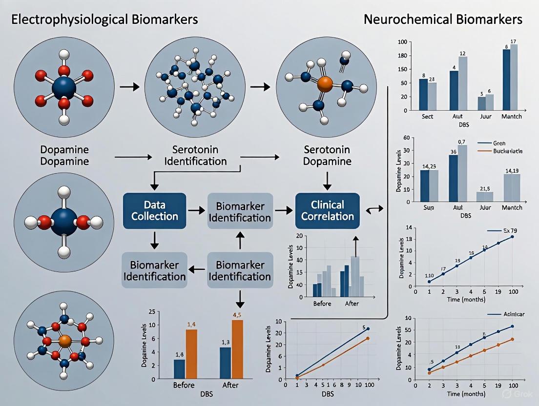
The following diagram illustrates the conceptual workflow for biomarker development and application in DBS research, integrating both electrophysiological and neurochemical data streams.
Diagram 1: Integrated biomarker development and application workflow in DBS research.
The Scientist's Toolkit: Essential Research Reagents and Materials
Table 3: Essential Research Reagents and Materials for Biomarker Studies in DBS
| Item | Function/Application | Specific Examples/Assays |
|---|---|---|
| High-Density EEG System | Recording cortical evoked potentials and oscillatory activity in response to DBS [7] | Intraoperative EEG recordings on the forehead during ALIC DBS surgery [7] [9] |
| Local Field Potential (LFP) Recording System | Acquiring subcortical neural signals directly from implanted DBS electrodes [4] | Sensing-enabled DBS systems for capturing STN power spectra (e.g., beta, gamma bands) [4] |
| Magnetoencephalography (MEG) | Non-invasive measurement of neural oscillations and coherence between deep brain structures and cortex [4] | Resting-state MEG combined with simultaneous LFP recordings [4] |
| Immunoassay Kits | Quantifying protein levels of neurochemical biomarkers in serum/plasma/CSF [6] | ELISA kits for sNfL, sGFAP, and sBDNF [6] |
| Probabilistic Tractography Software | Reconstructing white matter pathways connected to DBS targets for correlation with electrophysiology [7] | MRI-based tractography to link ALIC stimulation to connectivity with vmPFC/OFC and vlPFC [7] |
| Machine Learning Algorithms | Multivariate analysis of electrophysiological features to predict clinical outcomes [4] | Extreme Gradient Boosting (XGBoost) models to predict therapeutic window from STN power and coherence [4] |
Deep brain stimulation (DBS) has established itself as a transformative therapy for neurological and psychiatric disorders, yet the biological mechanisms underlying its therapeutic effects remain incompletely understood. In the broader context of biomarker research for DBS, two principal approaches have emerged: neurochemical biomarkers, which track neurotransmitter dynamics, and electrophysiological biomarkers, which measure rhythmic and evoked electrical activity in neural circuits. This review focuses on the latter category, examining how oscillatory rhythms and evoked potentials provide unique windows into brain network dynamics and therapeutic mechanisms. Unlike neurochemical approaches that often require invasive sampling or specialized imaging, electrophysiological biomarkers can be recorded directly from DBS electrodes themselves, creating opportunities for real-time therapy adjustment and personalized neuromodulation [10] [4].
The theoretical foundation for electrophysiological biomarkers rests on the principle that pathological brain states correlate with characteristic patterns of synchronized neural activity. In Parkinson's disease (PD), for instance, excessive beta oscillations (13-35 Hz) in the subthalamic nucleus (STN) associate with bradykinesia and rigidity, while in obsessive-compulsive disorder (OCD), low-frequency oscillations in cortico-striato-thalamo-cortical circuits reflect compulsive states [10] [4]. Similarly, evoked potentials generated by electrical stimulation provide insights into network connectivity and target engagement. The burgeoning research in this domain suggests that electrophysiological signatures offer superior temporal resolution and adaptive potential compared to static neurochemical measures, positioning them as critical tools for guiding DBS therapy in the evolving landscape of precision neurology.
Electrophysiological Biomarkers: Signaling Pathways and Experimental Workflows
Neural Circuit Dynamics and Biomarker Generation
The following diagram illustrates the fundamental signaling pathways through which electrophysiological biomarkers are generated and measured in DBS research, highlighting the relationship between pathological states, recording methodologies, and clinical applications:
This conceptual framework demonstrates how different pathological states generate distinct electrophysiological signatures that can be harnessed for specific clinical applications. The diagram highlights two primary categories of biomarkers: spontaneous oscillatory rhythms and stimulation-evoked potentials, both of which inform critical aspects of DBS therapy optimization.
Experimental Workflow for Biomarker Discovery
The translation of raw electrophysiological signals into clinically actionable biomarkers requires sophisticated experimental protocols and analytical pipelines. The following diagram outlines a generalized workflow for biomarker discovery and validation:
This experimental workflow highlights the multidisciplinary approach required for electrophysiological biomarker development, incorporating elements from signal processing, machine learning, and clinical neuroscience to transform raw recordings into validated clinical tools.
Comparative Analysis of Electrophysiological Biomarkers
Oscillatory Rhythm Biomarkers
Table 1: Oscillatory Rhythm Biomarkers in Neurological and Psychiatric Disorders
| Biomarker | Frequency Range | Primary Location | Clinical Correlation | Specificity/Accuracy |
|---|---|---|---|---|
| Beta Oscillations | 13-35 Hz | Subthalamic Nucleus (STN) | Bradykinesia, rigidity in PD [4] | 89.74% spatial accuracy in STN [11] |
| Delta/Alpha Power | 1-4 Hz / 8-13 Hz | External Globus Pallidus (GPe), ALIC | Compulsion states in OCD [10] | Universal across CSTC circuit structures [10] |
| High-Frequency Oscillations (HFOs) | 200-400 Hz | Subthalamic Nucleus (STN) | Therapeutic outcome in PD [11] | 82.05% spatial accuracy in STN [11] |
| Theta Oscillations | 4-8 Hz | Fornix, Hippocampus | Memory function in Alzheimer's [12] | Modulates alpha power during compulsions [10] |
Oscillatory rhythms represent endogenous patterns of synchronized neural activity that correlate with specific brain states and behaviors. In Parkinson's disease, beta oscillations in the STN have been extensively characterized as pathological signatures that normalize with both dopaminergic medication and effective DBS [4]. Recent research has demonstrated that beta power alone provides limited predictive value for therapeutic outcomes (ρ = -0.25), prompting investigation of multi-spectral approaches that incorporate additional frequency bands [11]. In OCD, a distinct pattern emerges with delta and alpha power increases observed across multiple basal ganglia structures during compulsive states, with GPe delta power specifically correlating with obsession severity (r = 0.77) [10]. This suggests that low-frequency oscillations may serve as transdiagnostic markers of pathological states across different neural circuits.
The spatial specificity of oscillatory biomarkers varies significantly by frequency band and anatomical location. Beta oscillations demonstrate particularly high spatial precision within the STN, with studies showing 89.74% accuracy for localizing sensorimotor regions compared to 82.05% for HFOs [11]. This spatial predictability makes oscillatory biomarkers valuable for both intraoperative targeting and postoperative programming, though their reliability can be affected by pharmacological state, arousal level, and disease progression. Unlike evoked potentials that require active stimulation, oscillatory rhythms can be recorded passively, enabling continuous monitoring of brain states for adaptive stimulation approaches.
Evoked Potential Biomarkers
Table 2: Evoked Potential Biomarkers in DBS Applications
| Biomarker | Components | Stimulation Parameters | Clinical Utility | Amplitude Correlation |
|---|---|---|---|---|
| DBS-Evoked Potentials (EPs) | ~35, ~75, ~120 ms peaks | 2 Hz monopolar, ALIC-EEG | Target engagement in OCD [7] [9] | Correlates with white matter connectivity (vmPFC/vlPFC) [7] |
| DBS-induced Local Evoked Potentials (DLEP) | 1-3 resonant peaks | Single-pulse or HF bursts, STN local | Contact selection in PD [11] | ρ = -0.33 with clinical outcomes [11] |
| Evoked Resonant Neural Activity (ERNA) | Stereotyped local response | Phase-locked bursts, STN | Biomarker for circuit reorganization [13] [14] | Modulated by beta phase stimulation [13] |
| Fornix-Evoked Potentials | Not specified | Fornix DBS, Alzheimer's | Hippocampal network modulation [12] | Associated with hippocampal volume preservation [12] |
Evoked potentials generated by electrical stimulation provide complementary information to spontaneous oscillations, reflecting the structural and functional connectivity of stimulated networks. DBS-evoked potentials (EPs) recorded via EEG during ALIC stimulation for OCD consistently demonstrate three oscillatory peaks at approximately 35, 75, and 120 ms, with amplitudes that vary significantly across contacts and correlate with optimal target engagement [7] [9]. These cortical responses reflect orthodromic activation of prefrontal circuits, with higher amplitudes predicting better white matter connectivity to ventromedial prefrontal cortex/orbitofrontal cortex and ventrolateral prefrontal cortex regions [7]. Non-responders to therapy exhibit less consistent EP waveforms across contacts, highlighting their potential predictive value.
Local evoked potentials, including DBS-induced local evoked potentials (DLEP) and evoked resonant neural activity (ERNA), provide even more direct measures of target engagement. DLEP recorded from adjacent contacts during STN stimulation shows superior spatial specificity (100% accuracy for single-pulse stimulation) and correlation with clinical outcomes (ρ = -0.33) compared to beta power [11]. These local responses are thought to reflect activation of reciprocal connections between basal ganglia structures, with amplitudes that mirror underlying tissue excitability. Recent advances in phase-locked DBS have demonstrated that ERNA amplitudes can be selectively modulated by stimulation delivered at specific beta phases, opening possibilities for finely tuned neuromodulation approaches that leverage these evoked responses as control signals [13].
Experimental Protocols and Methodologies
Intraoperative Evoked Potential Recording
The protocol for recording DBS-evoked potentials during electrode implantation surgery involves synchronized stimulation and recording systems [7] [9]. For ALIC DBS in OCD patients, researchers delivered monopolar stimulation at 2 Hz through each electrode contact while recording EEG responses from forehead electrodes. This approach capitalizes on the surgical access to directly assess target engagement by measuring downstream cortical responses. The recorded signals are processed through bandpass filtering and artifact rejection algorithms, with particular attention to consistent waveform morphology across trials. Evoked potential characteristics are then correlated with preoperative tractography data to verify alignment with desired white matter pathways. This method has demonstrated particular value for distinguishing treatment responders from non-responders based on waveform consistency across contacts [7].
For local evoked potential recordings such as DLEP/ERNA, the methodology shifts to local field potential recordings from adjacent DBS contacts during stimulation [11] [13]. Single-pulse or short-burst stimulation paradigms are typically employed to minimize adaptation effects and stimulation artifacts. Advanced artifact removal techniques, including template subtraction and Kalman filtering approaches, are essential for recovering the neural signals obscured by the massive stimulation artifact [13] [14]. The resulting waveforms are analyzed for characteristic components including latency, amplitude, and resonant properties, which are then mapped to anatomical positions and clinical outcomes. These local recordings provide immediate feedback on lead placement within the intended target structure, with higher amplitudes typically indicating optimal placement within the sensorimotor STN for PD applications [11].
Symptom-Provoked Oscillatory Recording
The investigation of oscillatory biomarkers in OCD employs a structured symptom provocation paradigm to link neural activity to specific disease states [10]. Patients implanted with sensing DBS devices undergo a standardized recording protocol comprising four sequential states: baseline (watching a neutral movie), obsession (personalized provocation of intrusive thoughts), compulsion (performance of ritualistic behaviors), and relief (subsidence of urge). Each phase lasts approximately 3 minutes, with continuous LFP recordings from multiple electrode pairs localized to specific basal ganglia structures. Patients provide periodic self-ratings of symptom severity using visual analog scales throughout the protocol, creating a temporal alignment between subjective experience and neural activity.
Analysis of the recorded signals focuses on time-frequency decomposition to quantify oscillatory power across standard frequency bands (delta, theta, alpha, beta, gamma) [10]. Statistical comparisons between behavioral states identify frequency-specific power changes associated with symptom expression, with particular emphasis on generalizable patterns across patients. For the identified oscillatory biomarkers, additional analyses examine cross-frequency coupling and correlation with symptom severity ratings. This approach has revealed that delta and alpha power increases during compulsions represent the most consistent transstructural biomarkers in OCD, observed across ALIC, GPe, NAc, and alAC [10]. Furthermore, the dissociation between motor and mental compulsions has helped identify which biomarkers reflect movement versus compulsive states more broadly.
Phase-Locked Stimulation Framework
Advanced closed-loop DBS approaches require precise timing of stimulation relative to ongoing brain rhythms, necessitating sophisticated phase-tracking methodologies [13] [14]. The implementation of phase-locked DBS involves a computer-in-the-loop system that continuously monitors oscillatory activity and delivers stimulation at predetermined phases. The technical pipeline begins with artifact suppression using Kalman filtering, which employs a blanking mechanism during stimulation artifacts and relies on an autoregressive model to reconstruct the underlying neural signal. This approach demonstrates superior performance compared to template subtraction or interpolation methods, particularly when stimulation duration is underestimated.
Following artifact removal, real-time phase estimation utilizes non-resonant oscillators to track instantaneous phase dynamics in targeted frequency bands [13] [14]. This method provides stable phase estimates without the latency issues associated with causal Hilbert transform approaches. The integrated system has demonstrated over 90% accuracy in delivering stimulation within ±π/2 radians of the target phase for STN beta rhythms. Validation experiments involve delivering stimulation at different phases and measuring both electrophysiological consequences (e.g., ERNA amplitude modulation) and behavioral effects (e.g., finger-tapping velocity in PD). This framework enables causal investigation of phase-dependent neuromodulation while establishing technical foundations for clinically viable closed-loop DBS systems.
The Scientist's Toolkit: Essential Research Reagents and Solutions
Table 3: Essential Methodologies and Analytical Tools for Electrophysiological Biomarker Research
| Category | Specific Tools/Methods | Primary Function | Key Advantages |
|---|---|---|---|
| Recording Platforms | Sensing DBS systems (Medtronic) | Simultaneous stimulation and LFP recording | Enables chronic biomarker investigation [10] |
| MEG-LFP simultaneous recording | Whole-brain coverage with STN signals | Correlates cortical and subcortical activity [4] | |
| Intraoperative EEG systems | Cortical evoked potential recording | Assesses target engagement during surgery [7] | |
| Stimulation Paradigms | Single-pulse stimulation | Evokes local and network responses | Minimal adaptation effects [11] |
| High-frequency burst stimulation | Activates resonant circuits | Mimics therapeutic DBS parameters [11] | |
| Phase-locked stimulation | Targets specific oscillation phases | Causal investigation of phase-effects [13] | |
| Artifact Management | Kalman filter artifact removal | Reconstructs neural signal during stimulation | Model-driven, handles variable artifacts [13] [14] |
| Template subtraction | Removes stereotyped artifacts | Effective for consistent artifact shapes [14] | |
| Blanking with interpolation | Replaces artifact-contaminated segments | Simple implementation for brief artifacts [14] | |
| Analytical Approaches | Time-frequency analysis | Quantifies oscillatory power | Links frequency-specific activity to behavior [10] |
| Tractography integration | Relates biomarkers to structural connectivity | Explains biomarker variability [7] | |
| Machine learning prediction | Predicts therapeutic windows from features | Multivariate biomarker integration [4] |
This methodological toolkit highlights the interdisciplinary nature of electrophysiological biomarker research, combining specialized hardware, stimulation paradigms, signal processing techniques, and analytical approaches. The selection of appropriate tools depends on the specific research question, with distinct advantages and limitations for each methodology.
Discussion: Integration with Neurochemical Biomarkers and Future Directions
The expanding evidence for electrophysiological biomarkers in DBS raises important questions about their relationship to neurochemical biomarkers and their respective roles in guiding therapy. While electrophysiological measures offer millisecond temporal resolution and direct links to circuit-level dysfunction, neurochemical biomarkers provide complementary information about synaptic function and neurotransmitter dynamics. The integration of these approaches remains largely unexplored but holds promise for a more comprehensive understanding of DBS mechanisms. For instance, the relationship between STN beta power and dopamine levels—two established biomarkers in PD—is complex and context-dependent, suggesting that multimodal biomarker integration may be necessary for optimal therapy personalization [4].
Future research directions should prioritize the validation of electrophysiological biomarkers in large, diverse patient cohorts and the development of standardized recording paradigms that enable cross-study comparisons. The translation of biomarkers from research tools to clinical decision aids requires demonstration of reliability, specificity, and practical utility in realistic clinical settings. Promisingly, machine learning approaches that integrate multiple electrophysiological features have already shown potential for predicting therapeutic windows and optimizing contact selection [4]. Additionally, the emergence of sensing-enabled DBS systems creates opportunities for chronic biomarker monitoring and adaptive stimulation, potentially revolutionizing neuromodulation therapy by making it responsive to the fluctuating brain states that characterize neurological and psychiatric disorders.
As the field progresses, electrophysiological biomarkers are poised to bridge the gap between abstract network-level theories of brain dysfunction and practical clinical management of DBS therapy. Their capacity to reflect both spontaneous and evoked activity across distributed networks provides a unique window into the neural circuits targeted by DBS, offering a path toward more precise, personalized, and effective neuromodulation approaches for a growing range of neurological and psychiatric conditions.
The pursuit of objective biological signatures, or biomarkers, is revolutionizing the application of Deep Brain Stimulation (DBS) for neurological and psychiatric disorders. Within this field, a central thesis is emerging: while electrophysiological biomarkers (e.g., local field potentials) have seen more rapid clinical integration, neurochemical biomarkers offer a profound, complementary window into the molecular mechanisms of neuromodulation. Electrophysiological biomarkers reflect the rhythmic, synchronized electrical activity of neuronal populations, such as beta-band oscillations (13-35 Hz) in the subthalamic nucleus of Parkinson's disease patients [15] [16]. In contrast, neurochemical biomarkers provide a direct measure of neurotransmitter dynamics—the chemical messengers that underlie synaptic communication, plasticity, and ultimately, brain function and behavior [17] [18]. Understanding the interplay between these two classes of biomarkers is critical for advancing from open-loop DBS systems, which deliver constant stimulation, to adaptive closed-loop systems that can respond in real-time to the brain's fluctuating neurochemical and electrical states [18].
This guide provides a comparative overview of the key neurotransmitter systems and signaling molecules that serve as neurochemical biomarkers in DBS research. We focus on the experimental data supporting their roles, the methodologies for their detection, and their potential for creating more personalized and effective neuromodulation therapies.
Key Neurotransmitter Systems in DBS
The therapeutic and side effects of DBS are mediated through the modulation of complex neurochemical networks. The table below summarizes the primary neurotransmitters involved, their documented changes with DBS, and their association with clinical outcomes.
Table 1: Key Neurochemical Biomarkers in Deep Brain Stimulation
| Neurotransmitter | Primary Role/System | Change with DBS (Key Brain Region) | Associated Clinical Outcome | Evidence Level |
|---|---|---|---|---|
| Dopamine | Nigrostriatal pathway, motor control, reward [17] [19] | Increased striatal release (STN-DBS) [19] [18] | Improvement in parkinsonian motor symptoms [19] [18] | Strong (Preclinical & Indirect Clinical) |
| Glutamate | Primary excitatory neurotransmitter [17] | Altered release; reduced excitotoxicity (STN-DBS) [19] | Motor improvement; potential neuroprotection [19] | Moderate (Preclinical) |
| GABA (γ-aminobutyric acid) | Primary inhibitory neurotransmitter [17] | Increased in pallidum (GPi-DBS); modulated balance [19] | Suppression of pathological network activity [19] | Moderate (Preclinical) |
| Serotonin | Mood, appetite, sleep [17] | Altered release (STN-DBS) [19] | Mood-related side effects [19] | Moderate (Preclinical) |
| Adenosine | Purinergic signaling, neuromodulation [18] | Increased in thalamus (VIM-DBS) [18] | Reduction in essential tremor [18] | Preliminary Clinical |
| Noradrenaline | Arousal, attention, stress response [17] | LC-NA system integrity required for STN-DBS efficacy [19] | Influences overall therapeutic response [19] | Moderate (Preclinical) |
Detailed Neurotransmitter Pathways and DBS Mechanisms
Dopaminergic Systems: The most robust neurochemical evidence involves dopamine. STN-DBS has been shown to induce phasic dopamine release in the striatum, a mechanism critical for its motor-improving effects [18]. This release is thought to help correct the dysfunctional balance between the direct and indirect pathways in the basal ganglia, a hallmark of Parkinson's pathology [19]. The efficacy of DBS may depend on the integrity of other systems, such as the noradrenergic locus coeruleus, highlighting the interconnected nature of neurotransmitter networks [19].
Glutamate and GABA Systems: DBS acts to rebalance excitatory and inhibitory tones. For instance, STN-DBS is thought to modulate the hyperdirect pathway, reducing excessive glutamatergic drive from the cortex to the STN and output nuclei [19]. Simultaneously, GPi-DBS may work by enhancing inhibitory GABAergic output from the pallidum to the thalamus, thereby suppressing aberrant motor commands [19].
Non-Canonical Signaling Molecules: Beyond classical neurotransmitters, molecules like adenosine have been identified as rapid-response biomarkers. Clinical studies using fast-scan cyclic voltammetry in the thalamus of essential tremor patients showed that DBS-induced tremor suppression correlated with a swift increase in adenosine, suggesting it as a potential feedback signal for closed-loop control [18].
Experimental Protocols for Neurochemical Analysis
Monitoring neurochemical dynamics in the context of DBS presents significant technical challenges. The following section outlines the primary methodologies employed in both preclinical and clinical research.
Primary Analytical Techniques
Table 2: Core Methodologies for Monitoring Neurochemical Biomarkers
| Method | Temporal Resolution | Spatial Resolution | Key Measurables | Main Advantage | Main Limitation |
|---|---|---|---|---|---|
| Microdialysis | Minutes | ~1 mm (probe size) | Glutamate, GABA, dopamine, serotonin metabolites | Measures a wide range of neurochemicals; well-established | Poor temporal resolution; large probe size |
| Fast-Scan Cyclic Voltammetry (FSCV) | Sub-second (ms) | μm around electrode | Dopamine, serotonin, adenosine | Excellent temporal resolution for "phasic" release | Limited to electroactive molecules; electrode fouling |
| Enzyme-Based Biosensors | Seconds to minutes | μm around sensor | Glutamate, GABA, lactate | Can target non-electroactive molecules | Stability and biocompatibility over long term |
| Positron Emission Tomography (PET) | Minutes | 3-5 mm (system dependent) | Synaptic dopamine release (via receptor ligands) | Translational; applicable in humans | Indirect measure; poor temporal resolution; radiation exposure |
Detailed Workflow for Preclinical FSCV During DBS
A common protocol for real-time monitoring of electroactive neurotransmitters like dopamine involves combining DBS with FSCV in animal models.
Figure 1: Experimental workflow for combining FSCV with DBS in preclinical models.
Key Steps:
- Electrode Implantation: A DBS electrode is stereotactically implanted into the target structure (e.g., STN). A carbon-fiber microelectrode for FSCV is implanted in the projection area of interest (e.g., striatum) [18].
- Stimulation and Recording: DBS is applied using parameters typical for the condition (e.g., high-frequency for Parkinson's disease). Concurrently, a triangular waveform (e.g., -0.4 V to +1.3 V and back, at 400 V/s) is applied to the FSCV electrode at 10 Hz [18].
- Data Acquisition and Analysis: The resulting current is measured. Background currents are subtracted to reveal Faradaic currents from the oxidation and reduction of neurotransmitters. The unique voltage signature (voltammogram) of dopamine or other molecules allows for its identification [18].
- Calibration: The electrode is calibrated post-hoc in a solution containing a known concentration of the analyte to convert the electrochemical current to a molar concentration.
The Scientist's Toolkit: Essential Research Reagents and Materials
Successful investigation of neurochemical biomarkers requires a suite of specialized tools and reagents.
Table 3: Essential Research Reagents and Materials for Neurochemical DBS Studies
| Category / Item | Specific Examples | Function in Research |
|---|---|---|
| DBS Electrodes | Medtronic 3387/3389, directional leads | Implanted into deep brain targets to deliver electrical stimulation. |
| Neurochemical Sensing Electrodes | Carbon-fiber microelectrodes (for FSCV), Enzyme-coated biosensors (e.g., glutamate oxidase for glutamate sensing) | Detect and measure real-time changes in specific neurotransmitter concentrations. |
| In Vivo Micropumps | Syringe pumps for microinfusion, Microdialysis pumps | Deliver drugs or artificial cerebrospinal fluid (aCSF) locally during experiments. |
| Key Reagents & Chemicals | Artificial Cerebrospinal Fluid (aCSF), Neurotransmitter standards (DA, 5-HT, Glu), Enzyme inhibitors (e.g., for GABA uptake) | Maintain physiological conditions; calibrate sensors; manipulate specific neurochemical systems. |
| Animal Models | 6-OHDA lesioned rats, MPTP-treated primates | Provide a pathophysiological model of human disorders (e.g., Parkinson's disease) for testing DBS mechanisms. |
| Data Acquisition Systems | Multichannel systems (e.g., from Tucker-Davis Technologies), Fast-stat potentiostats (for FSCV) | Record neural signals (LFPs) and perform electrochemical measurements synchronously with DBS. |
Comparative Analysis: Neurochemical vs. Electrophysiological Biomarkers
The choice between neurochemical and electrophysiological biomarkers is not a matter of superiority, but of application. Each offers distinct advantages and faces unique challenges.
Figure 2: Comparative strengths and challenges of neurochemical and electrophysiological biomarkers.
Current State of the Field:
- Electrophysiological biomarkers are currently more advanced in clinical translation. Beta-band power in the STN is a validated biomarker for Parkinsonian symptoms and has been successfully used to guide adaptive DBS systems, which adjust stimulation based on real-time neural feedback [18] [15] [16]. Algorithms for automatically detecting beta peaks are now highly sophisticated, achieving performance comparable to expert consensus [16].
- Neurochemical biomarkers remain primarily in the research domain but hold immense promise. They provide a more direct link to the synaptic actions of drugs and stimulation. The development of closed-loop DBS based on neurochemical feedback, such as dopamine or adenosine release, is an active area of investigation that could lead to highly personalized therapies [18].
Neurochemical biomarkers represent a frontier in the pursuit of precision neuromodulation. While the field has moved beyond the simplistic view of DBS as a mere "functional lesion," a deep understanding of its neurochemical mechanisms remains elusive. The future of DBS research lies in multi-modal biomarker integration, combining the high temporal resolution of electrophysiology with the molecular specificity of neurochemistry. This approach will be essential for developing the next generation of closed-loop systems that can adapt not only to the brain's electrical rhythms but also to its complex chemical dialogue. Overcoming the significant technical hurdles in long-term, stable neurochemical sensing will be critical. As these tools evolve, they will unlock a new era of personalized, responsive, and more effective DBS therapies for a broader range of neurological and psychiatric disorders.
Deep Brain Stimulation (DBS) has established itself as a powerful therapeutic intervention for a range of otherwise treatment-resistant neurological and neuropsychiatric disorders. Its clinical efficacy is well-established in Parkinson's disease (PD) and essential tremor, with growing evidence supporting its use in obsessive-compulsive disorder (OCD) and major depressive disorder (MDD) [20]. Despite its expanding clinical application, the neurobiological mechanisms through which DBS exerts its therapeutic effects remain incompletely understood. The prevailing hypothesis posits that DBS acts by modulating dysfunctional neural circuits and neurochemical systems, effectively "repairing" pathological brain activity [21]. Current research focuses on two primary categories of biomarkers to unravel these mechanisms: electrophysiological signatures (such as local field potentials) and neurochemical changes (in neurotransmitters like dopamine, GABA, and glutamate). Understanding these complementary aspects is crucial for optimizing stimulation parameters, enhancing efficacy, and developing next-generation DBS systems.
The history of mechanistic theories for DBS has evolved significantly. Initially, the "reversible lesion" hypothesis suggested that DBS simply inhibited overactive brain structures, similar to the effects of surgical lesions [22]. This was subsequently challenged and refined into the "informational lesion" concept, which proposes that high-frequency stimulation disrupts the transmission of pathological neural signals through the stimulated region without necessarily destroying tissue [23]. More recent network-based theories emphasize that DBS modulates distributed neural circuits, particularly those involving cortico-striato-thalamo-cortical (CSTC) pathways, which are implicated in multiple neurological and psychiatric conditions [23] [24].
Electrophysiological Biomarkers in DBS Research
Electrophysiological biomarkers provide real-time readouts of neuronal population activity and network dynamics. These biomarkers are typically derived from local field potentials (LFPs), which are extracellular electrical signals that reflect the synchronized synaptic activity of neuronal populations [10]. The frequency and amplitude of LFP oscillations have been functionally linked to cognitive processes, neuronal communication, and information processing throughout the brain [10].
Key Electrophysiological Findings in OCD
Groundbreaking research has identified specific LFP signatures associated with core symptoms of OCD. A 2025 study recording from sensing DBS electrodes in different basal ganglia structures during personalized symptom provocation identified two general markers of compulsion: delta (δ) and alpha (α) LFP power was significantly increased during compulsions across multiple brain regions, including the external globus pallidus (GPe), nucleus accumbens (NAc), and anterior limb of the internal capsule (ALIC) [10].
Table 1: Electrophysiological Biomarkers in OCD Compulsions
| Brain Region | Delta (δ) Power Change | Alpha (α) Power Change | Functional Correlation |
|---|---|---|---|
| External Globus Pallidus (GPe) | Significant increase | Significant increase | Correlated with OCD symptom severity |
| Anterior Limb of Internal Capsule (ALIC) | Significant increase | Significant increase | Modulated by theta phase during compulsions |
| Nucleus Accumbens (NAc) | Significant increase | Significant increase | Primarily motor component of compulsions |
| Anterior Lateral Anterior Commissure (alAC) | Significant increase | Significant increase | Action-dependent compulsion signals |
When researchers distinguished between motor and mental compulsions, they found that increased delta power during non-motor/mental compulsions persisted only in ALIC and GPe, suggesting these signals may represent universal biomarkers of compulsivity unconfounded by motor function [10]. Furthermore, a meaningful connection was established between these subcortical signals and clinical experience: GPe delta power correlated with OCD symptom severity [10].
Another electrophysiological approach involves DBS-evoked potentials (EPs) recorded with electroencephalography (EEG). Intraoperative studies in OCD patients undergoing ALIC DBS have revealed consistent EPs with three oscillatory peaks (approximately 35, 75, and 120 ms) across patients [9]. Importantly, EP amplitude varied across contacts, with the largest responses occurring when stimulation overlapped with preoperatively defined tractographic targets. Higher EP amplitudes correlated with greater white matter connectivity to prefrontal cortical regions, and treatment nonresponders exhibited less consistent EP waveforms [9].
Experimental Protocols for Electrophysiological Recording
The methodology for identifying these biomarkers typically involves:
- Patient Implantation: Surgical implantation of sensing DBS electrodes (e.g., Medtronic DBS systems) in target structures [10].
- Symptom Provocation: Structured experiments involving baseline recording, followed by personalized provocation of obsessions and compulsions while patients report symptom severity on visual analog scales [10].
- LFP Recording: Continuous LFP recordings from DBS electrodes across different behavioral states (baseline, obsession, compulsion, relief) [10].
- Signal Processing: Time-frequency analysis of LFP data using non-parametric randomization tests and ANOVA with multiple comparison corrections to identify significant power changes across frequency bands [10].
- Correlation Analysis: Relating LFP power changes to clinical symptom severity scores and behavioral states [10].
Diagram 1: Electrophysiological Biomarker Discovery Workflow. This flowchart illustrates the standardized experimental protocol for identifying local field potential (LFP) biomarkers in DBS research, from surgical implantation through data analysis to biomarker validation.
Neurochemical Mechanisms of DBS
While electrophysiological approaches capture neuronal population dynamics, neurochemical measurements provide complementary insight into neurotransmitter systems modulated by DBS. The leading hypothesis suggests that DBS induces its therapeutic effects not only by altering electrical activity but also by modifying the release of key neurotransmitters including dopamine, serotonin, glutamate, and GABA [20] [25].
Neurochemical Measurements in DBS Research
Advanced electrochemical sensing techniques have enabled researchers to measure neurotransmitter dynamics in both preclinical and clinical settings. The primary methods include:
Table 2: Neurochemical Measurement Techniques in DBS Research
| Technique | Temporal Resolution | Key Advantages | Limitations | Applications in DBS |
|---|---|---|---|---|
| Microdialysis | >1 minute (slow) | Broad molecular detection range; well-established | Significant tissue damage; poor temporal resolution | Baseline neurotransmitter levels [25] |
| Fast-Scan Cyclic Voltammetry (FSCV) | Sub-second (fast) | High sensitivity and selectivity; measures phasic release | Limited to stimulation-evoked release; electrode fouling | Real-time dopamine dynamics in PD [25] |
| Multiple-Cyclic Square Wave Voltammetry (M-CSWV) | Sub-second (fast) | Measures tonic neurotransmitter levels | More complex data analysis | Tonic vs. phasic neurotransmitter release [25] |
| Fiber Photometry with Genetically Encoded Sensors | Sub-second (fast) | Cell-type specific measurements; minimal tissue damage | Requires genetic manipulation | Glutamate/GABA release in STN [22] |
Key Neurochemical Findings Across Disorders
Research has revealed disorder-specific neurochemical changes in response to DBS:
Parkinson's Disease: A seminal study using spectrally resolved fiber photometry with genetically encoded sensors in PD mouse models demonstrated that high-frequency DBS of the subthalamic nucleus (STN) activates afferent axons while inhibiting STN neurons [22]. These contrasting presynaptic and postsynaptic effects arise from a decrease in local neurotransmitter release with a larger decrease in glutamate than GABA, shifting the excitation/inhibition balance toward inhibition [22]. Chemogenetic inhibition, but not excitation, of STN neurons mimics the therapeutic effects of DBS in PD models, suggesting that inhibition of STN is a key mechanism of therapeutic DBS [22].
Addiction Disorders: DBS targeting the nucleus accumbens (NAc) demonstrates that high-frequency stimulation significantly increases extracellular GABA without altering glutamate levels in brain areas implicated in addiction [21]. This effect appears mediated by DBS inhibiting the GABA uptake system rather than promoting vesicular GABA release. The increased GABA concentration subsequently calms hyperactive circuits linked to drug-seeking behaviors [21].
Obsessive-Compulsive Disorder: While specific neurochemical data for OCD is less extensive, the involvement of CSTC circuits suggests modulation of multiple neurotransmitter systems. The anterior limb of the internal capsule (ALIC), a common DBS target for OCD, contains fibers connecting prefrontal cortical regions with subcortical structures, involving dopaminergic, serotonergic, and glutamatergic pathways [23].
Direct Comparison: Electrophysiological vs. Neurochemical Biomarkers
The pursuit of DBS mechanisms has proceeded along two parallel tracks: electrophysiological measurements of neuronal oscillations and neurochemical assessments of neurotransmitter dynamics. Each approach offers distinct advantages and limitations for understanding DBS effects and developing improved therapies.
Table 3: Electrophysiological vs. Neurochemical Biomarkers in DBS Research
| Parameter | Electrophysiological Biomarkers | Neurochemical Biomarkers |
|---|---|---|
| Measured Signal | Local field potentials (LFPs), evoked potentials | Neurotransmitter concentrations (dopamine, GABA, glutamate, etc.) |
| Temporal Resolution | Millisecond to second range | Second to minute range (depending on technique) |
| Spatial Specificity | Regional (captures population activity) | Can be cell-type specific with advanced techniques |
| Clinical Translation | Already implemented in sensing DBS devices | Limited to research settings; technical challenges remain |
| Key Findings | Delta/alpha power increases during OCD compulsions [10] | Differential glutamate vs. GABA depression in STN DBS [22] |
| Advantages | Real-time symptom correlation; network-level insights | Direct measurement of neurochemical imbalances |
| Limitations | Indirect measure of neuronal activity; complex interpretation | Invasive measurement techniques; limited multiplexing |
Integrated Mechanisms: From Synapses to Networks
The most compelling mechanistic framework for DBS integrates both electrophysiological and neurochemical perspectives. The "differential synaptic depression" hypothesis emerging from recent research suggests that high-frequency DBS causes a decrease in local neurotransmitter release, with a larger decrease in glutamate than GABA, thereby shifting the excitation/inhibition balance toward inhibition [22]. This neurochemical change manifests electrophysiologically as altered oscillatory patterns across neural networks.
Diagram 2: Integrated DBS Mechanism: Differential Synaptic Depression. This diagram illustrates the current leading hypothesis of DBS mechanisms, integrating presynaptic activation with postsynaptic inhibition mediated by unequal reduction in neurotransmitter release.
The therapeutic effects of DBS appear to operate through multiple complementary mechanisms:
Immediate Neurotransmitter Modulation: DBS induces rapid changes in neurotransmitter release, particularly affecting GABA and glutamate systems, which can immediately alter circuit function [22] [21].
Disruption of Pathological Oscillations: By overriding abnormal firing patterns, DBS disrupts synchronized oscillations in disease-relevant circuits, such as excessive beta power in PD or low-frequency increases in OCD [10] [21].
Long-Term Neuroplasticity: With chronic stimulation, DBS induces neuroadaptations and structural changes that may reverse maladaptive plasticity associated with disease progression [21].
Network-Wide Effects: Through antidromic and orthodromic activation, DBS influences distributed brain networks beyond the immediate stimulation site, particularly prefrontal networks in psychiatric disorders [24].
Advancing DBS research requires specialized methodologies and reagents. The following table summarizes key resources for investigating DBS mechanisms:
Table 4: Essential Research Resources for DBS Mechanism Studies
| Resource Category | Specific Tools | Research Applications | Key Features |
|---|---|---|---|
| Electrophysiology Systems | Sensing DBS electrodes; EEG systems | LFP recording; evoked potentials | Multi-contact designs; sensing capabilities [10] [9] |
| Neurochemical Sensors | Fast-scan cyclic voltammetry; fiber photometry | Real-time neurotransmitter monitoring | Genetically encoded sensors (GCaMP, iGluSnFR) [22] |
| Animal Models | 6-OHDA PD model; OCD models | Preclinical therapeutic testing | Disease-relevant pathophysiology [22] |
| Neuromodulation Techniques | Chemogenetics (DREADDs); optogenetics | Circuit-specific manipulation | Cell-type specificity; reversible modulation [22] |
| Neural Tractography | Diffusion MRI; probabilistic tractography | Surgical targeting; connectivity analysis | Preoperative planning; target optimization [9] |
| Computational Tools | Signal processing algorithms; machine learning | Data analysis; closed-loop control | Feature extraction; adaptive stimulation [25] |
The leading hypothesis for DBS mechanisms has evolved from simplistic "inhibition" or "excitation" models toward a sophisticated understanding that integrates neurochemical and electrophysiological perspectives. The differential synaptic depression hypothesis, where DBS causes a greater reduction in glutamate release compared to GABA, thereby shifting the excitation/inhibition balance toward inhibition, represents the current frontier in mechanistic understanding [22]. Simultaneously, research has identified distinct electrophysiological biomarkers—particularly increased delta and alpha power during OCD compulsions—that correlate with symptom states and severity [10].
The future of DBS mechanism research lies in integrating these complementary perspectives. Electrophysiological biomarkers offer the temporal resolution and clinical practicality needed for adaptive DBS systems, while neurochemical measurements provide the molecular specificity to understand fundamental disease processes and treatment effects. The ongoing development of closed-loop DBS systems that respond to pathological neural signatures will likely incorporate both types of biomarkers to deliver more personalized and effective neuromodulation therapies [25]. As sensing technologies advance and our understanding of brain circuits deepens, the integration of electrophysiological and neurochemical approaches will continue to illuminate the complex mechanisms by which DBS modulates neural circuits to alleviate suffering from treatment-resistant neurological and psychiatric disorders.
The Critical Need for Biomarkers in an Expanding DBS Landscape
Deep Brain Stimulation (DBS) has evolved into an established therapy for neurological and psychiatric disorders, yet its application remains hampered by a fundamental challenge: the lack of objective, measurable biological indicators to guide treatment. The expanding landscape of DBS now includes targets for Parkinson's disease (PD), epilepsy, obsessive-compulsive disorder (OCD), and other conditions, intensifying the need for biomarkers to personalize therapy. Biomarkers—objective measures of biological processes—are revolutionizing DBS by enabling precise target engagement, predicting clinical response, and informing adaptive stimulation strategies. Within this context, a critical comparison emerges between two dominant biomarker categories: electrophysiological biomarkers, which capture electrical brain activity, and neurochemical biomarkers, which measure neurotransmitter dynamics. This guide provides researchers and drug development professionals with a comparative analysis of these biomarker classes, supported by experimental data and methodological protocols.
Electrophysiological Biomarkers for DBS
Electrophysiological biomarkers are derived from recordings of the brain's electrical activity, spanning scales from large-scale cortical oscillations to single-neuron firing. These signals provide real-time feedback on neural circuit dynamics and are increasingly integrated into clinical DBS systems.
Key Electrophysiological Biomarkers and Applications
Table 1: Electrophysiological Biomarkers in DBS Applications
| Biomarker Type | Recording Method | Primary Clinical Applications | Key Findings & Performance Data | References |
|---|---|---|---|---|
| Local Field Potentials (LFP) | Implanted DBS leads | Parkinson's disease (PD), Dystonia | - PD: Suppression of pathological beta band (13-35 Hz) oscillations in the Subthalamic Nucleus (STN) correlates with motor improvement. Closed-loop DBS using beta power improved UPDRS-III scores by 50% vs. 30% with open-loop.- Dystonia: Heightened low-frequency (4-12 Hz) oscillations in the Globus Pallidus internus (GPi) correlate with symptom severity and are suppressed by therapeutic DBS. | [26] [18] |
| Cortical Evoked Potentials (cEP) | Scalp Electroencephalography (EEG) | Obsessive-Compulsive Disorder (OCD), Depression | - OCD: Intraoperative ALIC DBS evokes potentials with three oscillatory peaks (~35, ~75, ~120 ms). Amplitude correlates with white matter connectivity to prefrontal targets and distinguished treatment responders from non-responders. | [27] [9] |
| Thalamic Stereotactic-EEG (sEEG) | Intracranial depth electrodes | Epilepsy | - Mapping thalamocortical effective connectivity via evoked potentials can predict engagement of seizure networks. Spectral features from thalamic sEEG can guide ambulatory seizure detection parameters for sensing-enabled DBS devices. | [28] |
Experimental Protocol: Intraoperative Evoked Potential Recording for ALIC DBS in OCD
Objective: To establish evoked potentials (EPs) as a biomarker for target engagement and clinical efficacy in Deep Brain Stimulation of the Anterior Limb of the Internal Capsule for Obsessive-Compulsive Disorder.
- Coholders: 10 patients with severe, treatment-resistant OCD undergoing awake ALIC DBS implantation.
- Tractography-Based Targeting: Preoperative surgical planning using probabilistic tractography to define the optimal target within the ALIC, maximizing connectivity to ventromedial prefrontal cortex/orbitofrontal cortex and ventrolateral prefrontal cortex.
- Stimulation: Monopolar, cathode-first symmetrical biphasic pulses were delivered successively through all four contacts and directional segments of the implanted DBS lead.
- Stimulation Parameters: 2 Hz frequency, 90 μs pulse width per phase.
- Amplitude: 3.5 mA for directional contacts, 5.5 mA for ring contacts.
- EEG Recording: Cortical responses were recorded using 4 forehead electrodes (FP1, FP2, AF7, AF8) at a 22 kHz sampling rate, referenced to the right mastoid.
- Signal Processing:
- Bipolar Referencing: AF7-AF8 for left hemisphere, AF8-AF7 for right hemisphere.
- Filtering: High-pass filter (0.5 Hz) to remove baseline drift, followed by a bandpass filter (5-50 Hz) to visualize evoked responses.
- Artifact Removal: The stimulation artifact was removed via linear interpolation between -1 ms and 4 ms.
- Epoching & Averaging: The signal was epoched time-locked to the stimulation artifact, and EPs were obtained by averaging across trials.
- Feature Extraction: The magnitude of the EP was quantified as the Area Under the Curve between 10 ms and 100 ms post-stimulus, normalized for stimulation amplitude.
Electrophysiological Biomarker Workflow
The following diagram illustrates the typical workflow for developing and utilizing electrophysiological biomarkers in DBS research and clinical application.
Neurochemical Biomarkers for DBS
Neurochemical biomarkers involve the measurement of neurotransmitter and neuromodulator dynamics in the brain. While technologically more challenging, they offer direct insight into the molecular mechanisms underlying DBS therapy.
Key Neurochemical Biomarkers and Applications
Table 2: Neurochemical Biomarkers in DBS Applications
| Biomarker | Measurement Technique | Primary Clinical Applications | Key Findings & Performance Data | References |
|---|---|---|---|---|
| Dopamine | Fast-Scan Cyclic Voltammetry (FSCV) | Parkinson's disease (PD) | - STN DBS induces striatal phasic dopamine release in animal models, suggesting dopaminergic modulation contributes to therapeutic effect. Real-time monitoring is a potential feedback mechanism for closed-loop systems. | [18] |
| Adenosine | Fast-Scan Cyclic Voltammetry (FSCV) | Essential Tremor (ET) | - VIM thalamus DBS in ET patients caused a rapid, significant increase in adenosine oxidation currents, corresponding to a reduction in hand tremor. Suggests adenosine as a potential feedback biomarker. | [18] |
| Serum Neurofilament Light (sNfL) | Immunoassay | Parkinson's disease (PD) | - sNfL is elevated in PD patients vs. healthy controls, indicating neuroaxonal damage. DBS surgery causes a transient increase in sNfL and GFAP, but levels normalize after 1 year, suggesting DBS does not promote chronic neurodegeneration. | [6] |
| Gamma-aminobutyric acid (GABA) | Microbiosensors | Essential Tremor (ET) | - Evidence for GABAergic dysfunction in the cerebellum in ET. A dual microbiosensor for GABA and glutamate has been described, though sensitivity for physiological concentrations remains a challenge. | [18] |
Experimental Protocol: Neurochemical Monitoring for Closed-Loop DBS
Objective: To utilize real-time neurochemical measurements as a feedback control signal for adaptive deep brain stimulation.
Methodology Summary [18]:
- Electrochemical Sensing: The core technology is Fast-Scan Cyclic Voltammetry (FSCV).
- Principle: A microelectrode is implanted in the target brain region (e.g., striatum for PD, thalamus for ET). A rapid, repeating triangular voltage waveform is applied to the electrode, oxidizing and reducing electroactive neurochemicals like dopamine and adenosine.
- Measurement: The resulting oxidation and reduction currents are measured, providing a "electrochemical fingerprint" that identifies the specific molecule and its concentration changes with high temporal (subsecond) and spatial (submillimeter) resolution.
- Synchronization: Neurochemical measurements are synchronized with behavioral or physiological readouts (e.g., accelerometry for tremor quantification in ET) and DBS stimulation parameters.
- Closed-Loop Control Algorithm: A feedback algorithm is designed to modulate DBS parameters (e.g., amplitude, frequency) based on predefined thresholds of neurochemical concentration. For example, stimulation amplitude might be increased when adenosine levels fall below a certain threshold in ET.
- Validation: The therapeutic efficacy of the neurochemical-guided closed-loop DBS is compared to open-loop, continuous stimulation using standardized clinical rating scales (e.g., UPDRS-III for PD).
Comparative Analysis: Electrophysiological vs. Neurochemical Biomarkers
Table 3: Direct Comparison of Electrophysiological and Neurochemical Biomarkers
| Feature | Electrophysiological Biomarkers | Neurochemical Biomarkers |
|---|---|---|
| Measured Entity | Electrical potentials from neural populations (oscillations, evoked potentials) | Concentration dynamics of neurotransmitters & neuromodulators (e.g., Dopamine, Adenosine) |
| Primary Technologies | EEG, MEG, Local Field Potentials (LFP), Microelectrode Recording (MER) | Fast-Scan Cyclic Voltammetry (FSCV), Amperometry, Microbiosensors |
| Temporal Resolution | Excellent (Milliseconds to microseconds) | Excellent (Subsecond to seconds) |
| Spatial Resolution | Variable (Poor for scalp EEG, high for LFP/MER) | High (Micrometer scale with FSCV) |
| Clinical Translation | More Advanced (Integrated into commercial DBS systems for PD) | Emerging (Predominantly in preclinical and early clinical research) |
| Key Advantage | Directly reflects network-level brain dynamics and pathology; suitable for rapid closed-loop control. | Provides insight into molecular mechanisms of DBS and disease; direct link to neuropharmacology. |
| Key Challenge | Specificity can be limited; signals can be contaminated by artifacts. | Limited to electroactive molecules; biofouling and long-term stability of sensors. |
| Exemplary Finding | ALIC DBS EPs predict clinical response in OCD [27] [9]. | VIM DBS releases adenosine to reduce tremor in ET [18]. |
The Scientist's Toolkit: Essential Research Reagents and Materials
Table 4: Key Reagents and Solutions for DBS Biomarker Research
| Reagent / Material | Function / Application | Specific Examples / Notes |
|---|---|---|
| Directional DBS Leads | Allows for precise steering of electrical current and assessment of different fiber pathways. Critical for mapping biomarker responses. | SenSight leads used to stimulate directional segments and record EPs in ALIC [27]. |
| High-Contrast MRI Sequences | Enables direct visualization of small subcortical targets for accurate lead placement and post-op localization. | FGATIR for ANT; MP2RAGE for CM nucleus; EDGE-MICRA for CM/Pf complex [28]. |
| Probabilistic Tractography Software | Reconstructs white matter pathways from diffusion MRI data. Used for pre-operative target planning and correlating biomarker responses with structural connectivity. | Used to define ALIC connectivity to vmPFC/OFC and vlPFC; correlates with EP amplitude [27] [9]. |
| Fast-Scan Cyclic Voltammetry (FSCV) Setup | For real-time, in vivo detection of electroactive neurochemicals. Includes carbon-fiber microelectrodes, potentiostat, and data acquisition software. | Key for measuring adenosine dynamics in ET and dopamine in PD models [18]. |
| Immunoassay Kits | For quantifying serum or CSF-based protein biomarkers of neurodegeneration and neuroplasticity. | Used to measure sNfL, sGFAP, and BDNF levels in PD patients pre- and post-DBS [6]. |
| Computational Modeling Software | For electric field modeling of DBS and estimating the volume of tissue activated. Correlates stimulation location with clinical and biomarker outcomes. | Used to define "sweet spots" and model overlap with tracts like the DRTt in PD [28] [29]. |
The expanding therapeutic landscape of DBS creates a critical need for biomarkers to guide personalized, effective neuromodulation. Both electrophysiological and neurochemical biomarkers offer powerful, complementary paths forward. Electrophysiological biomarkers, with their strong clinical foothold and capacity for real-time network monitoring, are currently leading the transition to adaptive DBS systems. Neurochemical biomarkers, while less mature, provide a unique window into the molecular underpinnings of disease and therapy, promising a future where DBS can be tailored based on specific neurotransmitter dysfunctions. The convergence of these approaches—integrating electrical brain signals with molecular dynamics—holds the greatest potential. Future research must focus on standardizing measurement protocols, validating biomarkers in large, diverse cohorts, and developing integrated, dual-sensing next-generation DBS platforms that can simultaneously record both electrical and chemical brain activity to fully realize the promise of precision neuromodulation.
Methodology and Clinical Translation: From Signal Acquisition to Therapeutic Application
In the pursuit of refining Deep Brain Stimulation (DBS) therapies, the choice of biomarkers is pivotal. While neurochemical biomarkers provide insights into molecular changes, electrophysiological biomarkers offer a direct, real-time window into neural circuit dynamics. This guide objectively compares three core electrophysiological signal modalities—Electroencephalography (EEG), Local Field Potentials (LFP), and the strategies enabling Adaptive Deep Brain Stimulation (aDBS). We detail their performance, experimental protocols, and integration into a responsive neuromodulation framework, providing a structured resource for researchers and drug development professionals.
Electrophysiological Signal Modalities at a Glance
The table below summarizes the key characteristics, applications, and data supporting the use of EEG, LFP, and aDBS in clinical research.
Table 1: Comparative Overview of Electrophysiological Signal Modalities in DBS Research
| Feature | Scalp EEG | Local Field Potentials (LFP) | Adaptive DBS (aDBS) |
|---|---|---|---|
| Spatial Resolution | Low (whole-brain macro-scale activity) | High (micro-scale activity near DBS electrode) | High (derived from LFP or other sensing inputs) |
| Temporal Resolution | Very High (milliseconds) | Very High (milliseconds) | High (real-time processing) |
| Primary Application in DBS | Monitoring cortical evoked potentials & network effects [7] [9] | Identifying symptom-specific pathophysiological oscillations [30] [31] | Delivering symptom-contingent, closed-loop stimulation [30] |
| Key Experimental Findings | ALIC DBS evoked 3 oscillatory peaks (~35, ~75, ~120 ms); amplitude correlated with target engagement and clinical response in OCD [7] [9]. | STN beta power (~13-30 Hz) correlates with bradykinesia/rigidity severity (explains ~17% of symptom variance) [30]. Beta bursts and other bands also informative [31]. | aDBS using beta-band LFP feedback shown to effectively suppress symptoms and reduce side effects compared to conventional stimulation [30]. |
| Quantitative Data | Evoked potential amplitude correlates with white matter connectivity to vmPFC/OFC and vlPFC [7]. | Pooled correlation between beta power and bradykinesia/rigidity: R ≈ 0.45 (range 0.3-0.84 across studies) [30]. | Machine learning models using LFP features can predict therapeutic window of electrode contacts (r=0.45, p<0.001) [4]. |
| Main Advantages | Non-invasive; excellent for capturing cortical network engagement. | Direct recording from deep brain targets; causal link to symptoms. | Potential for automated, personalized therapy; reduced energy use and side effects. |
| Main Limitations | Indirect measure of deep brain activity; susceptible to artifacts. | Invasive; signals are localized to implanted target region. | Complexity of algorithm development and validation; reliant on a reliable physiomarker. |
Experimental Protocols and Methodologies
Intraoperative EEG for Target Engagement
Objective: To validate DBS lead placement and predict clinical outcomes by recording cortical responses to intraoperative stimulation [7] [9].
Key Workflow Steps:
- Patient Preparation: 10 patients with treatment-resistant OCD undergoing ALIC-DBS surgery [7].
- Stimulation: Monopolar, 2 Hz stimulation delivered through each contact of the externalized DBS lead [7] [9].
- EEG Recording: Scalp EEG electrodes placed on the forehead to record evoked potentials (EPs) [7].
- Data Analysis: EPs are analyzed for latency and amplitude of characteristic peaks (~35, ~75, ~120 ms). Amplitude is correlated with:
- Outcome: Contacts producing larger EP amplitudes correlated with optimal target overlap and better clinical response, serving as a biomarker for intraoperative target verification [7].
Identifying Symptom-Specific LFP Correlates
Objective: To discover and validate LFP signatures ("physiomarkers") that correlate with specific symptom severity for use in aDBS [30].
Key Workflow Steps:
- Patient & Recording: LFP signals are recorded from implanted DBS electrodes (e.g., in the Subthalamic Nucleus - STN) in patients with Parkinson's disease, typically after overnight withdrawal of medication [30] [4].
- Clinical Assessment: Simultaneous to LFP recording, motor symptoms (bradykinesia, rigidity, tremor) are assessed using standardized clinical scales like the UPDRS-III [30].
- Signal Processing: LFPs are processed to extract signal features, most commonly spectral power in specific frequency bands (e.g., beta: 13-30 Hz) [30].
- Correlation Analysis: Statistical analyses (Pearson's R or Spearman's ρ) are performed to correlate the LFP feature magnitude with clinical symptom scores [30].
- Outcome: Beta power is established as a robust physiomarker for bradykinesia and rigidity, explaining a significant portion of symptom variance and forming the basis for aDBS control algorithms [30].
Machine Learning for aDBS Contact Selection
Objective: To predict the therapeutic window (the difference between clinical effect and side effect thresholds) of individual DBS contacts using electrophysiological features [4].
Key Workflow Steps:
- Data Collection: Resting-state magnetoencephalography (MEG) and STN-LFPs are recorded simultaneously from patients with externalized DBS leads [4].
- Feature Extraction: A broad set of features is computed, including STN power and STN-cortex coherence across multiple frequency bands (theta, alpha, beta, gamma, HFO) [4].
- Clinical Calibration: A monopolar review is conducted to determine the therapeutic window for each electrode contact [4].
- Model Training: An extreme gradient boosting model is trained to predict the therapeutic window using the electrophysiological features [4].
- Validation: Model performance is validated using leave-one-electrode-out cross-validation and tested on an independent patient cohort [4].
- Outcome: The model successfully predicted therapeutic windows (r=0.45, p<0.001), relying most heavily on fast (>35 Hz) subthalamic activity and STN-cortex coherence, demonstrating feasibility for automated contact selection [4].
Signaling Pathways and Experimental Workflows
EEG and LFP Signal Pathway in DBS
The following diagram illustrates the pathway from electrical stimulation to the recording and interpretation of electrophysiological signals.
Pathway of Electrophysiological Signals
aDBS Closed-Loop Protocol
This workflow details the operational cycle of an adaptive DBS system.
Adaptive DBS Closed-Loop Cycle
The Scientist's Toolkit: Essential Research Reagents and Materials
The table below lists key materials and tools essential for conducting research in electrophysiological DBS.
Table 2: Key Reagents and Solutions for Electrophysiological DBS Research
| Item Name | Function/Application | Specific Example/Context |
|---|---|---|
| Sensing-Enabled Implantable Pulse Generator (IPG) | Enables chronic recording of LFP signals from the DBS electrode during everyday life. | Medtronic Percept PC IPG [30]. |
| Externalized DBS Leads | Allows for acute, high-fidelity LFP and MEG recordings in a controlled research setting shortly after implantation surgery. | Used in studies to map electrophysiology to clinical outcomes [4]. |
| Electroencephalography (EEG) System | Records cortical evoked potentials and oscillatory activity in response to or during DBS. | Intraoperative EEG for measuring DBS-evoked potentials from the forehead [7] [9]. |
| Probabilistic Tractography Software | Reconstructs white matter pathways from preoperative MRI; used to correlate electrophysiological signals with structural connectivity. | Correlating EEG-EP amplitude with connectivity to vmPFC/OFC [7]. |
| Spectral Analysis Software | Processes raw LFP/EEG signals to extract frequency-domain features (e.g., power in beta band). | Essential for identifying symptom-correlated oscillatory biomarkers [30] [31]. |
| Machine Learning Libraries | For developing predictive models that integrate multiple electrophysiological features to optimize DBS parameters. | Extreme gradient boosting (XGBoost) for predicting therapeutic window from LFP features [4]. |
The comparative data and methodologies presented herein underscore a definitive trend in DBS research: the shift from static, open-loop stimulation towards dynamic, physiology-driven interventions. While EEG provides a critical view of cortical engagement, LFPs offer direct access to pathological deep brain circuits. The convergence of these signals within aDBS strategies, powered by sophisticated algorithms, represents the forefront of personalized neuromodulation. For drug development, these electrophysiological tools are indispensable for validating target engagement, defining physiological endpoints, and ultimately developing more effective therapeutic devices for neurological and psychiatric disorders.
The development of advanced neurostimulation therapies, particularly deep brain stimulation (DBS), has revolutionized treatment for neurological and psychiatric disorders. While electrophysiological biomarkers like local field potentials have traditionally informed DBS programming, a growing body of evidence highlights the crucial role of neurochemical signaling in therapeutic outcomes. DBS is known to evoke changes in neurotransmitter release that mirror normal physiology, which are associated with its therapeutic benefits [32] [33]. This recognition has driven the need for recording techniques that can monitor neurochemical dynamics with high temporal and spatial resolution, leading to the adoption of voltammetry and amperometry in neuroscience research.
The current clinical practice of DBS programming relies heavily on an open-loop, trial-and-error approach that requires multiple postoperative visits for parameter adjustment [32]. This process is limited by its inability to respond to dynamic changes in brain chemistry that occur due to disease progression, medication cycles, or behavioral states. Consequently, research has increasingly focused on developing closed-loop DBS systems that can automatically adjust stimulation parameters based on real-time feedback from neurochemical or electrophysiological biomarkers [18] [25]. Within this context, voltammetry and amperometry have emerged as powerful techniques capable of detecting neurotransmitter fluctuations at subsecond timescales, making them ideally suited for informing next-generation neuromodulation therapies [34].
This review provides a comprehensive comparison of voltammetry and amperometry techniques for monitoring neurochemical dynamics, with particular emphasis on their applications in DBS research. We examine the technical principles, experimental implementations, and comparative advantages of these methods while situating them within the broader framework of biomarker discovery for neuromodulation therapies.
Fundamental Principles: Voltammetry vs. Amperometry
Technical Foundations and Detection Methodologies
Amperometry operates on a relatively simple principle where a constant potential (voltage) is applied to the working electrode, and the resulting current is measured continuously over time [34]. This current is proportional to the concentration of electroactive molecules that are oxidized or reduced at the electrode surface. The key advantage of amperometry lies in its excellent temporal resolution (≤ 1 msec), which enables the detection of extremely rapid neurochemical events such as vesicular release from neurons [34]. However, a significant limitation of traditional amperometry is its poor chemical specificity, as it cannot distinguish between different electroactive compounds that oxidize at similar potentials. This limitation has been addressed through the development of enzyme-linked biosensors that confer specificity for non-electroactive neurotransmitters like glutamate and GABA [34] [18].
Fast-scan cyclic voltammetry (FSCV) employs a more complex approach where the applied potential is linearly scanned (ramped) back and forth between set limits, typically at rapid rates (100-1000 V/s) [35] [34]. As the voltage scans through the oxidation or reduction potential of specific neurotransmitters, electrons are transferred, generating measurable currents. The resulting data is displayed as a voltammogram (current vs. applied potential), which serves as a chemical signature to identify specific analytes [34]. For example, dopamine oxidation occurs at approximately +0.6 V during the positive scan, while reduction of the electro-formed dopamine-o-quinone back to dopamine occurs at -0.2 V during the negative scan [34]. This characteristic "fingerprint" enables FSCV to distinguish between different neurotransmitters and separate them from confounding signals such as pH changes [35].
Table 1: Comparison of Fundamental Detection Principles
| Feature | Amperometry | Fast-Scan Cyclic Voltammetry (FSCV) |
|---|---|---|
| Applied Potential | Constant voltage | Linearly scanned triangle waveform |
| Temporal Resolution | ≤ 1 msec | 100-1000 msec |
| Chemical Specificity | Low (unless coupled with enzyme-linked biosensors) | High (via characteristic voltammograms) |
| Primary Output | Current vs. time plot | Current vs. applied potential plot |
| Data Interpretation | Direct concentration measurements | Background-subtracted voltammograms |
| Detectable Analytes | Electroactive compounds; expanded range with biosensors | Dopamine, serotonin, adenosine, pH changes, oxygen |
Neurochemical Signaling Pathways in DBS
The following diagram illustrates key neurochemical signaling pathways implicated in neurological disorders and modulated by deep brain stimulation, highlighting neurotransmitters detectable via voltammetry and amperometry:
Neurochemical Pathways in DBS. Abbreviations: VTA/SNc (ventral tegmental area/substantia nigra pars compacta); NAc (nucleus accumbens); STN (subthalamic nucleus); DBS (deep brain stimulation). DBS modulates multiple neurotransmitter systems, including dopamine (yellow), glutamate (blue), adenosine (green), and GABA (gray), which can be monitored using electrochemical techniques.
Experimental Applications and Protocols
Standardized Experimental Setups for DBS Research
In vivo applications of voltammetry and amperometry in DBS research typically involve implanting a carbon-fiber microelectrode (CFM) into a specific brain region of interest, such as the striatum for dopamine measurements or the thalamus for adenosine detection [34] [36]. These electrodes, with diameters typically ranging from 5-30 μm, cause minimal tissue damage compared to larger probes like those used in microdialysis [35]. A reference electrode (typically Ag/AgCl for animal studies) completes the circuit, while a DBS electrode is implanted in the target stimulation site (e.g., subthalamic nucleus or ventral intermediate nucleus of the thalamus) [34].
Electrical stimulation is delivered through the DBS electrode using parameters tailored to the specific research objectives. Typical stimulation protocols include biphasic charge-balanced pulses delivered at frequencies ranging from 10-130 Hz, with pulse widths of 60-200 μs, and amplitudes of 50-200 μA [36]. The neurochemical measurements are synchronized with stimulation events, requiring sophisticated instrumentation to minimize stimulation artifacts. The WINCS Harmoni system represents an advanced research platform that addresses this challenge by integrating wireless multi-channel neurochemical sensing with synchronized stimulation capabilities, allowing for real-time closed-loop control of neurochemical responses [36].
Protocol for Measuring Stimulation-Evoked Dopamine Release
Electrode Preparation: Carbon-fiber microelectrodes are fabricated by sealing a carbon fiber (diameter: 5-30 μm) in a glass capillary and cutting to expose 50-200 μm of fiber. Electrodes are conditioned before use by applying the scanning waveform repeatedly [35] [34].
Surgical Preparation: Animals are anesthetized and positioned in a stereotactic frame. Electrode implantation follows stereotactic coordinates specific to the target region (e.g., dorsal striatum: AP +1.0 mm, ML ±2.0 mm, DV -4.5 mm from bregma for rats) [36].
Stimulation Parameters: Biphasic stimulation (typically 60-100 Hz, 100-200 μA, 100-200 μs pulse width) is applied to the DBS target (e.g., substantia nigra pars compacta/ventral tegmental area) for 1-5 seconds [36].
FSCV Recording Parameters: For dopamine detection, a triangular waveform scanning from -0.4 V to +1.3 V and back at 400 V/s is applied at 10 Hz frequency. Current is measured during both forward and reverse scans [34].
Data Processing: Background subtraction is performed to isolate faradaic currents from charging currents. Principal component analysis or machine learning algorithms may be applied to distinguish dopamine from pH changes or other confounding signals [35].
Kinetic Analysis: Stimulation-evoked dopamine transients are analyzed using the Michaelis-Menten model to extract parameters for dopamine release and uptake [35].
Table 2: Detection Parameters for Key Neurotransmitters
| Neuro-transmitter | Technique | Waveform | Scan Rate (V/s) | Oxidation Peak (V) | Reduction Peak (V) |
|---|---|---|---|---|---|
| Dopamine | FSCV | Triangle (-0.4→+1.3→-0.4) | 400 | +0.6 | -0.2 |
| Dopamine | Amperometry | Fixed potential | N/A | +0.8 | N/A |
| Adenosine | FSCV | Triangle (-0.4→+1.5→-0.4) | 400 | +1.4, +1.0, +0.5 | N/A |
| Adenosine | Amperometry | Fixed potential | N/A | +0.5→0.6 | N/A |
| Serotonin | FSCV | N-shape (+0.2→+1.0→-0.1→+0.2) | 1000 | +0.8 | 0 |
| Glutamate | Amperometry | Fixed potential | N/A | +0.5→0.6 | N/A |
Experimental Workflow for Neurochemical Monitoring
The following diagram illustrates a typical experimental workflow for closed-loop neurochemical monitoring and modulation in DBS research:
Experimental Workflow for Closed-Loop DBS. The process begins with sensor implantation and stimulation delivery, followed by neurochemical recording, signal processing, control system analysis, and parameter adjustment, creating a closed-loop feedback system for responsive neuromodulation.
Comparative Performance Analysis
Technical Capabilities and Limitations
When selecting between voltammetry and amperometry for DBS research, investigators must consider multiple performance characteristics aligned with their experimental objectives. The following comparative analysis highlights key technical differences:
Table 3: Comprehensive Technique Comparison
| Performance Characteristic | Amperometry | Fast-Scan Cyclic Voltammetry | Microdialysis |
|---|---|---|---|
| Temporal Resolution | Excellent (≤ 1 msec) | Very Good (100-1000 msec) | Poor (>1 minute) |
| Spatial Resolution | Excellent (μm scale) | Excellent (μm scale) | Poor (mm scale) |
| Chemical Specificity | Low (unless enzyme-linked) | High (characteristic voltammograms) | Excellent (with HPLC) |
| Measurement Type | Phasic and tonic (with biosensors) | Primarily phasic (stimulation-evoked) | Tonic (basal levels) |
| Tissue Damage | Minimal | Minimal | Significant |
| Simultaneous Multi-analyte | Limited | Possible with advanced processing | Excellent |
| In Vivo Implementation | Good (compatible with behaving animals) | Good (compatible with behaving animals) | Limited in freely moving |
| Clinical Translation Potential | High (with biosensors) | Moderate | Low |
The selection between these techniques depends heavily on the research questions. Amperometry excels when monitoring rapid kinetics of known analytes, particularly when coupled with enzyme-linked biosensors for specific neurotransmitters like glutamate or GABA [34]. In contrast, FSCV is ideal for identifying unknown electroactive compounds in complex biological environments or when studying multiple electroactive species simultaneously [35] [34]. For DBS research specifically, FSCV has been particularly valuable in characterizing stimulation-evoked dopamine and adenosine release, both of which have been implicated in the therapeutic mechanisms of DBS for movement disorders [32] [18].
Applications in Neurological Disorder Research
The application of these electrochemical techniques has yielded significant insights into the neurochemical mechanisms underlying various neurological disorders:
Parkinson's Disease (PD): FSCV studies have demonstrated that STN-DBS evokes striatal dopamine release in animal models, suggesting that dopaminergic modulation may contribute to the therapeutic effects of DBS in PD [18] [25]. These findings have been corroborated by human studies where FSCV was used to measure real-time dopamine dynamics in the parkinsonian brain during DBS surgery [25].
Essential Tremor (ET): Clinical studies using FSCV have detected rapid increases in adenosine concentrations in the ventral intermediate nucleus (VIM) of the thalamus during DBS, with corresponding reductions in hand tremor [18]. This suggests adenosine may serve as both a potential biomarker for closed-loop DBS and a mediator of therapeutic effects.
Substance Use Disorders (SUDs): FSCV has been extensively used to characterize drug-induced changes in dopamine transmission. Studies have shown that presentation of cocaine-related cues evokes phasic dopamine release in the nucleus accumbens of rats trained to self-administer cocaine [35]. Similarly, alcohol consumption has been shown to increase extracellular dopamine concentrations by 25% to 50% [37].
Obsessive-Compulsive Disorder (OCD): While most research has focused on electrophysiological biomarkers for OCD DBS [9], the development of serotonin detection methods using FSCV with N-shaped waveforms [36] opens possibilities for investigating serotonergic mechanisms in OCD neuromodulation.
Essential Research Reagent Solutions
The implementation of voltammetry and amperometry in DBS research requires specialized materials and equipment. The following table details key research reagent solutions and their functions:
Table 4: Essential Research Reagents and Equipment
| Item | Function | Example Applications |
|---|---|---|
| Carbon Fiber Microelectrodes | Working electrode for neurochemical detection; minimal tissue damage | In vivo detection of dopamine, serotonin, adenosine [34] |
| Enzyme-linked Biosensors | Confer specificity for non-electroactive analytes | Glutamate detection via glutamate oxidase; adenosine detection via multi-enzyme systems [34] |
| WINCS Harmoni System | Integrated wireless sensing and stimulation platform | Closed-loop control of neurochemical responses in large animal DBS models [36] |
| Ag/AgCl Reference Electrodes | Provide stable reference potential for electrochemical measurements | In vivo animal studies; required for accurate potential application [34] |
| Principal Component Analysis (PCA) | Statistical tool for distinguishing neurotransmitters from confounding signals | Separating dopamine transients from pH changes in FSCV data [35] |
| Michaelis-Menten Modeling | Kinetic analysis of neurotransmitter release and uptake | Quantifying changes in dopamine uptake in models of addiction or Parkinson's disease [35] |
Voltammetry and amperometry represent complementary techniques in the toolbox of neurochemical recording methods, each with distinct advantages for specific applications in DBS research. Amperometry offers unparalleled temporal resolution for monitoring rapid neurochemical events, particularly when combined with enzyme-linked biosensors for specific neurotransmitter detection. FSCV provides superior chemical specificity for identifying multiple electroactive species in complex biological environments, making it ideal for exploratory studies of stimulation-evoked neurotransmitter release.
The integration of these electrochemical techniques with DBS technology holds particular promise for the development of closed-loop neuromodulation systems that can automatically adjust stimulation parameters based on real-time neurochemical feedback [32] [18] [36]. Such "smart" neuroprosthetic systems would represent a significant advance over current open-loop DBS paradigms, potentially improving therapeutic efficacy while reducing side effects and extending battery life [37]. Future developments in this field will likely focus on creating miniaturized, fully implantable systems capable of long-term neurochemical monitoring, expanding the palette of detectable neurotransmitters, and refining control algorithms for more precise and adaptive neuromodulation therapies.
As research progresses, the combination of neurochemical and electrophysiological biomarkers will likely provide a more comprehensive understanding of DBS mechanisms and enable increasingly sophisticated approaches to personalized neuromodulation therapy for neurological and psychiatric disorders.
The optimization of Deep Brain Stimulation (DBS) programming is paramount for achieving maximal therapeutic efficacy in neurological disorders. Within this realm, electrophysiological biomarkers, particularly beta-band oscillations and DBS-evoked potentials, have emerged as powerful, data-driven tools to guide parameter selection. This review objectively compares the performance of these electrophysiological signals against each other and contextualizes them within the broader paradigm of electrophysiological versus neurochemical biomarkers for DBS research. We summarize experimental data quantifying their relationships with clinical outcomes, detail the methodologies for their acquisition, and delineate their specific applications in automating and personalizing DBS therapy.
Deep Brain Stimulation (DBS) is an established therapy for Parkinson's disease (PD), essential tremor, and dystonia. However, post-operative programming—the process of selecting the optimal electrode contact and stimulation parameters (amplitude, frequency, pulse width)—remains a significant challenge. Traditional methods rely on a time-consuming, trial-and-error clinical examination that can take hours to complete [4]. This process has become even more complex with advanced DBS systems offering thousands of possible parameter combinations [4]. The pursuit of biomarkers to objectively guide programming has therefore become a major focus in neuromodulation research. Two primary categories of biomarkers have emerged: electrophysiological signals, such as local field potentials (LFPs) measured from the DBS lead itself, and neurochemical biomarkers, involving real-time measurement of neurotransmitters like dopamine and adenosine [18]. This guide focuses on electrophysiology in action, providing a direct comparison of the performance and application of spontaneous beta-band oscillations and stimulation-evoked potentials for streamlining DBS programming.
Comparative Performance of Electrophysiological Biomarkers
The table below summarizes the key characteristics, supporting evidence, and performance metrics of the primary electrophysiological biomarkers used in DBS programming.
Table 1: Comparative Analysis of Electrophysiological Biomarkers for DBS Programming
| Biomarker | Definition & Physiological Correlation | Temporal Dynamics | Key Supporting Evidence | Advantages | Limitations |
|---|---|---|---|---|---|
| Beta-Band Oscillations (13-35 Hz) | Pathological synchronization in the subthalamic nucleus (STN) correlated with bradykinesia and rigidity severity [38]. | Suppressed within 0.5 seconds of DBS onset; remains stable over time [39]. | - Machine learning model using beta power predicted therapeutic window (r=0.45, p<0.001) [4].- Beta peak presence marks the "electrophysiological sweet spot" for lead placement in 92% of patients [40]. | - Strong correlation with akinetic-rigid symptoms.- Directly measurable from the DBS lead.- Validated for closed-loop DBS [18]. | - Not present in all patients.- Can be suppressed by anesthesia.- Subjective identification without algorithms [40]. |
| Evoked Resonant Neural Activity (ERNA) | A large-amplitude, decaying oscillation evoked by DBS pulses, believed to be of neural origin [39]. | Frequency and amplitude decrease before reaching a steady state after ~70 seconds of continuous DBS [39]. | - Serves as a potential marker for effective circuit modulation.- Amplitude diminishes with repeated stimulation blocks, indicating neural adaptation [39]. | - A direct measure of the circuit's response to stimulation.- Highly robust and reproducible. | - Long time constant may limit use in rapid adaptive DBS.- Its direct link to specific symptoms is less established than for beta. |
| DBS-Evoked Potentials (DBS-EPs) | Cortical or subcortical potentials time-locked to a DBS pulse, reflecting activation of specific pathways like the hyperdirect pathway [41] [42]. | Long-latency components (>7 ms) are predominantly in the beta frequency range and are modulated by pulse timing [42]. | - Short-latency cortical evoked potentials used to validate computational models of pathway activation [41].- Pulse intervals of 1.5-4.0 ms maximize beta-frequency evoked responses [42]. | - Provides direct insight into circuit engagement.- Can be used to validate and improve computational models for programming. | - Requires sophisticated setup (e.g., EEG).- Methodology is still primarily a research tool. |
Experimental Protocols and Methodologies
Protocol for Beta-Band Guided Contact Selection
The use of beta oscillations to identify the optimal DBS contact typically follows a structured protocol.
- Objective: To predict the therapeutic window (the difference between side-effect and therapeutic thresholds) of each DBS electrode contact based on resting-state electrophysiology [4].
- Patient State: Recordings are performed with the patient at rest, typically after an overnight withdrawal from dopaminergic medication (OFF-state) [4].
- Data Acquisition: Simultaneous Local Field Potentials (LFPs) are recorded from all DBS contacts in the STN. In research settings, magnetoencephalography (MEG) may be co-recorded to assess STN-cortex coherence [4].
- Signal Processing: Power spectral density (PSD) is calculated for the LFP signal from each contact. Beta power is quantified, often in specific sub-bands (low-beta: 13-20 Hz; high-beta: 20-35 Hz).
- Beta Peak Detection: Algorithms are employed to objectively identify the frequency of peak amplitude within the beta band. Studies have evaluated ten different algorithms, with several (e.g., Algorithm IX) achieving 75% accuracy against an expert consensus benchmark [40]. The use of a monopolar sensing montage, as opposed to bipolar, can improve the resolution of beta peak classification [40].
- Model Prediction: A machine learning model (e.g., extreme gradient boosting) is trained using features such as STN beta power and STN-cortical coherence across multiple frequency bands. This model predicts the normalized therapeutic window for each contact, successfully ranking contacts to speed up the search for the optimal one [4].
Protocol for Characterizing DBS-Evoked Potentials
Assessing the circuit's response to stimulation provides another layer of information.
- Objective: To understand how pulse timing influences the activation of the basal-ganglia-thalamocortical (BGTC) circuit, as measured by evoked potentials [42].
- Stimulation Paradigm: Paired-pulse stimulation is delivered through the DBS lead implanted in the STN. The paired pulses are delivered with varying inter-pulse intervals (IPIs), typically ranging from 0.2 ms to 10 ms [42].
- Data Acquisition: Neural responses are measured via two primary modalities:
- Local Field Potentials (LFPs): Recorded directly from the DBS lead itself.
- Scalp Electroencephalography (EEG): Recorded from multiple electrodes on the scalp, time-locked to the stimulation pulses.
- Signal Analysis: DBS-evoked potentials (DBS-EPs) are computed by averaging the responses. The wavelet transform is then applied to the evoked responses to analyze how the amplitude and frequency content (especially in the beta band) change as a function of the inter-pulse interval [42].
- Key Finding: Research shows that IPIs between 1.5 ms and 4.0 ms yield a significant increase in the beta-frequency content of the evoked response in both the STN and cortex, indicating more effective engagement of the circuit [42].
Visualizing Electrophysiology-Guided DBS Programming
The following diagrams illustrate the logical workflow for using these biomarkers and the pathways they modulate.
Biomarker Pathway and DBS Modulation
Experimental Workflow for Programming
The Scientist's Toolkit: Essential Research Reagents and Materials
The translation of these electrophysiological biomarkers from research to clinical application relies on a specific toolkit.
Table 2: Key Research Reagents and Solutions for Electrophysiological DBS Studies
| Item | Function in Research | Specific Examples & Notes |
|---|---|---|
| Sensing-Enabled DBS Systems | Allows for simultaneous stimulation and recording of LFPs from implanted leads. | Medtronic Activa PC+S [18]; Systems from Newronika and PINS [40]. |
| Externalized DBS Leads | Temporary externalization of the implanted lead enables sophisticated intra- and post-operative research recordings and stimulation paradigms. | Used in studies investigating ERNA dynamics [39] and paired-pulse evoked potentials [42]. |
| Microelectrodes | For intraoperative microelectrode recording (MER) to map single-neuron firing patterns and identify target structures like the STN. | Used to characterize sensorimotor regions of the STN based on firing patterns [38] [26]. |
| Algorithmic Peak Detection | Software tools to objectively identify beta peaks from power spectral densities, removing subjective bias. | Algorithms using algebraic dynamic peak amplitude thresholding showed high accuracy (>75%) against expert consensus [40]. |
| Computational Modeling Software | Creates patient-specific models to predict the activation of neural pathways (e.g., hyperdirect pathway) by DBS. | "Driving Force" models in native imaging space showed highest accuracy in predicting experimental cortical evoked potentials [41]. |
| Multimodal Recording Systems | Systems that integrate EEG/MEG with LFP recordings to correlate cortical and subcortical activity. | Combined MEG-LFP recordings used to train models predicting therapeutic windows [4] [26]. |
Discussion: Electrophysiology in the Broader Biomarker Context
Electrophysiological biomarkers offer distinct advantages for DBS programming. Their primary strength lies in their high temporal resolution, allowing for real-time monitoring and feedback, as exemplified by their use in adaptive DBS systems [18]. Beta oscillations, in particular, provide a direct correlate of pathological motor state. However, the electrophysiological approach is not without limitations. The significant heterogeneity in beta oscillations across patients and the complexity of evoked response analyses present challenges for standardization.
When contrasted with neurochemical biomarkers, the differences are notable. Neurochemical monitoring, such as measuring dopamine or adenosine release using fast-scan cyclic voltammetry (FSCV), provides insight into the neurochemical imbalances that underlie disease states [18]. For instance, adenosine release in the thalamus has been correlated with tremor suppression in essential tremor [18]. However, electrochemical sensing technology is less mature for chronic clinical use, and many key neurotransmitters, like GABA, are not directly electroactive, posing a detection challenge [18]. The choice between electrophysiological and neurochemical biomarkers is not necessarily mutually exclusive; a multi-modal approach that captures both circuit dynamics and neurochemical release may ultimately provide the most comprehensive picture for optimizing neuromodulation therapies.
Beta-band oscillations and DBS-evoked potentials represent a powerful arsenal of electrophysiological tools that are actively transforming DBS programming from a subjective art into a data-driven science. Quantitative comparisons show that beta power can successfully predict therapeutic windows, while ERNA and cortical evoked potentials provide critical insights into the neural circuit's response to stimulation. The experimental protocols for their measurement are well-established and are increasingly being enhanced by machine learning and computational modeling. As research continues to refine these techniques and integrate them with other biomarker modalities, the future of DBS programming points toward highly personalized, automated, and effective therapies for patients with neurological disorders.
The quest for objective biological signatures, or biomarkers, to guide therapies for neurological and psychiatric disorders represents a major frontier in neuroscience. Within this pursuit, deep brain stimulation (DBS) has emerged as a powerful therapeutic tool that also provides a unique window into the working brain. The ongoing debate centers on which type of biomarker—electrophysiological signals of brain rhythms or neurochemical concentrations of signaling molecules—provides the most insightful window for understanding disease states and optimizing treatments. This guide focuses on two key neurochemical players: dopamine and adenosine. These neurotransmitters are not merely passive indicators; they are active participants in a complex signaling dance that regulates motor function, motivation, and reward. Their interplay, particularly within the basal ganglia, is fundamental to both health and disease [43] [44]. This article objectively compares the roles of electrophysiological and neurochemical biomarkers in the context of DBS research, providing a detailed examination of the supporting experimental data, methodologies, and tools that are driving this field forward.
Dopamine and Adenosine: Masters of Neural Circuit Balance
Dopamine and adenosine exert powerful, often opposing, influences on neural circuits. Understanding their individual roles and their receptor interactions is crucial for deciphering their value as biomarkers.
Dopamine: The Signal for Action and Reward
Dopamine is a catecholamine neurotransmitter produced primarily in the ventral tegmental area (VTA) and the substantia nigra pars compacta. Its pathways project to critical regions like the nucleus accumbens and prefrontal cortex, modulating functions including motor control, motivation, cognition, and reward-seeking behavior [25]. Dysregulation of dopaminergic signaling is a hallmark of several conditions. For instance, the degeneration of dopaminergic neurons in the substantia nigra is the cardinal pathological feature of Parkinson's disease, leading to bradykinesia, tremor, and rigidity [25]. Conversely, hyperactive dopaminergic activity in mesolimbic pathways is strongly implicated in schizophrenia and can influence the motor tics of Tourette syndrome [25].
Adenosine: The Modulator of Stability
Adenosine is an endogenous nucleoside that functions as a key neuromodulator throughout the central nervous system. Its extracellular concentration is closely tied to cellular metabolic activity and energy demand [43]. The two major adenosine receptors in the brain are the A1 receptor (A1R), which is widely distributed, and the A2A receptor (A2AR), which has a more restricted distribution and is highly concentrated in the striatum, particularly on the GABAergic striato-pallidal neurons that also express dopamine D2 receptors [43]. This co-localization is critical for the functional interaction between the two systems.
Receptor-Receptor Interactions and Their Functional Consequences
The antagonistic relationship between dopamine and adenosine is not merely a matter of opposing signals converging on the same neuron; it is often mediated by direct physical and functional interactions between their receptors at the plasma membrane. A key concept is the formation of receptor mosaics (RMs), which are higher-order complexes of multiple receptors (e.g., A2A-D2, A1-D1, or even A2A-CB1-D2) [43]. These complexes allow for integrative signal processing through allosteric receptor-receptor interactions.
Table 1: Key Dopamine and Adenosine Receptor Interactions in the Striatum
| Receptor Complex | Primary Neuron Type | Functional Interaction | Pathophysiological Relevance |
|---|---|---|---|
| A2AR-D2R Heteromer | Striato-pallidal (Indirect Pathway) | Antagonistic: A2AR activation reduces D2R recognition and Gi/o coupling [43]. | Parkinson's Disease, L-DOPA-induced dyskinesias, drug addiction [43]. |
| A1R-D1R Heteromer | Striato-nigral (Direct Pathway) | Antagonistic: A1R activation modulates D1R signaling [43]. | Motor control, potential role in dyskinesias [43]. |
| PKA/Rap1 Pathway | Medium Spiny Neurons (D2R-MSNs) | Balance of signals: A2AR activation stimulates PKA; D2R activation inhibits it. Basal dopamine tone in vivo typically blocks the A2AR effect [44]. | Regulation of neuronal excitability and synaptic plasticity; relevant to motivational states [44]. |
The following diagram illustrates the complex and antagonistic interactions between dopamine and adenosine receptor systems within the striatal medium spiny neurons (MSNs), which are critical for maintaining circuit balance.
Diagram 1: Dopamine-Adenosine Receptor Interactions in Striatal Neurons. The diagram highlights the antagonistic A2A-D2 receptor interaction within a receptor mosaic on the D2-MSN, a key node for circuit balance.
Electrophysiological vs. Neurochemical Biomarkers: A Comparative Analysis
Biomarkers for DBS can be broadly categorized, each with distinct strengths, limitations, and measurement techniques. The following table provides a high-level comparison of the two major categories.
Table 2: Comparative Overview of Electrophysiological and Neurochemical Biomarkers for DBS
| Feature | Electrophysiological Biomarkers | Neurochemical Biomarkers (Dopamine/Adenosine) |
|---|---|---|
| Nature of Signal | Neural oscillations (local field potentials), neuronal spiking, evoked potentials [26] [45]. | Tonic and phasic concentrations of specific neurotransmitters in the extracellular space [25]. |
| Primary Insights | Network-level dysfunction, synchronization of neural populations, real-time brain state [26]. | Molecular-level signaling, neuromodulator balance, synaptic and volume transmission [43] [25]. |
| Key Example in PD | Exaggerated beta-band (13-30 Hz) oscillations in the STN, suppressed by DBS and levodopa [26] [45]. | Dopamine deficiency in the striatum; altered adenosine A2A receptor signaling in the striatum and STN [43] [46]. |
| Key Example in OCD/Depression | Evoked potentials from ALIC DBS correlated with white matter connectivity and clinical response [9]. | Specific patterns of cingulate dynamics tracked recovery in treatment-resistant depression with DBS [47]. |
| Measurement Techniques | LFP, EEG, MEG, microelectrode recording (MER) [26]. | Fast-scan cyclic voltammetry (FSCV), multiple-cyclic square wave voltammetry (M-CSWV), microdialysis [25]. |
| Temporal Resolution | Excellent (milliseconds to seconds) [26]. | Good (FSCV: sub-second; microdialysis: minutes) [25]. |
| Spatial Resolution | LFP/MER: Excellent (local circuits); EEG: Poor [26]. | Excellent (micrometer scale with FSCV) [25]. |
| Invasiveness | Ranges from non-invasive (EEG) to fully invasive (LFP/MER). | Typically requires invasive implantation of sensing electrodes. |
| Current Clinical Translation | More advanced; used for target localization and closed-loop DBS [26] [45]. | Predominantly experimental; used in research to understand mechanisms [25]. |
Experimental Data and Protocols
To move from theoretical comparison to practical application, a detailed look at the experimental data and methodologies is essential.
Measuring Neurochemical Dynamics: Key Protocols
Fast-Scan Cyclic Voltammetry (FSCV) FSCV is a powerful electrochemical technique for monitoring rapid changes in neurotransmitter concentrations in vivo.
- Principle: A carbon-fiber microelectrode is implanted into the brain region of interest. A triangular waveform voltage (e.g., -0.4 V to +1.3 V and back, at 400 V/s) is applied to the electrode. As the voltage scans, neurotransmitters like dopamine are oxidized and reduced at specific potentials, generating a characteristic current profile [25].
- Workflow:
- Electrode Preparation: A carbon-fiber microelectrode (5-7 µm diameter) is prepared and calibrated.
- Surgical Implantation: The electrode is stereotactically implanted into the target brain region (e.g., striatum, STN) in an anesthetized or behaving animal, or during human DBS surgery [25].
- Stimulation & Recording: Neural activity is evoked (electrically or behaviorally), and the voltage waveform is applied multiple times per second.
- Data Acquisition: The resulting oxidation and reduction currents are recorded.
- Analysis: Currents are background-subtracted and compared to calibration data to identify and quantify the neurotransmitter with high temporal (sub-second) and spatial resolution [25].
Multiple-Cyclic Square Wave Voltammetry (M-CSWV) M-CSWV is an emerging alternative to FSCV that offers distinct advantages.
- Principle: This technique applies a series of square-wave steps rather than a triangular scan. This waveform can be optimized to increase signal-to-noise ratio and, crucially, allows for the measurement of both slow, tonic (baseline) and fast, phasic (evoked) neurotransmitter levels, which FSCV cannot do [25].
- Workflow: The implantation and surgical procedures are similar to FSCV. The key difference lies in the applied waveform and the subsequent data analysis, which is tailored to deconvolve tonic and phasic signals from the recorded current [25].
The experimental workflow for integrating these neurochemical measurements into a therapeutic research loop is summarized below.
Diagram 2: Closed-Loop DBS Workflow. This diagram outlines the Observation-Orientation-Decision-Action (OODA) loop, a framework for conceptualizing biomarker-driven adaptive DBS systems that can use both neurochemical and electrophysiological inputs [25].
Quantitative Data in Disease and Treatment
The value of dopamine and adenosine as biomarkers is underscored by quantitative data linking their dynamics to disease states and treatment effects.
Table 3: Experimental Neurochemical Data in Disease States and DBS
| Neurochemical / Receptor | Experimental Context | Key Quantitative Finding | Source / Technique |
|---|---|---|---|
| Dopamine (Tonic & Phasic) | Parkinson's Disease (Human) | Real-time dopamine fluctuations in the striatum were measured during a decision-making task, encoding evaluations of outcomes (relief/regret) [25]. | Intraoperative FSCV [25] |
| A2A and D2 Receptor Distribution | Human Post-Mortem STh Study | A2AR and D2R showed a "peculiar topographical distribution" and colocalization in the dorsal/medial STh, suggesting possible A2AR-D2R heteromers [46]. | Immunohistochemistry and Immunofluorescence [46] |
| PKA/Rap1 Pathway Activity | Striatal Medium Spiny Neurons (Rodent) | D2R antagonist increased PKA activity (via Rap1gap phosphorylation) in D2-MSNs in vivo; this effect was blocked by an A2AR antagonist, demonstrating adenosine's role when dopamine tone is low [44]. | In vivo pharmacology & Western Blot [44] |
| Cingulate Dynamics Biomarker | Treatment-Resistant Depression (Human) | A specific brain activity pattern from SCC DBS recordings tracked recovery; 90% of patients improved, 70% remitted. The biomarker provided an early warning of potential relapse [47]. | DBS-sensed LFP & Explainable AI [47] |
The Scientist's Toolkit: Essential Research Reagents and Materials
Translating the theoretical interplay of dopamine and adenosine into tangible data requires a specific set of research tools. The following table details key reagents and their applications in this field.
Table 4: Key Research Reagents and Materials for Dopamine/Adenosine Biomarker Investigation
| Reagent / Material | Function / Application | Example Use in Context |
|---|---|---|
| Carbon-Fiber Microelectrodes | The sensing element for in vivo voltammetry (FSCV, M-CSWV). Provides high spatial and temporal resolution for neurotransmitter detection [25]. | Measuring phasic dopamine release in the striatum during behavioral tasks or electrical stimulation [25]. |
| A2AR Agonist (e.g., CGS21680) | Selective pharmacological activation of adenosine A2A receptors to probe their function. | In vitro: Demonstrating A2AR-mediated increase in PKA activity in striatal slices [44]. |
| A2AR Antagonist (e.g., SCH58261) | Selective pharmacological blockade of adenosine A2A receptors to investigate their role in signaling pathways. | In vivo: Blocking the increase in PKA activity in D2-MSNs induced by a D2R antagonist, confirming adenosine's active role [44]. |
| D2R Agonist (e.g., Quinpirole) & Antagonist (e.g., Eticlopride) | Tools to activate or block dopamine D2 receptors, respectively, to dissect dopaminergic signaling. | In vitro: Quinpirole used to block A2AR-mediated PKA activation. In vivo: Eticlopride used to reveal tonic dopamine action [44]. |
| Anti-A2AR / Anti-D2R Antibodies | Immunohistochemistry and immunofluorescence to visualize receptor localization, density, and distribution in brain tissue. | Mapping the topography of A2AR and D2R expression and their colocalization in the human subthalamic nucleus [46]. |
| Dual-Sensing DBS Systems | Implantable neurostimulators with the capability to both deliver stimulation and record local field potentials (LFPs) and neurochemical signals. | Streaming electrophysiological data from the subcallosal cingulate to identify biomarkers of depression recovery [47]. |
The comparison between electrophysiological and neurochemical biomarkers for DBS is not a contest with a single winner. Electrophysiological signals, such as beta oscillations, provide a robust, network-level view of brain state that has already enabled the development of closed-loop DBS systems for movement disorders [26] [45]. In contrast, neurochemical biomarkers like dopamine and adenosine offer a unique, molecular-level perspective on the neuromodulatory imbalances that underlie disease pathophysiology [43] [25] [44]. The antagonistic dance between these two systems, mediated by receptor mosaics in the striatum and beyond, represents a critical layer of complexity and a rich target for therapeutic intervention.
Current evidence suggests that the future of precision neuromodulation lies not in choosing one over the other, but in their integration. The most powerful DBS systems will likely be those that can simultaneously sense both the electrical rhythms of neural networks and the chemical language of neuromodulation. As sensing technologies like M-CSWV advance and dual-capability implants become more sophisticated, researchers and clinicians will be equipped to build a more complete, dynamic picture of the diseased brain. This will pave the way for truly personalized therapies that can adapt to a patient's unique neurochemical and electrophysiological signature, offering new hope for those with severe neurological and psychiatric disorders.
Deep Brain Stimulation (DBS) has established itself as a transformative therapy for neurological and psychiatric disorders, but conventional open-loop systems deliver continuous electrical stimulation independent of the patient's fluctuating symptomatic state. The emergence of adaptive DBS (aDBS) represents a paradigm shift toward personalized neuromodulation, where stimulation parameters are dynamically adjusted based on real-time physiological feedback [48]. This closed-loop approach relies critically on detecting and interpreting biomarkers—objective, quantifiable biological signals that correlate with symptom severity or disease state. The development of aDBS systems has created a fundamental research dichotomy between electrophysiological biomarkers (primarily local field potentials [LFPs] recorded from DBS electrodes) and neurochemical biomarkers (measuring dynamic neurotransmitter release), each with distinct advantages, technical challenges, and clinical applications [49] [50]. This guide provides a comparative analysis of these biomarker approaches, examining their implementation in current experimental protocols and their potential to shape the next generation of intelligent neuromodulation therapies.
Comparative Analysis of Biomarker Modalities
The selection of appropriate biomarkers is foundational to effective aDBS system design. The table below summarizes the core characteristics, strengths, and limitations of electrophysiological and neurochemical biomarker approaches.
Table 1: Comparative Analysis of Biomarker Modalities for Adaptive DBS
| Feature | Electrophysiological Biomarkers | Neurochemical Biomarkers |
|---|---|---|
| Primary Signals | Local Field Potentials (LFPs), cortical evoked potentials (cEPs), oscillatory power (e.g., beta bursts) | Tonic/phasic dopamine, serotonin, and other neurotransmitter dynamics measured via voltammetry |
| Temporal Resolution | High (millisecond scale) | Very High (sub-second to millisecond for phasic changes) |
| Spatial Specificity | Moderate (circuit-level activity) | High (precise chemical monitoring at electrode tip) |
| Clinical Validation | Established for Parkinson's disease (e.g., subthalamic beta power) [50] | Preclinical and early-phase human trials (e.g., dopamine in addiction models) [49] |
| Key Advantage | Clinically translated, correlates with motor symptoms, integrated into commercial devices | Provides mechanistic insight into neurochemical basis of disorders and therapy |
| Primary Challenge | Signal robustness, artifact rejection, relevance to non-motor symptoms [51] | Technical complexity of biosensors, limited long-term stability of electrodes, clinical translation |
| Exemplar Technology | Implantable pulse generators with sensing capabilities (e.g., Medtronic Percept RC) [50] | Multimodal platforms like MAVEN for concurrent neurochemical and electrophysiological monitoring [49] |
Experimental Protocols for Biomarker Investigation
Protocol for Electrophysiological Biomarker Validation
The establishment of electrophysiological biomarkers relies on rigorous clinical protocols that correlate neural signals with clinical states.
- Objective: To identify and validate subcortical beta oscillations as a control signal for aDBS in Parkinson's disease (PD) [50].
- Patient Preparation: Patients with implanted DBS systems are assessed in both OFF and ON medication states. Sensing-compatible DBS leads and a pulse generator capable of chronic LFP recording (e.g., Medtronic Percept TM) are used.
- Signal Acquisition: Local field potentials are streamed continuously from the DBS electrodes in the subthalamic nucleus (STN) using the device's "BrainSense Streaming" or "Timeline" features. Data is collected over several days in ambulatory, real-world settings.
- Biomarker Identification: Power spectral density of the LFPs is computed. A distinct peak in the beta frequency range (13-30 Hz) is identified. The 25th and 75th percentiles of the daytime beta power are typically set as the lower and upper thresholds for aDBS control, respectively [50].
- Clinical Correlation: Patients concurrently complete ecological momentary assessments (EMAs) on a smartphone, rating symptoms like overall well-being, general movement, dyskinesia, and tremor. Statistical models (e.g., linear mixed-effects models) correlate beta power fluctuations with the EMA scores.
- aDBS Programming: The aDBS algorithm is programmed to increase stimulation amplitude when beta power exceeds the upper threshold (indicating a hypokinetic state) and decrease amplitude when beta power falls below the lower threshold (to conserve battery and potentially avoid side effects) [50].
Protocol for Neurochemical Biomarker Sensing
Investigating neurochemical biomarkers requires advanced platforms that can measure neurotransmitters with high temporal resolution.
- Objective: To concurrently measure phasic and tonic neurotransmitter dynamics alongside electrophysiology during DBS [49].
- Platform: The Multifunctional Apparatus for Voltammetry, Electrophysiology, and Neuromodulation (MAVEN) is employed. This is a compact, battery-powered platform designed for intraoperative use.
- Sensor Integration: Carbon-fiber microelectrodes are implanted in the target brain region (e.g., striatum for dopamine). These electrodes are capable of both electrophysiological recording and neurochemical sensing.
- Neurochemical Sensing:
- Fast-scan cyclic voltammetry (FSCV): Used for detecting rapid, phasic fluctuations in neurotransmitter concentrations (e.g., dopamine release following a stimulus) at millisecond resolution.
- Multiple cyclic square wave voltammetry (MCSWV): Used for measuring slower, basal (tonic) neurotransmitter levels, which reflect the extracellular homeostatic environment.
- Data Integration: MAVEN interleaves the voltammetric measurements with electrophysiological recordings (LFPs) and programmable neurostimulation. Sophisticated software (WincsWare) is used to minimize stimulation artifacts and enable near-simultaneous, real-time multimodal readouts [49].
- Validation: The platform is validated in animal models (rodent, swine) by measuring changes in dopamine and serotonin dynamics in response to electrical neuromodulation or pharmacological interventions (e.g., administration of addictive substances).
Visualization of aDBS Workflows and Systems
The following diagrams illustrate the core logical and technical workflows involved in implementing biomarker-driven aDBS.
Generalized Closed-Loop DBS Workflow
General aDBS Workflow - This diagram shows the continuous feedback loop of sensing, decoding, and stimulating that defines a closed-loop DBS system.
Multimodal Sensing Platform Architecture
Multimodal Platform - This diagram shows the architecture of integrated platforms like MAVEN, which enable concurrent electrophysiological recording, neurochemical sensing, and stimulation.
The Scientist's Toolkit: Key Research Reagents & Solutions
Successful investigation of biomarkers for aDBS requires a suite of specialized tools and analytical methods. The table below details essential components of the research toolkit.
Table 2: Essential Research Toolkit for Biomarker-Driven DBS Investigation
| Tool/Reagent | Function/Role | Application Context |
|---|---|---|
| Sensing-Enabled IPG (e.g., Percept RC) | Enables chronic recording of local field potentials (LFPs) from DBS leads in ambulatory patients. | Electrophysiological biomarker discovery and validation in real-world settings [50]. |
| Carbon-Fiber Microelectrodes | Serves as a biosensor for voltammetric measurements of neurotransmitters like dopamine and serotonin. | Neurochemical biomarker sensing in pre-clinical and intra-operative human studies [49]. |
| Multimodal Platform (e.g., MAVEN) | Integrates voltammetry, electrophysiological recording, and programmable neurostimulation in one device. | Investigating the interplay between neurochemical and electrical activity in neural circuits [49]. |
| Computational DBS Models (e.g., Driving Force model) | Predicts activation of specific white matter pathways (e.g., hyperdirect pathway) by stimulation. | Validating target engagement and optimizing programming based on individual anatomy [52]. |
| Structural Connectome Atlases (e.g., Horn, Yeh, Petersen) | Provides a map of brain wiring used to simulate and analyze the network effects of DBS. | Connectomic DBS analysis to understand therapy mechanisms and improve targeting [53]. |
| Machine Learning Classifiers (e.g., LDA) | Decodes symptom state from neural features (spectral power) to enable closed-loop control. | Translating complex neural signals into actionable control signals for aDBS [54]. |
Discussion and Future Directions
The integration of biomarkers into adaptive DBS systems marks a significant advancement toward personalized neuromodulation. Electrophysiological biomarkers, particularly subthalamic beta oscillations in Parkinson's disease, are currently more mature in their clinical translation, with commercially available systems already leveraging them for aDBS [50] [51]. However, challenges remain regarding signal robustness, the relevance of these signals to non-motor symptoms, and the complexity of programming [50] [51]. In contrast, neurochemical biomarker sensing offers a more direct window into the molecular mechanisms of both disease and therapy, with the potential to address a wider range of neuropsychiatric disorders [49]. Its path to clinical routine is hindered by technical hurdles related to biosensor stability and the computational demands of real-time analysis.
Future research will focus on overcoming these limitations through the development of multimodal platforms that combine the strengths of both approaches [48] [49]. The fusion of electrophysiological and neurochemical data, powered by artificial intelligence for advanced neural decoding, is anticipated to yield more robust and comprehensive control signals [48] [54]. Furthermore, the creation of highly specific structural and functional connectomes will enhance our ability to personalize targets and stimulation parameters based on individual brain network architecture [52] [53]. As expressed by international experts, aDBS is expected to become clinical routine within the next decade, but this will require concerted efforts to simplify implantation and programming procedures, define optimal patient populations, and validate new adaptive algorithms across a broader spectrum of neurological and psychiatric conditions [51].
Troubleshooting and Optimization: Overcoming Technical and Biological Challenges
Algorithmic Solutions for Standardizing Electrophysiological Biomarker Detection
The development of deep brain stimulation (DBS) therapies relies heavily on identifying reliable biomarkers to guide target engagement, optimize stimulation parameters, and monitor treatment response. Within this pursuit, a fundamental dichotomy exists between electrophysiological biomarkers, which measure patterns of electrical brain activity, and neurochemical biomarkers, which measure neurotransmitter dynamics [25]. While neurochemical approaches provide direct insight into molecular signaling pathways involving dopamine, serotonin, glutamate, and adenosine, they often require invasive sampling techniques like microdialysis or voltammetry, which can pose challenges for widespread clinical standardization and real-time monitoring [25].
Electrophysiological biomarkers, measured through electroencephalography (EEG) or local field potentials, offer a non-invasive or minimally invasive window into neural circuit dynamics with millisecond-level temporal resolution [55]. However, the high-dimensional nature of electrophysiological data creates significant challenges for standardization across research sites and patient populations. Algorithmic solutions are increasingly critical for extracting reproducible, objective signatures from complex EEG recordings, enabling their transition from research tools to clinically actionable biomarkers [56] [57]. This guide compares the performance of emerging computational approaches that aim to standardize the detection and interpretation of these biomarkers, with a specific focus on applications in DBS research and development.
Comparative Analysis of Algorithmic Performance Across Disorders
Different algorithmic approaches have demonstrated varying levels of accuracy in detecting electrophysiological biomarkers across neurological and psychiatric disorders. The table below summarizes the documented performance of various machine learning and analytical methods in standardizing biomarker detection for specific conditions.
Table 1: Performance Comparison of Algorithmic Approaches for Electrophysiological Biomarker Detection
| Disorder/Application | Algorithmic Approach | Key Biomarker Features | Reported Accuracy | Experimental Context |
|---|---|---|---|---|
| Schizophrenia (SCZ) | Support Vector Machine (SVM) | Channel-wise sink index dynamics from MEA | 95.8% (2DNs, PES) | Classification of patient-derived cerebral organoids & 2D cortical interneuron cultures [58] |
| Bipolar Disorder (BD) | Support Vector Machine (SVM) | Sink index features (mean, autocorrelation, skewness) | 91.6% (COs, PES) | Classification of cerebral organoids; improved from 83.3% at baseline [58] |
| Disorders of Consciousness (DoC) | Machine Learning (unspecified) | EEG-based functional connectivity | 83.3% (nontraumatic) | Prediction of clinical outcome 6 months post-injury [56] |
| DoC (Traumatic) | Machine Learning (unspecified) | Functional connectivity + dominant frequency | 80% (traumatic) | Clinical outcome prediction [56] |
| Alzheimer's Disease (AD) | Multiple ML Algorithms | EEG signal complexity, functional connectivity | 70-90%+ range | Diagnosis and differentiation from other dementias [57] |
| Obsessive-Compulsive Disorder (OCD) | Probabilistic Tractography | DBS-evoked potential amplitude (~35, ~75, ~120 ms peaks) | N/A (correlative) | Correlation with white matter connectivity to prefrontal regions [9] |
Detailed Experimental Protocols and Methodologies
Cerebral Organoid Electrophysiological Phenotyping
The high-accuracy classification of schizophrenia and bipolar disorder using SVM classifiers [58] relies on a sophisticated experimental workflow for extracting electrophysiological features from patient-derived neural cultures:
Cell Culture Preparation: Human induced pluripotent stem cells (iPSCs) are differentiated into either two-dimensional cortical interneuron cultures (2DNs) or three-dimensional cerebral organoids (COs) using standardized protocols, maintaining consistent differentiation efficiency across experimental batches.
Multi-Electrode Array (MEA) Recording: Cells are plated on MEA plates containing 4×4 microelectrode grids. Baseline electrophysiological activity is recorded for a standardized duration prior to stimulation protocol initiation.
Electrical Stimulation Protocol: Cultures undergo controlled electrical stimulation using predefined parameters delivered through the MEA system. Post-electrical stimulation (PES) recordings are then captured to assess stimulus-evoked network dynamics.
Feature Extraction: The sink index (a measure of network activity convergence) is computed dynamically for each recording channel. A feature map is generated containing statistical descriptors (mean, median, range, kurtosis, skewness, autocorrelation) of sink index dynamics across all channels.
Dimensionality Reduction and Classification: The Minimum Redundancy Maximum Relevance (MRMR) feature selection algorithm identifies the most discriminative features for cohort separation. These features are used to train a Support Vector Machine classifier with cross-validation to distinguish disease-specific electrophysiological signatures [58].
Intraoperative DBS Evoked Potential Acquisition
The correlation between DBS-evoked potentials and treatment response in OCD [9] utilizes this specific intraoperative protocol:
Patient Population: Patients with severe, treatment-resistant OCD undergoing DBS electrode implantation in the anterior limb of the internal capsule (ALIC).
Recording Setup: Intraoperative EEG recordings are obtained from forehead placements during surgical lead implantation, providing real-time feedback during the procedure.
Stimulation Parameters: Monopolar stimulation at 2 Hz is delivered through all electrode contacts in a systematic testing sequence, with careful documentation of contact locations.
Response Analysis: Evoked potentials are analyzed for consistent oscillatory peaks at approximately 35, 75, and 120 milliseconds post-stimulation. Peak amplitudes are quantified and correlated with both preoperative tractography data (measuring white matter connectivity to prefrontal regions) and postoperative reduction in OCD symptom severity measured by the Yale-Brown Obsessive Compulsive Scale [9].
Figure 1: Experimental workflows for cerebral organoid electrophysiological phenotyping and intraoperative DBS evoked potential acquisition.
Electrophysiological vs. Neurochemical Biomarkers: An Integrated Framework
The pursuit of standardized biomarkers for DBS research encompasses two parallel yet complementary approaches: electrophysiological and neurochemical. Each offers distinct advantages and faces unique standardization challenges that algorithmic solutions attempt to address.
Table 2: Comparative Analysis of Electrophysiological vs. Neurochemical Biomarkers in DBS Research
| Characteristic | Electrophysiological Biomarkers | Neurochemical Biomarkers |
|---|---|---|
| Measured Quantity | Neural oscillations, evoked potentials, functional connectivity | Neurotransmitter concentrations (dopamine, serotonin, glutamate, adenosine) |
| Primary Measurement Techniques | EEG, MEA, local field potentials | Fast-scan cyclic voltammetry (FSCV), microdialysis, multiple-cyclic square wave voltammetry (M-CSWV) |
| Temporal Resolution | Millisecond range | Sub-second to minutes (depending on technique) |
| Spatial Resolution | Moderate (scalp EEG) to high (MEA, depth electrodes) | High (with miniaturized sensors) |
| Standardization Challenges | Inter-site variability, signal artifacts, individual anatomical differences | Sensor calibration, tissue reactivity, chemical stability |
| Algorithmic Solutions | Machine learning classification, feature selection, connectivity mapping | Chemometric analysis, principal component analysis, kinetic modeling |
| Clinical Feasibility | High (non-invasive to minimally invasive) | Moderate to low (requires invasive implantation) |
| Therapeutic Relevance | Direct circuit engagement assessment | Molecular mechanism insight |
Electrophysiological biomarkers provide direct insight into the information processing dynamics of neural circuits that DBS aims to modulate. The rhythmic oscillatory patterns and evoked potential signatures reflect the integrated output of excitatory and inhibitory synaptic activity across neuronal populations [55]. Algorithmic standardization of these signals enables researchers to move beyond qualitative assessment to quantitative biomarkers of circuit engagement. For instance, the consistent observation of three oscillatory peaks (~35, ~75, and ~120 ms) in ALIC DBS for OCD provides a temporally precise signature that can be algorithmically detected and quantified across patients [9].
In contrast, neurochemical biomarkers offer direct measurement of neurotransmitter release and modulation, providing molecular-level insight into DBS mechanisms of action. Techniques like FSCV can track dopamine fluctuations with sub-second temporal resolution in human patients during DBS surgery [25]. However, standardization challenges include maintaining sensor calibration across recording sessions and accounting for individual variations in blood-brain barrier properties and tissue reactivity. Algorithmic approaches to neurochemical standardization include chemometric analysis for resolving overlapping signals and kinetic modeling for distinguishing release and reuptake dynamics.
Figure 2: Complementary framework for electrophysiological and neurochemical biomarker standardization in DBS research.
The Scientist's Toolkit: Essential Research Reagent Solutions
Successful implementation of standardized electrophysiological biomarker detection requires specific research tools and analytical solutions. The following table details key components of the methodological pipeline for algorithmically-driven biomarker research.
Table 3: Essential Research Toolkit for Electrophysiological Biomarker Standardization
| Tool/Category | Specific Examples | Function in Workflow |
|---|---|---|
| Cell Culture Models | Patient-derived iPSCs, Cerebral organoids (COs), 2D cortical interneuron cultures (2DNs) | Provide physiologically relevant human neural tissue for in vitro biomarker discovery and validation [58] |
| Electrophysiology Platforms | Multi-electrode array (MEA) systems, EEG recording equipment, Intraoperative neuromonitoring systems | Acquire raw electrophysiological signals under baseline and stimulated conditions [9] [58] |
| Feature Extraction Algorithms | Sink index dynamics, Functional connectivity metrics, Oscillatory power analysis, Signal complexity measures | Quantify relevant characteristics from raw electrophysiological data for downstream analysis [56] [58] |
| Dimensionality Reduction Methods | Minimum Redundancy Maximum Relevance (MRMR), Principal Component Analysis (PCA), Independent Component Analysis (ICA) | Identify most discriminative features while reducing computational complexity and overfitting [58] [57] |
| Classification Algorithms | Support Vector Machines (SVM), Random Forest, Deep Learning Networks | Distinguish disease states or treatment responses based on electrophysiological features [58] [57] |
| Validation Frameworks | Cross-validation, Hold-out validation, Independent cohort validation | Assess generalizability and clinical translation potential of biomarker algorithms [56] [57] |
Algorithmic solutions are fundamentally transforming the standardization of electrophysiological biomarker detection, enabling reproducible, quantitative assessment of neural circuit function across diverse patient populations and research sites. The comparative data presented in this guide demonstrates that machine learning approaches, particularly Support Vector Machines and feature selection algorithms like MRMR, can achieve classification accuracies exceeding 90% for certain neuropsychiatric disorders when applied to well-controlled electrophysiological datasets [58].
While electrophysiological biomarkers offer superior temporal resolution and clinical feasibility for real-time applications, neurochemical biomarkers provide complementary molecular insights that remain crucial for understanding the fundamental mechanisms of DBS therapies [25]. The ongoing development of closed-loop DBS systems that can dynamically adjust stimulation parameters based on biomarker feedback will likely benefit from integrated approaches that incorporate both electrophysiological and neurochemical sensing modalities [25].
For researchers and drug development professionals, the strategic implementation of the algorithmic solutions and experimental protocols outlined in this guide can accelerate the translation of electrophysiological biomarkers from research tools to clinically validated endpoints for DBS optimization. As standardization methodologies continue to evolve, electrophysiological biomarkers are poised to play an increasingly central role in personalizing neuromodulation therapies for neurological and psychiatric disorders.
Deep brain stimulation (DBS) has evolved into a powerful therapeutic intervention for neurological and psychiatric disorders, with ongoing research paradigms increasingly emphasizing biomarker-driven approaches. The scientific community is actively exploring two primary biomarker categories: electrophysiological signals (such as local field potentials [LFPs] and EEG) and neurochemical signatures (including neurotransmitters like dopamine and adenosine) [18] [26]. While electrophysiological biomarkers like beta oscillations in Parkinson's disease have successfully informed target identification and adaptive DBS systems [18] [26], the integration of neurochemical sensing presents unique technical challenges that must be overcome to realize closed-loop systems responsive to chemical brain activity.
Neurochemical sensing for DBS applications primarily utilizes electrochemical techniques like fast-scan cyclic voltammetry (FSCV), which provides sub-second temporal resolution and nanomolar sensitivity for detecting electroactive neurochemicals [59] [60]. However, translating these measurements from research tools to reliable clinical components requires addressing three fundamental technical hurdles: achieving selectivity against interfering species, ensuring long-term stability for chronic implantation, and mitigating biofouling from protein adsorption and immune responses [61] [59] [60]. This guide systematically compares current electrode technologies and their performance in addressing these challenges, providing researchers with experimental data and methodologies to advance DBS biomarker discovery.
Table 1: Core Technical Hurdles in Neurochemical Sensing for DBS Applications
| Technical Challenge | Impact on DBS Applications | Current Status |
|---|---|---|
| Selectivity | Ability to distinguish specific neurotransmitters (e.g., dopamine vs. adenosine) for disorder-specific feedback | Limited discrimination between structurally similar analytes and interference from ascorbic acid, DOPAC |
| Stability | Long-term reliability for chronic implantation and continuous monitoring | Conventional carbon fiber microelectrodes (CFMEs) suffer from mechanical degradation and signal drift over weeks |
| Biofouling | Protein adsorption and glial encapsulation that diminish sensor sensitivity and accuracy | Significant signal attenuation occurs within hours to days post-implantation due to biological responses |
Electrode Platforms: Comparative Performance Analysis
Carbon Fiber Microelectrodes (CFMEs)
Experimental Protocol: Traditional CFMEs are fabricated by aspirating a single carbon fiber (7-10 μm diameter) into a glass capillary, which is then pulled to form a sealed microelectrode with a small exposed carbon surface [60]. Electrodes are typically preconditioned before dopamine detection using FSCV sweeps (−0.4–1.5 V at 400 V/s, 30 Hz) to establish stable electrochemical properties [61]. For dopamine sensing, a standard FSCV waveform (−0.4–1.3 V sweep; 10 Hz) is applied, and the resulting current is measured against known dopamine concentrations in Tris buffer (pH 7.4) or artificial cerebrospinal fluid [61].
Table 2: Performance Metrics of Standard Carbon Fiber Microelectrodes
| Parameter | Performance | Experimental Conditions |
|---|---|---|
| Dopamine Sensitivity | 12.2 ± 4.9 pA/μm² (7 μm CFMEs) [61] | In vitro measurement in Tris buffer, 1 μM dopamine |
| Limit of Detection | ~50 nM dopamine [59] | FSCV with −0.4 to 1.3 V sweep at 400 V/s |
| Mechanical Stability | Prone to breakage during insertion; limited chronic stability [61] [59] | In vivo rodent models showing signal degradation over weeks |
| Biofouling Resistance | Moderate; signal attenuation of 30-60% within hours post-implantation [60] | In vivo measurements showing reduced sensitivity due to protein adsorption |
Advanced Electrode Designs and Material Innovations
Experimental Protocol for 30 μm Cone-Shaped CFMEs: To address mechanical limitations, researchers have developed larger diameter CFMEs (30 μm) with cone-shaped tips created via electrochemical etching [61]. The fabrication involves applying a direct current voltage of 10 V to a 1 mm segment of carbon fiber submerged in Tris buffer for 20 seconds, while a linear actuator moves the electrode upward at constant speed to form the conical shape [61]. Biocompatibility is assessed through immunofluorescence analysis of brain tissue for glial markers (Iba1 for microglia, GFAP for astrocytes) post-implantation [61].
Experimental Protocol for Carbon-Coated Microelectrodes (CCMs): A novel approach transforms standard gold microelectrodes into CCMs through electroplating via potentiostatic deposition of graphene oxide onto gold surfaces, followed by mild annealing at 250°C for 1 hour in an N₂ environment [62]. The resulting carbon coating (~100 nm thick) is characterized using transmission electron microscopy and grazing incidence X-ray diffraction to confirm structural properties [62]. Electrochemical stability is tested through prolonged soaking in phosphate-buffered saline with daily impedance measurements [62].
Table 3: Advanced Electrode Designs for Enhanced Neurochemical Sensing
| Electrode Type | Innovation | Performance Advantages | Limitations |
|---|---|---|---|
| 30 μm Cone-Shaped CFMEs [61] | Larger diameter with tapered tip geometry | 3.7-fold improvement in in vivo dopamine signals (47.5 ± 19.8 nA); 4.7-fold increased lifespan; reduced glial activation | Requires specialized fabrication equipment; larger footprint may cause more tissue displacement |
| Carbon-Coated Microelectrodes (CCMs) [62] | Graphene-based coating on gold electrodes via electroplating and annealing | High sensitivity (125.5 nA/μM dopamine); low LOD (5 nM); scalable to 100-channel arrays with high uniformity | Coating stability dependent on annealing process; long-term in vivo performance requires validation |
| Boron-Doped Diamond (BDD) [59] | sp³-hybridized carbon with boron doping for conductivity | Extreme potential window; low background current; excellent biocompatibility; resistant to biofouling | Lower sensitivity for neurotransmitters compared to CFMEs; complex fabrication process |
Diagram 1: Neurochemical Sensing Platform Relationships. This diagram illustrates how different electrode platforms address key technical challenges and enable specific DBS application requirements.
Methodological Approaches to Addressing Technical Hurdles
Enhancing Selectivity Through Waveform Optimization and Surface Modification
Experimental Protocol for Waveform Optimization: Researchers have developed specialized FSCV waveforms to improve selectivity between neurotransmitters with similar redox potentials. The standard dopamine waveform (−0.4 to 1.3 V at 400 V/s) can be modified by adjusting the switching potential, scan rate, or incorporating holding potentials to favor adsorption of specific analytes [59]. For example, serotonin detection often uses a waveform ranging from −0.2 to 1.0 V to prevent fouling while maintaining sensitivity [59]. Validation involves testing electrode response in solutions containing target analytes and common interferents like ascorbic acid and DOPAC at physiological concentrations [60].
Experimental Protocol for Surface Modifications: Electrode surfaces can be functionalized with various coatings to enhance selectivity. Nafion coating (a perfluorinated polymer) is applied via dip-coating or electrochemical deposition to create a negatively charged surface that repels ascorbic acid and DOPAC while attracting cations like dopamine [59] [60]. Carbon nanospikes and carbon nanotube yarns increase surface area and electron transfer rates, improving sensitivity and selectivity through enhanced adsorption properties [59]. Performance is quantified by measuring the current ratio between target analytes and interferents compared to unmodified electrodes [60].
Improving Stability and Mitigating Biofouling
Experimental Protocol for Stability Testing: Electrode longevity is assessed through accelerated aging tests involving continuous FSCV scanning in buffered solutions [61]. The Voltammetry Instrument for Neurochemical Applications (VINA) system enables simultaneous testing of multiple electrodes, with stability quantified by tracking sensitivity changes to standard dopamine concentrations over thousands of scan cycles [61]. For in vivo stability assessment, electrodes are implanted in rodent models, and dopamine signals are monitored during electrical stimulation or behavioral tasks over several weeks [61].
Experimental Protocol for Biofouling Resistance: Biofouling is evaluated through both in vitro and in vivo models. In vitro testing involves incubating electrodes in protein-rich solutions (e.g., 10% fetal bovine serum) and measuring sensitivity changes over time [62]. In vivo assessment combines electrochemical measurements with post-implantation immunohistochemical analysis of brain tissue for glial markers (Iba1, GFAP) to correlate signal attenuation with the degree of glial encapsulation [61]. Electrodes with smaller diameters (7 μm) and improved biocompatibility (cone-shaped designs) demonstrate significantly reduced immune responses [61].
Table 4: Experimental Protocols for Addressing Neurochemical Sensing Challenges
| Challenge | Experimental Approach | Key Metrics | Typical Results |
|---|---|---|---|
| Selectivity | FSCV with modified waveforms; surface coatings (Nafion, PEDOT) | Current ratio between target and interferent; peak separation in cyclic voltammograms | Nafion coating improves dopamine/ascorbic acid selectivity by >10:1 ratio [60] |
| Stability | Accelerated aging tests; chronic in vivo implantation | Signal retention (%); sensitivity change over time; number of scans until failure | 30 μm cone-shaped CFMEs show 4.7-fold increased lifespan vs. 7 μm CFMEs [61] |
| Biofouling | Protein incubation tests; immunofluorescence post-explanation | Signal attenuation (%); glial cell density at implantation site | Cone-shaped CFMEs show significantly lower Iba1 and GFAP expression [61] |
Diagram 2: Experimental Workflow for Neurochemical Sensor Development. This diagram outlines the key stages in developing and validating neurochemical sensors, highlighting where specific technical challenges are addressed throughout the process.
The Scientist's Toolkit: Essential Research Materials and Methods
Table 5: Research Reagent Solutions for Neurochemical Sensing
| Research Tool | Function | Application Notes |
|---|---|---|
| Carbon Fibers (AS4, PAN-based) [61] [60] | Core electrode material for CFMEs | PAN-based fibers offer faster electron transfer; pitch-based fibers handle larger currents but have higher background |
| Tris Buffer (pH 7.4) [61] | Electrochemical testing medium | Provides stable pH environment; contains 15 mM Trizma phosphate, 3.25 mM KCl, 140 mM NaCl, 1.2 mM CaCl₂, 1.25 mM NaH₂PO₄, 1.2 mM MgCl₂, 2.0 mM Na₂SO₄ |
| Graphene Oxide Dispersion [62] | Precursor for carbon-coated electrodes | Enables room-temperature electroplating; requires subsequent annealing at 250°C for 1 hour in N₂ environment |
| Nafion Coatings [59] [60] | Cation-selective membrane | Repels ascorbic acid and DOPAC; applied via dip-coating or electrochemical deposition |
| Fast-Scan Cyclic Voltammetry Setup [61] [59] | Primary measurement technique | Requires waveform generator, potentiostat, and data acquisition system; typical parameters: -0.4 to 1.3 V at 400 V/s, 10 Hz repetition |
| Immunofluorescence Staining Kits (Iba1, GFAP markers) [61] | Biocompatibility assessment | Quantifies microglial and astrocytic response to implanted electrodes; critical for evaluating biofouling |
Implications for DBS Research: Electrophysiological vs. Neurochemical Biomarkers
The technical advancements in neurochemical sensing directly impact the development of next-generation DBS systems. While electrophysiological biomarkers like beta oscillations in Parkinson's disease have demonstrated clinical utility for adaptive DBS [18] [26], they primarily reflect neuronal firing patterns rather than chemical signaling. Neurochemical biomarkers offer complementary information about neurotransmitter dynamics that may provide more specific insights into disease states and treatment mechanisms [18] [33].
Evidence suggests that DBS efficacy is linked to neurotransmitter release—studies show that striatal dopamine release occurs following STN stimulation in PD models, while adenosine release correlates with tremor reduction in essential tremor [18]. However, leveraging these neurochemical biomarkers for closed-loop DBS requires sensors that overcome the selectivity, stability, and biofouling challenges detailed in this guide. Current research focuses on developing hybrid systems that integrate both electrophysiological and neurochemical sensing to create more comprehensive adaptive DBS platforms [18] [33].
The timeline for sensor stabilization is particularly relevant for DBS applications, as research indicates that electrophysiological signals undergo significant changes in the acute postoperative phase, with beta power and complexity stabilizing approximately 22-29 days after implantation [63]. This stabilization period aligns with the clinical observation of the "microlesion effect"—transient improvement followed by symptom recurrence—highlighting the importance of understanding both electrical and chemical biomarker dynamics during this critical period [63].
Neurochemical sensing platforms have made significant strides in addressing the core challenges of selectivity, stability, and biofouling. Advanced electrode designs including cone-shaped CFMEs, carbon-coated microelectrodes, and boron-doped diamond electrodes demonstrate improved performance characteristics compared to conventional CFMEs. However, no single platform currently addresses all challenges optimally, necessitating continued research into material science, surface engineering, and implantation methodologies.
For DBS researchers, selection of appropriate neurochemical sensing platforms should be guided by specific application requirements: acute versus chronic monitoring, target neurotransmitters, and integration capabilities with existing DBS hardware. As these technologies mature, they will increasingly enable the development of sophisticated closed-loop DBS systems that respond to both electrophysiological and neurochemical biomarkers, ultimately providing more personalized and effective neuromodulation therapies for neurological and psychiatric disorders.
In deep brain stimulation (DBS) research, a critical challenge lies in disentangling the biological effects arising from the surgical implantation of electrodes from those resulting from chronic electrical neurostimulation. This distinction is vital for optimizing therapeutic outcomes, accurately interpreting research data, and developing personalized DBS therapies. The scientific community is addressing this challenge by leveraging two primary classes of biomarkers: neurochemical biomarkers measured in the blood, which reflect structural cellular damage or plasticity, and electrophysiological biomarkers recorded from the brain itself, which reflect dynamic neural circuit activity. This guide provides a direct comparison of these biomarker classes, supported by experimental data and methodologies, to inform research and development efforts.
Comparative Biomarker Profiles: Surgery vs. Stimulation
The table below summarizes the temporal dynamics and clinical correlations of key biomarkers, highlighting their distinct origins.
Table 1: Comparative Profiles of DBS Biomarkers
| Biomarker Class | Specific Biomarker | Response to Surgical Injury | Response to Chronic Stimulation | Temporal Dynamics | Key Clinical Correlations |
|---|---|---|---|---|---|
| Neurochemical (Serum) | sGFAP (Glial Fibrillary Acidic Protein) | Sharp increase, indicating acute astrocyte reactivity to surgical trauma [64] [65]. | No sustained change; returns to baseline [64]. | Peaks at 1 week, returns to baseline within weeks [64] [65]. | Associated with preoperative cognitive status; potential link to early brain injury [65]. |
| sNfL (Neurofilament Light Chain) | Delayed increase, indicating neuroaxonal damage from electrode insertion [64] [65]. | No sustained change; returns to baseline [64]. | Peaks at 1 month, returns to baseline after several months [64] [65]. | Long half-life makes it less ideal for monitoring acute perioperative damage [65]. | |
| sBDNF (Brain-Derived Neurotrophic Factor) | Transient, non-significant decrease post-surgery [64]. | No clear sustained increase at 1-year follow-up [64]. | Decrease at 1 week, returns to baseline by 1 year [64]. | Not clearly associated with clinical motor improvement from STN-DBS [64]. | |
| Electrophysiological | DBS-Evoked Potentials (EPs) | Not applicable (measured post-implantation). | Amplitude varies with active stimulation contact; reflects target engagement [7] [9]. | Measured intra- or post-operatively during stimulation [7] [9]. | Higher amplitude correlates with better white matter connectivity and clinical response in OCD [7] [9]. |
| Therapeutic Window (TW) Model Predictions | Not applicable. | Predicts the therapeutic window of stimulation contacts based on resting-state electrophysiology [4]. | Predictive model applied during programming [4]. | Relies on STN power (e.g., gamma, HFO) and STN-cortex coherence; predicts optimal contact for chronic stimulation [4]. |
Experimental Protocols for Biomarker Interrogation
Protocol for Longitudinal Serum Biomarker Analysis
This protocol is designed to track the trajectory of surgical injury and recovery [64] [65].
- Objective: To quantify the acute and long-term effects of DBS electrode implantation surgery on biomarkers of neural injury (sNfL, sGFAP) and neuroplasticity (sBDNF).
- Patient Cohort: Patients with Parkinson's disease (PD) newly undergoing STN-DBS implantation (PD-nDBS). A comparison with patients on best medical therapy (PD-BMT) and healthy controls (HC) is optimal [64].
- Sample Collection: Serial blood samples are collected at multiple time points:
- Sample Analysis: Serum is isolated from blood samples. Concentrations of sNfL, sGFAP, and sBDNF are quantified using highly sensitive immunoassays (e.g., Single molecule array (Simoa) technology) [64] [65].
- Data Correlation: Biomarker levels are statistically correlated with simultaneous clinical assessments, such as the Movement Disorder Society Unified Parkinson's Disease Rating Scale (MDS-UPDRS), to determine any relationship with motor symptom improvement [64].
Protocol for Electrophysiological Biomarker Recording
This protocol assesses the quality of target engagement and predicts the response to chronic stimulation [7] [9] [4].
- Objective: To identify electrophysiological signatures that reflect optimal DBS target engagement and predict the therapeutic window of individual electrode contacts.
- Patient Cohort: Patients undergoing DBS surgery (e.g., for OCD or PD) with implanted, externalized DBS leads [7] [4].
- Intraoperative Evoked Potentials:
- Stimulation: During surgical lead placement, deliver monopolar, low-frequency (2 Hz) stimulation through each electrode contact [7] [9].
- Recording: Simultaneously record cortical responses using electroencephalography (EEG) electrodes placed on the forehead [7] [9].
- Analysis: Analyze the amplitude and waveform of the evoked potentials (EPs). EPs with larger amplitudes and consistent waveforms, particularly when the stimulation site overlaps with pre-defined tractographic targets, indicate superior target engagement [7] [9].
- Postoperative Resting-State Local Field Potentials (LFPs) and Connectivity:
- Recording: Post-operatively, record resting-state LFPs from the implanted DBS contacts and simultaneously record magnetoencephalography (MEG) to capture cortical activity [4].
- Feature Extraction: Calculate features from the recordings, including:
- STN power across multiple frequency bands (theta, alpha, beta, gamma, high-frequency oscillations [HFO]).
- STN-cortex coherence (functional connectivity) in the same frequency bands [4].
- Model Training: Employ machine learning (e.g., extreme gradient boosting) to build a model that predicts the clinically determined therapeutic window of each contact based on the extracted electrophysiological features [4].
Signaling Pathways and Experimental Workflows
The following diagram illustrates the distinct temporal pathways and methodological approaches for the two biomarker classes.
The Scientist's Toolkit: Research Reagent Solutions
Table 2: Essential Materials and Tools for DBS Biomarker Research
| Item | Function & Application | Specific Examples / Notes |
|---|---|---|
| High-Sensitivity Immunoassay Platform | Quantifies low-abundance neurochemical biomarkers (sNfL, sGFAP, BDNF) in serum/plasma. | Single molecule array (Simoa) technology is widely used for its high sensitivity [64] [65]. |
| DBS Electrodes with Externalized Leads | Allows for post-operative electrophysiological recordings (LFPs, EPs) prior to internal pulse generator implantation. | Used in research settings to capture STN power and coherence for predictive model training [4]. |
| Simultaneous MEG-LFP Recording System | Enables measurement of synchronized subthalamic and cortical activity to compute STN-cortex coherence. | Critical for extracting features that predict the therapeutic window of DBS contacts [4]. |
| Probabilistic Tractography Software | Reconstructs white matter pathways from pre-operative MRI; defines connectivity-based surgical targets. | Used to correlate EP amplitude with connectivity to prefrontal cortical regions [7] [9]. |
| Machine Learning Framework | Integrates multimodal data (electrophysiology, imaging) to predict optimal stimulation parameters. | Extreme gradient boosting (XGBoost) models can predict the therapeutic window from LFP/MEG features [4]. |
The distinction between surgery-induced and stimulation-induced effects is fundamental to advancing DBS science. Neurochemical biomarkers like sGFAP and sNfL are unequivocal indicators of the transient structural injury caused by electrode implantation, with distinct kinetic profiles that can confound the interpretation of chronic stimulation effects. In contrast, electrophysiological biomarkers, such as stimulation-evoked potentials and models based on STN-cortex coherence, are direct measures of target engagement and network modulation by electrical stimulation. The future of DBS optimization lies in the integrated use of both biomarker classes: using serum biomarkers to control for and understand the impact of surgical trauma, and employing electrophysiological biomarkers to guide personalized, data-driven stimulation therapy.
Deep brain stimulation (DBS) represents a transformative therapeutic approach for a spectrum of neurological and psychiatric disorders, from Parkinson's disease to obsessive-compulsive disorder. Its efficacy hinges on the precise engagement of neural circuits, a process increasingly guided by electrophysiological and neurochemical biomarkers [66]. These biomarker streams provide complementary insights into brain function: electrophysiological signals reveal the brain's intricate timing and rhythmic patterns, while neurochemical data expose the molecular messengers underlying neural communication. The integration of these multi-modal data streams presents both unprecedented opportunities and significant challenges for researchers and clinicians [67] [68].
The clinical necessity for such integration is starkly evident in the limitations of current DBS practice. Traditional approaches often rely on anatomical targeting and symptom observation, with stimulation parameters adjusted through lengthy clinical visits. This process can yield suboptimal outcomes if the engaged neural circuits or neurochemical dynamics are not fully characterized. As research advances, the vision of personalized, adaptive DBS has emerged, where stimulation parameters automatically adjust to the patient's changing physiological state [66]. Realizing this vision requires not only the simultaneous capture of multiple biomarker types but also sophisticated computational frameworks to interpret their complex interrelationships amidst substantial technical and analytical hurdles [67] [69].
Comparative Analysis: Electrophysiological vs. Neurochemical Biomarkers
Technical Specifications and Performance Metrics
Table 1: Comparative technical specifications of electrophysiological and neurochemical biomarker modalities.
| Parameter | Electrophysiological Biomarkers | Neurochemical Biomarkers |
|---|---|---|
| Temporal Resolution | Millisecond to sub-millisecond [26] | Seconds to minutes (MCSWV) [66] |
| Spatial Resolution | Microns (MER) to centimeters (EEG) [26] | Microns (CFM) [66] |
| Primary Signals Detected | Local field potentials, single-unit activity, oscillatory patterns [9] [26] | Tonic neurotransmitter concentrations (e.g., dopamine) [66] |
| Key Measurement Platforms | EEG, MEG, LFP, MER [26] | Multiple cyclic square wave voltammetry (MCSWV) [66] |
| Invasiveness Level | Non-invasive (EEG/MEG) to fully invasive (LFP/MER) [26] | Fully invasive (carbon fiber microelectrodes) [66] |
| Clinical Translation Stage | Advanced (routinely used intraoperatively) [9] [26] | Experimental (large-animal models) [66] |
| Representative Findings | Beta oscillations in PD (13-30 Hz); evoked potentials in OCD (35, 75, 120 ms) [9] [26] | Tonic dopamine increases in NAc following fentanyl administration [66] |
Functional Correlates and Clinical Applications
Table 2: Functional correlates and clinical applications of multi-modal DBS biomarkers.
| Biomarker Category | Functional Correlates | Clinical/Research Applications |
|---|---|---|
| Electrophysiological - Oscillatory Patterns | Pathological beta oscillations (13-30 Hz) in STN correlate with PD motor symptoms [26] | DBS target identification; closed-loop stimulation triggers [26] |
| Electrophysiological - Evoked Potentials | ALIC DBS evokes triphasic EEG potentials (35, 75, 120 ms) reflecting prefrontal target engagement [9] | Intraoperative confirmation of optimal lead placement in OCD DBS [9] |
| Neurochemical - Tonic Dopamine | Mesolimbic dopamine signaling in reward processing and addiction pathophysiology [66] | Tracking opioid effects in addiction models; biomarker for closed-loop DBS [66] |
| Multimodal Integration | LFP beta power coupled with neurotransmitter release dynamics [66] | Comprehensive circuit-level understanding for advanced DBS algorithms |
Experimental Protocols for Multi-Modal Biomarker Acquisition
Intraoperative Electrophysiological Biomarker Recording for OCD DBS
The protocol for acquiring electrophysiological biomarkers during DBS surgery for obsessive-compulsive disorder involves precise intraoperative recording techniques [9]. Patients undergoing ALIC DBS implantation receive monopolar stimulation at 2 Hz through all electrode contacts. Concurrently, electroencephalography (EEG) recordings are obtained from forehead electrodes to capture DBS-evoked potentials. The resulting waveforms are analyzed for three characteristic oscillatory peaks at approximately 35, 75, and 120 milliseconds post-stimulation. The amplitude of these evoked potentials is quantified and correlated with probabilistic tractography data obtained preoperatively, specifically measuring white matter connectivity between the stimulation site and prefrontal cortical regions of interest, including the ventromedial prefrontal cortex/orbitofrontal cortex and ventrolateral prefrontal cortex. This integrated approach allows researchers to verify target engagement and optimize lead placement based on objective electrophysiological biomarkers rather than anatomical estimates alone [9].
Simultaneous Neurochemical and Electrophysiological Recording in Preclinical Models
The Multifunctional Apparatus for Voltammetry, Electrophysiology, and Neuromodulation (MAVEN) platform enables simultaneous acquisition of neurochemical and electrophysiological biomarkers in large-animal models [66]. In a swine model of tractography-guided ventral tegmental area DBS, a carbon fiber microelectrode is stereotactically implanted in the nucleus accumbens. This configuration allows concurrent measurement of tonic dopamine concentrations and local field potentials. Neurochemical monitoring employs multiple cyclic square wave voltammetry with specific parameters: initial potential of -0.2 V, staircase increment of +25 mV, square wave amplitude of ±0.4 V, pulse duration of 1.0 ms, five cyclic square waves per scan, and a scan rate of 0.1 Hz. Baseline recordings are followed by post-fentanyl administration measurements to capture opioid-induced neuroadaptive changes. This protocol successfully demonstrates correlated increases in tonic dopamine concentrations with shifts in lower-frequency LFP band power, providing a multi-modal signature of opioid exposure relevant to addiction research [66].
Figure 1: Simultaneous neurochemical and electrophysiological biomarker acquisition workflow using the MAVEN platform in a swine model.
Data Integration Challenges and Computational Approaches
Technical and Analytical Hurdles
The path to meaningful multi-modal biomarker integration is fraught with technical and analytical challenges that complicate clinical translation. Data heterogeneity stands as a primary obstacle, with electrophysiological and neurochemical data streams exhibiting dramatically different temporal resolutions, signal-to-noise ratios, and physical dimensionalities [67] [68]. Electrophysiological signals operate on millisecond timescales, while neurochemical measurements may require seconds to complete a single voltammetric scan, creating fundamental alignment problems [66] [26]. Furthermore, the clinical environment introduces substantial artifact contamination from various sources, including electrical interference, motion, and stimulation itself, which can obscure delicate biomarker signals [9].
Beyond technical acquisition issues, significant computational and modeling challenges emerge when attempting to derive clinically actionable insights from integrated data. Current machine learning approaches struggle with the curse of dimensionality when processing high-frequency neural data alongside lower-dimensional neurochemical measures [67]. Model interpretability remains another critical barrier, as clinicians reasonably demand transparent reasoning behind algorithmic recommendations before incorporating them into therapeutic decisions [67] [68]. Perhaps most fundamentally, the biological relationship between electrophysiological patterns and neurochemical dynamics is incompletely understood for many neural circuits, making it difficult to construct accurate models that can predict one modality from another or explain their interactions in health and disease [66].
Emerging Solutions and Integration Strategies
Innovative computational and engineering approaches are emerging to address these multi-modal integration challenges. Adaptive filtering techniques show promise for real-time artifact removal, particularly for distinguishing stimulation-induced evoked potentials from background neural activity [9]. For data fusion, multimodal AI systems are being developed specifically to handle healthcare's complex data landscape, though their implementation requires addressing significant standardization and interoperability hurdles [67] [68]. These systems increasingly employ dimensionality reduction algorithms to identify latent features that capture the shared variance between electrophysiological and neurochemical modalities while discarding irrelevant noise [67].
On the architectural front, federated learning approaches offer a potential solution to data privacy concerns by training algorithms across decentralized datasets without transferring sensitive patient information [68]. Meanwhile, the adoption of FAIR data principles (Findable, Accessible, Interoperable, Reusable) provides a framework for standardizing data representation across institutions, though substantial work remains to implement these standards consistently [68]. The creation of specialized multimodal data platforms like TileDB, Flywheel, and Owkin represents another strategic approach, offering tailored solutions for organizing, analyzing, and sharing complex biomarker datasets while maintaining compliance with healthcare regulations [68].
Figure 2: Key challenges in multi-modal biomarker data integration and corresponding emerging solutions.
The Scientist's Toolkit: Essential Research Reagent Solutions
Table 3: Essential research reagents and platforms for multi-modal biomarker investigation in DBS research.
| Tool/Platform | Primary Function | Modality Support | Key Features |
|---|---|---|---|
| MAVEN Platform [66] | Simultaneous neurochemical and electrophysiological recording | Neurochemical, Electrophysiological | Integrated voltammetry and LFP recording; compatible with carbon fiber microelectrodes; real-time signal processing |
| Carbon Fiber Microelectrodes [66] | High-resolution neurochemical sensing | Neurochemical | Micron-scale spatial resolution; biocompatible; sensitive to tonic neurotransmitter concentrations |
| Multiple Cyclic Square Wave Voltammetry [66] | Tonic neurotransmitter quantification | Neurochemical | Measures dopamine dynamics; parameters: -0.2 V initial potential, +25 mV increment, ±0.4 V amplitude |
| High-Density EEG Systems [9] [26] | Scalp recording of evoked potentials and oscillations | Electrophysiological | Non-invasive; millisecond temporal resolution; source localization capabilities when combined with structural MRI |
| Microelectrode Recording Systems [26] | Single-unit and LFP recording during DBS surgery | Electrophysiological | Intraoperative target verification; identification of functional borders (e.g., STN sub-territories) |
| Probabilistic Tractography [9] | White matter pathway reconstruction | Structural Connectivity | Preoperative target planning; correlates with electrophysiological biomarker efficacy |
| TileDB Platform [68] | Multimodal data management and analysis | Both | Handles high-dimensional biomedical data; supports secure data sharing; enables federated learning |
The integration of electrophysiological and neurochemical biomarker streams represents a formidable but essential frontier in advancing DBS therapy. While both modalities offer unique and complementary insights into neural circuit function, their combined potential far exceeds their individual contributions. Electrophysiological biomarkers provide the temporal precision needed to understand neural dynamics, while neurochemical biomarkers reveal the molecular underpinnings of neural communication. The technical challenges of data heterogeneity, temporal alignment, and artifact management remain significant, but emerging computational approaches—particularly multimodal AI systems and specialized data platforms—offer promising pathways forward [67] [68].
The future of multi-modal biomarker integration points toward closed-loop neuromodulation systems capable of adapting to a patient's changing physiological state across multiple dimensions of neural function [66]. Realizing this vision will require not only technical innovations but also improved cross-disciplinary collaboration between engineers, data scientists, and clinicians. Furthermore, the adoption of standardized data formats and shared analytical frameworks will be crucial for accelerating discovery and translation [68]. As these multi-modal approaches mature, they hold the potential to transform DBS from a broadly calibrated intervention to a precisely targeted therapy that dynamically responds to the complex, interconnected signatures of brain function and dysfunction.
Optimizing Biomarker Selection for Individualized Patient Therapy
Deep brain stimulation (DBS) has emerged as a transformative therapy for severe neurological and psychiatric disorders, yet its optimization remains challenging due to interindividual variability in neuroanatomy and disease pathophysiology. The selection of appropriate biomarkers is paramount for guiding target engagement, parameter programming, and developing adaptive systems. Within this context, a fundamental distinction exists between electrophysiological biomarkers, which capture patterns of neural electrical activity, and neurochemical biomarkers, which measure fluctuations in neurotransmitter systems. This review provides a comparative analysis of these biomarker classes, evaluating their respective strengths, validation status, and applicability for personalizing DBS therapy across different disease states. We synthesize current experimental data and methodologies to inform biomarker selection strategies for researchers and clinicians working in therapeutic development.
Biomarker Classification and Functional Roles
Biomarkers in DBS can be categorized into distinct types, each providing unique information for diagnosis, target selection, and treatment monitoring. A comprehensive overview of the primary biomarker classes is provided in Table 1.
Table 1: Classification of Biomarkers in Deep Brain Stimulation
| Biomarker Category | Measured Parameters | Primary Applications in DBS | Key Advantages |
|---|---|---|---|
| Electrophysiological | Local field potentials (LFPs), cortical oscillations (EEG/MEG), evoked potentials | Target localization, closed-loop control, outcome prediction [8] [18] | High temporal resolution, real-time feedback capability |
| Neurochemical | Dopamine, serotonin, glutamate, adenosine concentrations | Understanding therapeutic mechanisms, feedback for closed-loop systems [18] | Direct measurement of neuropharmacological effects |
| Neuroimaging | Structural/functional connectivity (DTI, fMRI), tractography | Surgical targeting, lead placement optimization [9] [70] | Preoperative planning, individual connectome mapping |
| Molecular & Serum | Neurofilament light chain (NfL), GFAP, BDNF [6] | Monitoring disease progression, surgical effects | Minimally invasive, tracks neurodegeneration/neuroplasticity |
| Clinical/Behavioral | Motor scores (UPDRS-III), symptom diaries, ecological momentary assessments [71] | Outcome measurement, parameter titration | Direct clinical relevance, easy to implement |
Electrophysiological and neurochemical biomarkers are particularly crucial for developing adaptive DBS systems. Electrophysiological biomarkers reflect the dynamic oscillatory activity of neural networks, such as beta-band oscillations in the subthalamic nucleus (STN) for Parkinson's disease or low-frequency oscillations (4-12 Hz) in the globus pallidus internus (GPi) for dystonia [26] [72]. These signals provide millisecond-resolution feedback on pathological states. In contrast, neurochemical biomarkers track neurotransmitter release, such as dopamine in the striatum following STN stimulation or adenosine release in the thalamus during essential tremor treatment [18]. These measurements directly interface with the neuropharmacological mechanisms underlying symptom expression.
Comparative Analysis of Electrophysiological and Neurochemical Biomarkers
Performance and Application Across Disorders
The utility of biomarker classes varies significantly across different neurological and psychiatric conditions. Table 2 summarizes quantitative data on biomarker performance for major DBS indications.
Table 2: Biomarker Performance Across Neurological and Psychiatric Disorders
| Disorder | DBS Target | Electrophysiological Biomarkers | Neurochemical Biomarkers | Clinical Correlation |
|---|---|---|---|---|
| Parkinson's Disease | STN, GPi | Beta power (13-35 Hz) correlates with bradykinesia/rigidity; Gamma/HFO power predicts therapeutic window [4] [71] | Striatal dopamine release post-stimulation [18] | UPDRS-III improvements: 27% with beta-guided aDBS vs. 30% with cDBS [18]; 50% improvement in aDBS with 44% stimulation reduction [18] |
| Obsessive-Compulsive Disorder | ALIC, VC/VS | Evoked potentials (∼35, ∼75, ∼120 ms) correlate with target engagement [9] | Serotonin, glutamate dynamics in corticostriatal circuits [18] | EP amplitude correlates with white matter connectivity to prefrontal targets; nonresponders show less consistent EPs [9] |
| Essential Tremor | VIM, DRTt | Coherence in tremor frequency range (4-8 Hz) [26] | Adenosine release during stimulation; GABAergic dysfunction [18] | Tremor reduction corresponds to adenosine increase; up to 89% hand tremor improvement with VIM DBS [18] [70] |
| Dystonia | GPi | Low-frequency oscillations (4-12 Hz) correlate with symptom severity [26] [72] | GABA, dopamine dysfunction in basal ganglia [72] | GPi oscillations suppressed by therapeutic DBS with proportional clinical improvement [26] |
| Depression | SCC, Area 25 | Theta/gamma oscillations in limbic-cortical circuits [73] | Striatal dopamine/serotonin dynamics during decision-making [73] | EEG-derived biomarkers track depression recovery; ongoing trials for response prediction [73] |
Methodological Approaches and Experimental Protocols
Electrophysiological Biomarker Protocols
Local Field Potential (LFP) Recording from Deep Brain Structures: This protocol involves recording oscillatory activity directly from implanted DBS electrodes [26]. For Parkinson's disease, patients are typically recorded in the OFF-medication state after overnight withdrawal of dopaminergic drugs. The recorded signals are amplified, filtered, and digitized for analysis. Key steps include:
- Surgical Implantation: DBS electrodes are placed stereotactically in the target nucleus (e.g., STN for PD, GPi for dystonia).
- Signal Acquisition: Bipolar recordings are obtained from adjacent contacts, sampled at ≥1000 Hz.
- Spectral Analysis: Power spectral density is computed using Fast Fourier Transform, focusing on disease-relevant frequency bands.
- Clinical Correlation: Oscillatory features are correlated with clinical symptom scales.
Evoked Potential Mapping for Target Engagement: This technique, used for OCD and depression, measures cortical responses to single-pulse DBS [9] [73]. The methodology includes:
- Stimulation: Monopolar stimulation at low frequencies (e.g., 2 Hz) is delivered through each electrode contact.
- Recording: EEG electrodes on the forehead capture far-field potentials.
- Averaging: Responses to hundreds of pulses are averaged to extract consistent waveform components.
- Connectivity Analysis: EP characteristics are correlated with structural connectivity from preoperative diffusion MRI tractography.
Neurochemical Biomarker Protocols
Fast-Scan Cyclic Voltammetry (FSCV) for Real-Time Neurotransmitter Monitoring: FSCV enables subsecond measurement of electroactive neurotransmitters like dopamine and adenosine [18]. The experimental workflow involves:
- Electrode Preparation: Carbon-fiber microelectrodes are prepared and calibrated for target analytes.
- Waveform Application: A triangular waveform (-0.4 to +1.3 V and back) is applied at 400 V/s, repeated at 10 Hz.
- Implantation: Electrodes are implanted in target regions (e.g., striatum for dopamine, VIM for adenosine).
- Current Measurement: Oxidation and reduction currents are measured, creating characteristic cyclic voltammograms for neurotransmitter identification.
- Stimulation Coupling: Measurements are synchronized with DBS delivery or symptom provocation.
Serum Biomarker Analysis for Surgical Effects and Neuroplasticity: This minimally invasive approach tracks molecular markers of neural damage and plasticity [6]. The protocol includes:
- Blood Sampling: Serial blood draws are collected preoperatively and at multiple postoperative intervals.
- Sample Processing: Serum is separated by centrifugation and stored at -80°C.
- Immunoassay Analysis: Single molecule array (Simoa) or ELISA techniques quantify proteins including:
- Neurofilament light chain (NfL): Marker of axonal damage
- Glial fibrillary acidic protein (GFAP): Marker of astrocyte activation
- Brain-derived neurotrophic factor (BDNF): Marker of neuroplasticity
- Clinical Correlation: Biomarker levels are correlated with surgical events and clinical outcomes.
Visualization of Biomarker Pathways and Workflows
Electrophysiological vs. Neurochemical Biomarker Pathways
Experimental Workflow for Biomarker Development
The Scientist's Toolkit: Essential Research Reagents and Solutions
Table 3: Essential Research Tools for DBS Biomarker Investigation
| Tool/Technology | Primary Application | Key Function in Biomarker Research |
|---|---|---|
| Medtronic RC+S System | Adaptive DBS research [73] | Enables simultaneous sensing and stimulation with bidirectional communication |
| Fast-Scan Cyclic Voltammetry | Neurochemical monitoring [18] | Provides subsecond measurements of electroactive neurotransmitters (dopamine, adenosine) |
| High-Density EEG Systems | Cortical signal acquisition [26] | Records evoked potentials and cortical oscillations with high temporal resolution |
| Magnetoencephalography (MEG) | Non-invasive network mapping [4] [26] | Maps whole-brain oscillatory activity with superior spatial resolution for EEG |
| Diffusion Tensor Imaging | Structural connectivity [9] [70] | Visualizes white matter tracts for connectomic targeting and analysis |
| Simoa/HD-1 Analyzer | Ultrasensitive protein assays [6] | Quantifies serum biomarkers (NfL, GFAP, BDNF) at sub-femtomolar concentrations |
| Machine Learning Algorithms | Predictive modeling [4] | Identifies multimodal biomarker patterns to predict optimal stimulation parameters |
Discussion: Integration Strategies and Future Directions
The evidence suggests that electrophysiological biomarkers currently hold advantages for immediate clinical translation, particularly for movement disorders. Their high temporal resolution and established correlations with symptom states make them ideal for closed-loop DBS systems. The commercial implementation of beta-guided adaptive DBS for Parkinson's disease demonstrates this translational pathway [71]. In contrast, neurochemical biomarkers face greater technical challenges for chronic implementation but provide unparalleled insight into neurotransmitter dynamics and therapeutic mechanisms [18].
Future optimization of DBS therapy will likely involve multimodal biomarker integration rather than reliance on a single biomarker class. Research indicates that combining STN power features with STN-cortex coherence provides superior prediction of therapeutic windows compared to either measure alone [4]. Similarly, correlating electrophysiological biomarkers with structural connectivity from DTI tractography enhances target engagement validation [9]. Emerging approaches include combining electrophysiological recordings with serum biomarker monitoring to distinguish stimulation effects from disease progression [6].
For researchers designing individualized DBS therapies, electrophysiological biomarkers offer more immediately actionable data for parameter optimization, while neurochemical biomarkers provide critical pathophysiological insights for novel target development. The choice between these biomarker classes should be guided by the specific clinical question, with electrophysiological measures favoring real-time adaptive control and neurochemical measures offering deeper mechanistic understanding for therapeutic innovation.
Validation and Comparative Analysis: Assessing Clinical Utility and Future Potential
Deep brain stimulation (DBS) has evolved from a reversible lesioning alternative to a sophisticated brain interface technology capable of interacting with defective neuronal circuit activity in specific spatial and temporal domains [74]. This evolution has created an pressing need for robust validation frameworks that bridge preclinical discovery and clinical application. The development of DBS therapies currently navigates a critical divide between two prominent biomarker classes: electrophysiological biomarkers, which capture real-time neuronal signaling patterns, and neurochemical/structural biomarkers, which reflect biochemical and anatomical changes. This review objectively compares these approaches through the lens of validation frameworks that translate laboratory discoveries into clinically actionable endpoints, providing researchers with a structured comparison of their performance characteristics, technical requirements, and clinical correlations.
The translational pathway for DBS biomarkers has historically relied on animal models, particularly MPTP-treated non-human primates for Parkinson's disease, which successfully replicated human parkinsonian symptoms and pathological changes including dopaminergic neuron loss in the substantia nigra [74]. These models enabled the fundamental discovery of increased tonic discharge in the GPi and STN, forming the pathophysiological basis for STN-DBS [74]. This historical success story established a template for biomarker validation that continues to inform contemporary research across neurological disorders.
Electrophysiological Biomarkers: Real-Time Circuit Engagement Signals
Electrophysiological biomarkers leverage electrical signals from neuronal populations to guide DBS targeting, programming, and adaptive control. These signals provide millisecond-scale temporal resolution of circuit dynamics, enabling real-time assessment of target engagement.
Key Biomarkers and Experimental Protocols
Table 1: Electrophysiological Biomarkers in DBS Applications
| Biomarker | Neural Source | Recording Method | Clinical Correlation | Therapeutic Context |
|---|---|---|---|---|
| Beta Peak Power Ratio | Subthalamic nucleus | Local field potentials (LFP) | 23.7% variance explained in therapeutic window [75] | Parkinson's disease |
| DBS-Evoked Potentials | Anterior limb internal capsule | Intraoperative EEG | Amplitude correlates with white matter connectivity to prefrontal cortex [7] [9] | Obsessive-compulsive disorder |
| Beta Band Activity (12-35 Hz) | Subthalamic nucleus | Chronic sensing DBS systems | Correlates with bradykinesia/rigidity severity [71] | Adaptive DBS for Parkinson's |
Beta Peak Power Ratio Methodology: The experimental protocol for assessing subthalamic beta peak power ratio involves acquiring local field potentials from DBS electrodes in patients with Parkinson's disease in both OFF and ON dopaminergic medication states [75]. Resting activity is measured in three consecutive 10-minute blocks with eyes open. Signals are processed using MATLAB with the Brainstorm toolbox, re-referenced to the mean of all LFP channels, and filtered with a 1 Hz high-pass filter to remove motion artifacts and a notch filter for line noise. Power spectra are generated using Welch's method with a 4000 ms window length and 50% overlap. The "fitting oscillations & one over f" algorithm identifies up to three peaks between 10-40 Hz, with Gaussian peak modeling between 0.5-12.0 Hz width, minimum peak height of 3 dB, and proximity threshold of two standard deviations [75]. The power ratio is calculated as the highest beta peak amplitude in the ON medication state divided by the OFF medication state.
DBS-Evoked Potential Protocol: For obsessive-compulsive disorder applications, researchers obtain intraoperative EEG recordings on the forehead during ALIC DBS surgery [7] [9]. Monopolar stimulation at 2 Hz is delivered through all electrode contacts, and EEG evoked potentials are analyzed in relation to stimulation contact location, white matter connectivity to prefrontal regions (assessed via probabilistic tractography), and reduction in Yale-Brown Obsessive Compulsive Scale scores [9]. Consistent DBS evoked potentials with three oscillatory peaks (∼35, ∼75, and ∼120 ms) demonstrate amplitude variations across contacts, with largest responses occurring when stimulation overlaps with preoperatively defined tractographic targets [7].
Experimental Workflow for Electrophysiological Biomarker Validation
The following diagram illustrates the standard workflow for developing and validating electrophysiological biomarkers from preclinical discovery through clinical correlation:
Neurochemical and Structural Biomarkers: Molecular and Anatomical Correlates
Neurochemical and structural biomarkers provide complementary information about biochemical and anatomical changes associated with DBS therapy, offering insights into potential disease-modifying effects.
Key Biomarkers and Measurement Approaches
Table 2: Neurochemical and Structural Biomarkers in DBS Applications
| Biomarker | Measurement Method | Time Scale | Clinical Correlation | Therapeutic Context |
|---|---|---|---|---|
| Hippocampal Volume | Structural MRI (T1-weighted) | 12 months | Reduced atrophy in DBS patients vs. matched controls [12] | Alzheimer's disease (fornix DBS) |
| Amyloid Accumulation | Flutemetamol PET | 6-12 months | Reduced Aβ PET binding [12] | Alzheimer's disease (fornix DBS) |
| CSF Aβ/tau Ratio | Lumbar puncture | 6-12 months | Increased Aβ/total-tau ratio [12] | Alzheimer's disease (fornix DBS) |
| White Matter Connectivity | Probabilistic tractography | Preoperative | Correlated with EP amplitude [7] | OCD (ALIC DBS) |
Structural MRI Volumetry Protocol: For hippocampal volume assessment in Alzheimer's disease trials, researchers acquire ≤1 mm slice thickness T1-weighted structural MRI scans preoperatively and at approximately 12 months post-implantation [12]. In the ADvance trial, 36 patients with fornix DBS were compared to 40 matched untreated AD patients from the Alzheimer's Disease Neuroimaging Initiative. Volumetric analysis employs automated pipelines (e.g., FreeSurfer) to quantify hippocampal atrophy rates between groups, with statistical adjustments for age, sex, and intracranial volume [12].
CSF Biomarker Methodology: The biomarker assessment protocol for Alzheimer's disease involves lumbar puncture for cerebrospinal fluid collection at baseline, 6 months, and 12 months post-implantation [12]. Samples are analyzed for amyloid-beta and total-tau concentrations using standardized immunoassays, with the ratio calculated as Aβ/total-tau. Concurrent flutemetamol PET imaging assesses amyloid plaque binding, with standardized uptake value ratios calculated relative to reference regions [12].
Structural Biomarker Validation Framework
The validation pathway for neurochemical and structural biomarkers follows a distinct trajectory from anatomical assessment to correlation with functional outcomes:
Comparative Performance: Electrophysiological vs. Neurochemical Biomarkers
Table 3: Direct Comparison of Biomarker Classes Across Validation Parameters
| Parameter | Electrophysiological Biomarkers | Neurochemical/Structural Biomarkers |
|---|---|---|
| Temporal Resolution | Millisecond to second scale [75] [71] | Months to years [12] |
| Invasiveness | Requires implanted electrodes or intraoperative monitoring [7] [75] | Varies (CSF: high; MRI: low) [12] |
| Therapeutic Guidance | Real-time adaptive DBS programming [71] | Patient selection and target planning [76] |
| Correlation Strength | 23.7% variance in therapeutic window [75] | Group-level differences in atrophy rates [12] |
| Clinical Implementation | Commercially available aDBS systems [71] | Preoperative screening and outcome prediction [76] |
| Mechanistic Insight | Circuit engagement and modulation [7] [74] | Structural connectivity and disease pathology [12] [76] |
The Scientist's Toolkit: Essential Research Reagents and Solutions
Table 4: Key Research Materials and Analytical Tools for DBS Biomarker Development
| Tool/Reagent | Function | Representative Use Case |
|---|---|---|
| DBS Systems with Sensing Capability | Simultaneous stimulation and local field potential recording | Beta peak detection in Parkinson's disease [75] [71] |
| Probabilistic Tractography | Visualization of white matter pathways connected to DBS target | Target identification for ALIC DBS in OCD [7] |
| FreeSurfer Pipeline | Automated volumetric analysis of structural MRI data | Hippocampal volume measurement in Alzheimer's trials [12] [76] |
| Brainstorm Toolbox | Analysis of electrophysiological signals in MATLAB environment | Beta peak detection and power spectral analysis [75] |
| Electroencephalography (EEG) | Recording cortical responses to DBS stimulation | Evoked potential measurement during ALIC DBS surgery [7] [9] |
| Amyloid PET Tracers | Quantification of amyloid plaque burden | Assessment of disease-modifying effects in AD [12] |
Integrated Validation Framework: Bridging Preclinical and Clinical Domains
The most robust validation framework integrates both electrophysiological and neurochemical/structural biomarkers across the therapeutic development pipeline. This integrated approach leverages the temporal precision of electrophysiological signals for real-time optimization while employing structural and molecular biomarkers for patient selection and assessment of disease-modifying effects.
The translation of coordinated reset DBS (crDBS) exemplifies this integrated framework [74]. Starting from computational models demonstrating theoretical feasibility, crDBS advanced through in vitro studies showing enduring desynchronization between hippocampal neuronal populations, followed by in vivo validation in MPTP-treated non-human primates demonstrating acute and long-lasting motor benefits, and finally to human studies showing significant reduction of peak beta power in PD patients [74]. This systematic progression across validation stages exemplifies the structured pathway from preclinical discovery to clinical endpoint correlation.
For disorders of consciousness, integrated MRI analysis combining both qualitative assessment (cortical atrophy, thalamic degeneration, corpus callosum damage) and quantitative volumetric analysis (striatal volume, total gray matter, ventricular volume) achieved high predictive accuracy (AUC = 0.88) for DBS candidate selection [76]. This integration of complementary biomarker approaches substantially improves therapeutic decision-making, particularly when advanced functional imaging is unavailable.
The validation of DBS therapies requires a multimodal approach that strategically employs both electrophysiological and neurochemical/structural biomarkers throughout the development pipeline. Electrophysiological biomarkers excel in providing real-time guidance for target engagement and adaptive stimulation, with beta power ratios explaining 23.7% of variance in therapeutic window [75] and evoked potentials reflecting connectivity to key prefrontal regions in OCD [7]. Neurochemical and structural biomarkers offer crucial insights into disease-modifying potential, with fornix DBS demonstrating reduced hippocampal atrophy and altered CSF Aβ/tau ratios in Alzheimer's disease [12].
The most effective validation frameworks leverage the complementary strengths of both approaches: electrophysiological biomarkers for their temporal precision and capacity for real-time adaptive control, and neurochemical/structural biomarkers for their ability to track slower disease processes and anatomical changes. This synergistic approach enables both optimization of immediate therapeutic efficacy and assessment of long-term disease modification, providing a comprehensive validation pathway from preclinical discovery to clinical endpoint correlation. As DBS technology continues to evolve toward closed-loop systems and earlier disease intervention, these integrated biomarker frameworks will become increasingly essential for validating novel therapeutic paradigms across neurological and psychiatric disorders.
In the pursuit of optimizing deep brain stimulation (DBS) for neurological and psychiatric disorders, researchers rely on two principal classes of biomarkers: electrophysiological, which offer exceptional temporal resolution for tracking neural circuit dynamics, and neurochemical, which provide molecular specificity for identifying neurotransmitter activity. This guide provides a structured comparison of these approaches, detailing their respective strengths, limitations, and experimental underpinnings. We synthesize current data and methodologies to inform the selection of appropriate biomarkers for DBS research and development, framing this within the broader objective of advancing personalized neuromodulation therapies.
Deep brain stimulation has established itself as a transformative therapy for movement disorders like Parkinson's disease and is increasingly investigated for psychiatric conditions such as obsessive-compulsive disorder (OCD) and major depression [77] [78]. A significant challenge limiting its efficacy and expansion is the lack of objective, measurable biomarkers to guide patient selection, target engagement, and therapy optimization [77] [8]. Biomarkers serve as quantifiable indicators of biological processes, both normal and pathological, and can be leveraged to tailor treatments to individual patients—a core tenet of precision psychiatry [78] [8].
Two dominant biomarker paradigms have emerged in DBS research. Electrophysiological biomarkers capture the brain's electrical activity, ranging from large-scale cortical rhythms measured by electroencephalography (EEG) to localized oscillations recorded as local field potentials (LFPs) from implanted DBS electrodes [27] [10]. Their primary strength lies in their superb temporal resolution, enabling millisecond-scale tracking of neural dynamics. In contrast, neurochemical biomarkers measure the release and fluctuation of neurotransmitters such as dopamine, serotonin, and glutamate [25]. Their defining advantage is molecular specificity, allowing researchers to pinpoint the specific neurochemical alterations that underlie disease states and therapeutic effects [25]. The following sections will objectively compare these two approaches, providing the experimental data and methodologies that underpin their use in modern DBS research.
Comparative Analysis: Electrophysiological vs. Neurochemical Biomarkers
The table below summarizes the core characteristics of electrophysiological and neurochemical biomarkers, directly comparing their key performance metrics.
Table 1: Core Characteristics of Electrophysiological and Neurochemical Biomarkers
| Feature | Electrophysiological Biomarkers | Neurochemical Biomarkers |
|---|---|---|
| Primary Strength | Excellent temporal resolution (milliseconds) [25] | High molecular specificity [25] |
| Spatial Resolution | Moderate (scalp EEG) to High (LFP, iEEG) [25] | High (with invasive techniques) [25] |
| Measured Signal | Neuronal population firing and synchronization [10] | Concentration and dynamics of specific neurotransmitters (e.g., dopamine, serotonin) [25] |
| Key Measurement Techniques | Electroencephalography (EEG), Local Field Potentials (LFP), Magnetoencephalography (MEG) [27] [4] | Fast-Scan Cyclic Voltammetry (FSCV), Multiple-Cyclic Square Wave Voltammetry (M-CSWV), Microdialysis [25] |
| Temporal Capability | Records signals in real-time (milliseconds) [25] | FSCV: measures phasic release (sub-second); Microdialysis: slow (minutes) [25] |
| Molecular Information | Indirect; infers neurochemical processes via signal patterns [79] | Direct measurement of specific neurotransmitters [25] |
| Invasiveness | Non-invasive (EEG) to highly invasive (LFP) [25] | Typically highly invasive (requires implanted sensors) [25] |
| Exemplary Findings | Increased delta/alpha LFP power in basal ganglia during OCD compulsions [10]; Evoked potentials predict DBS target engagement [27] | Real-time dopamine fluctuations in the human striatum during behavioral tasks [25] |
Experimental Data and Protocols
Electrophysiological Biomarkers in Action
Experimental Protocol for Intraoperative Evoked Potentials (EPs): A 2025 study established a protocol for recording DBS-evoked potentials (EPs) to optimize targeting for OCD. In 10 patients undergoing ALIC DBS surgery, researchers delivered monopolar stimulation at 2 Hz through all electrode contacts while recording EEG from forehead electrodes [27]. The signal was processed by removing baseline drift with a high-pass filter, segmenting into epochs time-locked to the stimulation artifact, and applying a bandpass filter (5-50 Hz) to visualize evoked responses. The analysis revealed consistent EPs with three oscillatory peaks at approximately 35, 75, and 120 ms. The amplitude of these EPs correlated with optimal lead placement and stronger white matter connectivity to prefrontal regions, and non-responders exhibited less consistent waveforms, highlighting its potential as a biomarker for target engagement [27].
Experimental Protocol for Local Field Potential (LFP) Recording: In a study investigating neural correlates of OCD, LFPs were recorded from sensing DBS electrodes implanted in basal ganglia structures (e.g., ALIC, nucleus accumbens, globus pallidus) in 11 patients [10]. Patients underwent a symptom provocation task, progressing through baseline, obsession, compulsion, and relief states while LFP data was collected. Time-frequency analysis of the continuous recordings revealed that compulsion states were marked by a significant increase in delta and alpha band power across all recorded brain structures. This increase was identified as a general electrophysiological biomarker of compulsivity [10].
Neurochemical Biomarkers in Action
Experimental Protocol for Fast-Scan Cyclic Voltammetry (FSCV): FSCV is a key technique for measuring phasic neurotransmitter release with high temporal and molecular specificity. It employs a carbon-fiber microelectrode implanted into the brain region of interest (e.g., the striatum) [25]. A triangular waveform voltage (e.g., -0.4 V to +1.3 V and back) is applied to the electrode at a high rate (10 Hz). As the voltage scans, molecules like dopamine undergo oxidation and reduction reactions at specific voltages, generating a current that is measured. This current produces a unique cyclic voltammogram that serves as a "fingerprint" to identify the neurotransmitter and quantify its concentration, allowing for sub-second monitoring of dopamine dynamics in response to stimuli or DBS [25].
Key Experimental Findings: A landmark study demonstrated the feasibility of real-time dopamine measurement in the human brain using FSCV [25]. Subsequent research showed that sub-second dopamine fluctuations in the striatum encode information related to decision-making and the experience of regret during behavioral tasks [25]. This establishes dopamine as a viable neurochemical biomarker for reward-based processing, with direct implications for developing closed-loop DBS for disorders involving motivational deficits.
Visualization of Workflows and Pathways
The following diagrams illustrate the core workflows for these biomarker classes and their integration into closed-loop DBS systems.
Electrophysiological Biomarker Workflow
Neurochemical Biomarker Workflow
The Closed-Loop DBS Framework (OODA Loop)
Both biomarker types can be integrated into a unified framework for closed-loop DBS, often conceptualized as an OODA (Observe–Orient–Decide–Act) loop [25].
The Scientist's Toolkit: Essential Research Reagents and Materials
Successful experimentation in this field requires specialized tools. The table below lists key reagents and materials used in the featured experiments.
Table 2: Key Research Reagent Solutions for DBS Biomarker Research
| Item | Function/Description | Experimental Context |
|---|---|---|
| Directional DBS Leads (e.g., Medtronic SenSight) | Implantable electrodes with segmented contacts allowing for directional current steering and localized recording of LFPs. | Used for both stimulation and recording of LFPs in basal ganglia structures in OCD and Parkinson's studies [27] [10]. |
| Carbon-Fiber Microelectrode | A thin, cylindrical carbon fiber used as a working electrode for voltammetry. Provides high sensitivity and temporal resolution for detecting electroactive neurotransmitters like dopamine. | The core sensing component in FSCV for measuring real-time, phasic dopamine release in the brain [25]. |
| Fast-Scan Cyclic Voltammetry (FSCV) Setup | An integrated system comprising a potentiostat, data acquisition hardware, and software for applying voltage waveforms and measuring subsequent currents. | Enables sub-second detection of neurotransmitter dynamics, as used in human studies of decision-making [25]. |
| EEG Recording System | A set of scalp electrodes, amplifiers, and high-speed data acquisition systems for recording electrical activity from the cortex. | Used intraoperatively to record DBS-evoked potentials from forehead electrodes to assess target engagement [27]. |
| Tractography Software | Neuroimaging software that uses Diffusion Tensor Imaging (DTI) to visualize white matter pathways in the brain. | Used preoperatively for surgical planning to define the optimal DBS target based on structural connectivity [27]. |
The choice between electrophysiological and neurochemical biomarkers in DBS research is not a matter of selecting a superior option, but rather of aligning tools with specific research questions and clinical goals. Electrophysiological techniques are unparalleled for capturing the rapid dynamics of neural circuits and have shown strong correlations with clinical states and stimulation efficacy [27] [10] [4]. Neurochemical techniques, while often more invasive, provide an irreplaceable layer of molecular specificity, directly quantifying neurotransmitter interactions that are central to many disease pathologies [25].
The future of DBS lies in closed-loop systems that can adapt stimulation in real-time based on biomarker feedback [25] [37]. The most robust systems will likely integrate both approaches, leveraging the high temporal resolution of LFPs to track brain state and the molecular specificity of neurochemical sensors to confirm engagement of the intended therapeutic pathway. As these technologies mature, the comparative guide presented here will aid researchers in designing experiments that effectively harness the strengths of each biomarker class to advance the field of precision neuromodulation.
This guide provides an objective comparison between two distinct classes of biomarkers used in Deep Brain Stimulation (DBS) research and clinical practice: electrophysiological (EP) biomarkers and serum neurodegeneration markers. While EP biomarkers offer real-time, target-specific feedback for surgical and therapeutic optimization, serum biomarkers provide a systemic, minimally invasive measure of neuronal and glial injury. The selection between these biomarker classes is not a question of superiority but of application context, as they serve complementary roles in refining DBS therapy, predicting clinical response, and monitoring procedural safety.
Quantitative Data Comparison
The tables below summarize key performance characteristics and kinetic profiles of the featured biomarkers, based on recent clinical studies.
Table 1: Biomarker Performance Characteristics in DBS Applications
| Characteristic | Electrophysiological (EP) Biomarkers | Serum Neurofilament Light (sNfL) | Serum GFAP (sGFAP) |
|---|---|---|---|
| Primary Role | Predicts optimal electrode placement and stimulation contacts [9] [4] | Monitors neuronal/axonal damage from DBS surgery [65] [80] | Monitors acute glial injury from DBS surgery [65] |
| Temporal Resolution | Milliseconds to seconds (Real-time feedback) [9] | Slow (Peaks at 6-8 weeks, baseline in months) [65] [80] | Fast (Peaks at ~1 day, baseline in weeks) [65] |
| Invasiveness | Invasive (Intraoperative or implanted device) [9] [4] | Minimally invasive (Blood sample) | Minimally invasive (Blood sample) |
| Key Predictive Finding | EP amplitude correlated with white matter connectivity to prefrontal targets; waveform consistency predicted clinical response in OCD [9] | Transient increase post-surgery confirms "microlesion effect"; chronic stimulation does not elevate levels [80] [6] | Rapid acute rise indicates surgical trauma; higher levels linked to lower pre-op cognitive performance [65] |
Table 2: Post-DBS Surgery Kinetics of Serum Biomarkers
| Time Point | sNfL Level (Pattern) | sGFAP Level (Pattern) |
|---|---|---|
| Pre-operative (Baseline) | Normal [65] [80] | Normal [65] |
| ~1 Day Post-Surgery | Increased [65] [80] | Sharply Increased (Peak) [65] |
| ~1-2 Months Post-Surgery | Peak Level [65] [80] | Returned to near baseline [65] |
| ~6-12 Months Post-Surgery | Returned to baseline [65] [80] [6] | Remained at baseline [65] [6] |
Detailed Experimental Protocols
Protocol for Intraoperative EP Biomarker Recording
This methodology, derived from a study on DBS for Obsessive-Compulsive Disorder (OCD), details the acquisition of evoked potentials to refine targeting [9].
- Patient Population: 10 patients with severe, treatment-resistant OCD undergoing ALIC DBS surgery.
- Stimulation Parameters:
- Location: Monopolar stimulation was delivered through each contact on the implanted DBS electrode.
- Signal: 2 Hz stimulation frequency.
- Data Acquisition:
- Recording Tool: Electroencephalography (EEG) recordings were obtained from electrodes placed on the forehead.
- Analysis: Evoked potentials (EPs) were analyzed for consistent oscillatory peaks occurring at approximately 35, 75, and 120 milliseconds.
- Correlation with Anatomy & Outcome:
- Tractography: Pre-operative diffusion MRI tractography defined the white matter pathways connecting the ALIC to prefrontal cortical regions (ventromedial prefrontal cortex/orbitofrontal cortex and ventrolateral prefrontal cortex).
- Clinical Assessment: Symptom severity was assessed using the Yale-Brown Obsessive Compulsive Scale (Y-BOCS).
- Outcome Correlation: EP amplitude was correlated with the stimulation contact's overlap with the tractographic target and with post-operative symptom reduction. Non-responders exhibited less consistent EP waveforms [9].
Protocol for Serum Biomarker Analysis Post-DBS
This protocol outlines the longitudinal measurement of serum proteins to monitor brain injury dynamics following DBS surgery [65] [80].
- Cohort Design:
- A prospective cohort of 58 patients undergoing DBS for movement disorders (Parkinson's disease, essential tremor, dystonia).
- Longitudinal blood sampling was performed at multiple time points: pre-operative baseline, post-operative day 1, and several follow-ups over six months.
- Sample Processing & Analysis:
- Blood Collection: Venous blood samples are collected and processed to obtain serum.
- Assay Technology: Serum concentrations of Glial Fibrillary Acidic Protein (sGFAP) and Neurofilament Light Chain (sNfL) are quantified using highly sensitive immunoassays.
- Clinical Correlation:
- Biomarker levels are plotted over time to establish kinetic profiles.
- Statistical models are used to correlate post-operative biomarker levels with clinical variables such as preoperative cognitive performance and the occurrence of complications like peri-lead edema [65].
Biomarker Pathways and Workflows
The following diagrams illustrate the fundamental signaling pathways for each biomarker class and their respective experimental workflows in a DBS context.
Serum Biomarker Release Pathway
Electrophysiological Biomarker Workflow
The Scientist's Toolkit
This table details essential reagents and materials required for implementing the described biomarker assays in a research setting.
Table 3: Key Research Reagent Solutions for DBS Biomarker Analysis
| Tool / Reagent | Primary Function | Specific Application Example |
|---|---|---|
| High-Density EEG System | Records electrical potentials from the scalp with high temporal resolution. | Capturing DBS-evoked potentials (EPs) with millisecond precision during surgery [9]. |
| DBS Electrodes & Stimulator | Delivers precise electrical stimulation to deep brain targets. | Intraoperative monopolar stimulation to elicit measurable EP biomarkers [9]. |
| Diffusion MRI & Tractography Software | Visualizes white matter pathways for surgical targeting. | Defining the tractographic target in the ALIC and correlating it with EP amplitude [9]. |
| sNfL & sGFAP Immunoassay Kits | Quantifies protein concentrations in serum/plasma via antibody binding. | Measuring longitudinal dynamics of neuronal (sNfL) and glial (sGFAP) injury in patient serum [65] [80]. |
| U-PLEX Assay (Electrochemiluminescence) | Multiplexed measurement of multiple cytokines from a single sample. | Analyzing inflammatory proteins like MIF in cerebrospinal fluid of essential tremor patients [81]. |
| Machine Learning Algorithms (e.g., XGBoost) | Builds predictive models from complex, multivariate datasets. | Predicting the therapeutic window of DBS contacts from electrophysiological features [4]. |
The quest to understand the mechanisms of Deep Brain Stimulation (DBS) and optimize its therapeutic applications has reached a critical juncture. While traditionally studied through isolated methodological approaches, recent research demonstrates that no single technique can fully capture the complexity of neural circuits and their modulation. The integration of imaging, electrophysiology, and neurochemistry has emerged as a transformative paradigm, enabling unprecedented insights into brain function in both health and disease. This multimodal approach is particularly vital for advancing the fundamental debate surrounding electrophysiological versus neurochemical biomarkers for DBS, as it moves beyond simplistic either-or comparisons to reveal how these signal types interact and complement one another.
The limitations of unimodal approaches have become increasingly apparent. Traditional electrophysiological techniques, while offering exquisite temporal resolution, lack essential neurochemical context [49]. Conversely, neurochemical measurement techniques like microdialysis suffer from poor temporal resolution, yielding only static snapshots rather than continuous, real-time data [49]. Similarly, neuroimaging methods often infer neural activity indirectly, lacking the spatiotemporal resolution required for understanding DBS mechanisms [49]. This methodological fragmentation has hindered progress in elucidating how DBS exerts its therapeutic effects and limited the development of optimized, personalized stimulation paradigms.
This review examines the synergistic potential of combined methodological approaches, comparing their relative strengths and limitations for advancing DBS research. We provide a comprehensive analysis of technological platforms, experimental protocols, and biomarker characteristics, with particular emphasis on how integrated systems are reshaping our understanding of neural circuit dynamics and paving the way for next-generation closed-loop neuromodulation therapies.
Technological Platforms for Multimodal Integration
Integrated Hardware Solutions
Substantial progress has been made in developing unified platforms that simultaneously capture multiple neural signals. The most advanced systems integrate neurochemical sensing, electrophysiological recording, and neuromodulation capabilities within a single device.
Table 1: Comparison of Multimodal Neural Sensing Platforms
| Platform | Key Capabilities | Neurochemical Sensing | Electrophysiological Recording | Stimulation Capabilities | Validation Status |
|---|---|---|---|---|---|
| MAVEN [49] | Concurrent electrophysiology, phasic/tonic neurochemical sensing, programmable neurostimulation | FSCV (phasic), MCSWV (tonic) for dopamine, serotonin | Local field potentials, single-unit activity | Programmable electrical neurostimulation | Preclinical validation in small and large animal models |
| Combined FSCV-MRI [82] | Quasi-simultaneous electrochemical recording and structural imaging | FSCV for dopamine and serotonin | Not specified | Not specified | Protocol development in animal models, human application during DBS procedures |
| Medtronic Percept [83] | Wireless LFP recording with integrated sensing | Not specified | Local field potentials | DBS with brain sensing capabilities | Clinical use in movement disorder patients |
The MAVEN (Multifunctional Apparatus for Voltammetry, Electrophysiology, and Neuromodulation) platform represents a particularly advanced implementation, enabling near-simultaneous, real-time acquisition of electrophysiological and comprehensive neurochemical (both tonic and phasic) signals in vivo, alongside delivery of programmable electrical stimulation [49]. This compact, battery-powered system minimizes stimulation artifacts and provides stable multimodal readouts, establishing feasibility for real-time monitoring in both basic and clinical neuroscience applications.
Complementary Imaging and Recording Combinations
Beyond fully integrated hardware platforms, researchers have developed sophisticated protocols for combining established techniques:
FSCV-MRI Integration: A groundbreaking protocol from UNC researchers enables combining fast scan cyclic voltammetry (FSCV) with MRI, overcoming technical challenges related to MRI gradient switching and radio frequency pulses that create artifacts in voltammetry signals [82]. This is achieved by timing MRI data collection during gaps in voltammetry measurements, allowing researchers to capture both chemical and structural brain data in near real-time.
EEG-MEG Fusion: Simultaneous electroencephalography (EEG) and magnetoencephalography (MEG) recording provides benefits that neither method possesses in isolation, offering improved spatial accuracy for source imaging [26]. This enhanced localization stems from the distinct signals detectable with each modality, with MEG's capability for more accurate source imaging complementing EEG's sensitivity in detecting activity of deeper subcortical areas.
Multimodal Human Intraoperative Recording: The unique opportunity to record from consenting humans undergoing diagnostic brain monitoring or receiving neurotechnology for clinical applications enables research on human brain function that would otherwise be impossible [84]. These integrated human brain research networks operate according to the highest ethical standards of clinical care and research while collecting invaluable multimodal data.
Experimental Protocols and Methodologies
Integrated Neurochemical-Electrophysiological Recording
The MAVEN platform employs a sophisticated experimental workflow that enables truly simultaneous measurement across modalities:
Preclinical Validation: The platform has been tested in small- (rodent) and large-animal (swine) models using addictive substances to elicit robust changes in dopamine and serotonin dynamics while delivering programmable neurostimulation [49].
Stimulation Artifact Mitigation: specialized interleaving techniques minimize stimulation artifacts that typically corrupt sensitive electrophysiological and neurochemical measurements, allowing clean signal acquisition even during active stimulation delivery.
Real-time Signal Processing: The system employs advanced signal processing to separate different components of neural signals, enabling discrimination between phasic and tonic neurotransmitter dynamics alongside traditional electrophysiological metrics.
Data Integration: The WincsWare software provides an intuitive interface for visualizing and analyzing the complex, multimodal datasets generated by the platform, facilitating interpretation of relationships between neurochemical, electrophysiological, and stimulation parameters.
Diagram 1: Integrated experimental workflow for multimodal DBS research combining neurochemical and electrophysiological approaches.
Image-Guided DBS Programming with Electrophysiological Validation
Clinical DBS programming has evolved from purely symptom-based adjustment to image-guided approaches that incorporate anatomical and electrophysiological data:
Anatomical Mapping: Preoperative thin-slice T1- and T2-weighted MR images are fused with postoperative CT images showing DBS leads using software such as Stimview XT [85]. This enables visualization of the STN, red nucleus, substantia nigra, and other relevant structures in relation to the implanted lead.
Lead Localization: The directional lead orientation is identified automatically by assessing CT artifacts of radiopaque markers, determining the precise direction of the lead [85].
Stimulation Field Optimization: Current steering (both horizontal and vertical) is used to create a stimulation field that matches the shape of the target structure (e.g., the dorsolateral STN), minimizing current spread to adjacent regions that might cause side effects [85].
Electrophysiological Correlation: Local field potential recordings from the implanted DBS leads provide feedback on neural activity patterns, allowing correlation between anatomical positioning, stimulation parameters, and physiological effects [83].
Non-Invasive Neuromodulation with Invasive Recording
A particularly powerful approach combines non-invasive neuromodulation with invasive recording capabilities:
Transcranial Ultrasound Stimulation (TUS) Protocol: Researchers have applied TUS to the basal ganglia, specifically the globus pallidus internus (GPi), to assess direct neuromodulatory effects on neural activity in DBS-implanted patients [83].
Acoustic Simulation and Safety: Prior to human application, extensive acoustic simulations model the transmitted acoustic pressure field and thermal rise in brain tissue, accounting for skull distortions [83]. Safety parameters are strictly maintained according to FDA guidelines and International Transcranial Ultrasonic Stimulation Safety Standards Consortium recommendations.
LFP Recording During TUS: Wireless local field potential recordings via implanted Medtronic Percept devices capture neural activity changes during and after TUS application, providing direct electrophysiological evidence of target engagement [83].
Behavioral Correlation: In healthy participants, TUS effects on behavior are assessed using tasks known to rely on the targeted structure (e.g., stop-signal task for GPi), establishing links between neural modulation and functional outcomes [83].
Comparative Analysis of Biomarker Characteristics
Electrophysiological vs. Neurochemical Biomarkers
The debate between electrophysiological and neurochemical biomarkers for DBS requires careful consideration of their respective characteristics, strengths, and limitations.
Table 2: Biomarker Characteristics for DBS Monitoring and Control
| Characteristic | Electrophysiological Biomarkers | Neurochemical Biomarkers |
|---|---|---|
| Temporal Resolution | Millisecond to second scale [26] | Millisecond (phasic) to minute (tonic) scale [49] |
| Spatial Specificity | Local field potentials: ~1-2 mm; Single-unit: cellular level [26] | Microelectrode surface: ~5-10 μm; Tissue volume: ~100 μm [25] |
| Representative Signals | Beta oscillations (13-30 Hz), Theta rhythms (4-8 Hz), LFP power spectra [26] [83] | Dopamine, serotonin, glutamate, adenosine [25] |
| DBS Correlation | Beta power reduction correlates with motor improvement in PD [26] | Tonic dopamine levels reflect chronic disease state; phasic changes linked to stimulation effects [49] |
| Measurement Techniques | EEG, MEG, LFP, MER [26] | FSCV (phasic), MCSWV (tonic), microdialysis [49] [25] |
| Clinical Translation | Approved sensing DBS systems (Medtronic Percept) [83] | Intraoperative human use demonstrated; chronic implantation under investigation [49] [82] |
| Therapeutic Relevance | Direct correlation with symptom severity [26] | Links to neurotransmitter imbalances in disease pathophysiology [25] |
Multimodal Biomarker Integration in Closed-Loop Systems
The most advanced approaches to biomarker development recognize that electrophysiological and neurochemical signals provide complementary information, and their integration offers superior capabilities for closed-loop DBS systems:
OODA Loop Framework: Closed-loop DBS systems can be conceptualized using the Observation–Orientation–Decision–Action (OODA) loop [25]. In this framework, the Observation phase encompasses acquisition of both electrochemical and electrophysiological signals, which are processed in the Orientation phase to extract relevant features (e.g., neurotransmitter concentrations, oscillation power). The Decision phase involves a controller system that selects appropriate stimulation parameters based on pre-set thresholds, which are then applied in the Action phase.
Cross-Modal Correlation: Research has revealed important relationships between electrophysiological and neurochemical signals. For example, alterations in oscillatory activity in the basal ganglia of patients with PD are normalized by dopamine therapy [26], demonstrating a direct link between neurochemical manipulation and electrophysiological readouts.
Complementary Timescales: The different temporal characteristics of electrophysiological and neurochemical biomarkers make them suitable for different aspects of closed-loop control. Fast electrophysiological signals (millisecond scale) can detect transient state changes, while slower neurochemical fluctuations (second to minute scale) reflect broader neurobiological states that may require different stimulation strategies [49] [26].
Diagram 2: Closed-loop DBS system framework integrating electrophysiological and neurochemical biomarkers within the OODA (Observation-Orientation-Decision-Action) loop.
The Scientist's Toolkit: Essential Research Reagents and Materials
Successful implementation of multimodal DBS research requires specialized tools and materials that enable precise measurement, stimulation, and analysis across modalities.
Table 3: Essential Research Materials for Multimodal DBS Investigations
| Category | Specific Tools/Materials | Key Function | Representative Examples |
|---|---|---|---|
| Multimodal Platforms | MAVEN system [49] | Integrated voltammetry, electrophysiology, and neuromodulation | Custom research platform |
| Directional DBS leads [85] | Precise current steering for targeted stimulation | Boston Scientific Vercise Cartesia | |
| Electrophysiology Tools | Microelectrode recording systems [26] | Intraoperative mapping and single-unit recording | Medtronic Leadpoint, FHC microelectrodes |
| LFP sensing neurostimulators [83] | Chronic brain sensing and stimulation | Medtronic Percept RC | |
| Neurochemical Sensors | Carbon-fiber microelectrodes [49] [25] | In vivo neurotransmitter detection via voltammetry | Custom fabricated electrodes |
| MRI-compatible flow cells [82] | Voltammetry calibration and reduced animal use | UNC-developed flow cell | |
| Imaging & Navigation | Surgical planning software [85] | Preoperative targeting and trajectory planning | Frame Link (Medtronic) |
| Image-guided programming software [85] | Visualization of anatomy relative to DBS leads | Stimview XT (Boston Scientific) | |
| Non-Invasive Modulation | Transcranial ultrasound systems [83] | Precise non-invasive deep brain stimulation | Custom TUS setups with neuronavigation |
| Computational Tools | Acoustic simulation software [83] | Modeling ultrasound propagation and thermal effects | BabelBrain |
| Signal processing algorithms [49] | Artifact removal and feature extraction | Custom MATLAB/Python scripts |
Critical Assessment of Methodological Limitations
Despite substantial progress, significant challenges remain in the full integration of imaging, electrophysiology, and neurochemistry for DBS research.
Technical and Analytical Challenges
Connectome Variability: The choice of structural connectome used in DBS simulations results in notably distinct pathway activation predictions, raising substantial concerns regarding the general reliability of connectomic DBS studies [53]. Quantitative analysis indicates little congruence in predicted patterns of brain network connectivity when using different atlases (Horn normative connectome, Yeh tract-to-region pathway atlas, Petersen histology-based pathway atlas, Majtanik histology-based pathway atlas).
Spatiotemporal Resolution Trade-offs: Each modality offers different advantages in spatial and temporal resolution, creating inherent tensions in data integration. While electrophysiological methods provide millisecond temporal resolution, their spatial specificity varies considerably [26]. Neurochemical techniques face their own resolution constraints, with different approaches optimized for either phasic (millisecond) or tonic (minute-scale) measurements [49].
Artifact Rejection: Simultaneous acquisition across modalities introduces complex artifacts, particularly when combining electrical stimulation with sensitive recording. Stimulation artifacts can overwhelm neurochemical and electrophysiological signals, requiring sophisticated interleaving and signal processing approaches to maintain data quality [49].
Clinical Translation Barriers
Invasiveness vs. Information Trade-off: More comprehensive multimodal assessment typically requires greater invasiveness, creating ethical and practical barriers for human studies. While the MAVEN platform offers unprecedented multimodal capabilities, its translation to human intraoperative use requires additional safety testing and regulatory approval [49].
Biomarker Validation: Establishing causal relationships between specific biomarkers and clinical outcomes remains challenging. While electrophysiological biomarkers like beta oscillations correlate with motor symptoms in PD, their utility as control signals for closed-loop DBS requires further validation [26]. Similarly, while neurochemical changes measured intraoperatively provide insight into circuit function, their chronic monitoring presents substantial technical hurdles [25].
The field of multimodal DBS research stands at a pivotal point, with integrated approaches poised to transform both basic neuroscience and clinical practice. Several promising directions emerge from current research:
First, the ongoing miniaturization and integration of sensing technologies will enable increasingly comprehensive neural monitoring. Platforms like MAVEN demonstrate the feasibility of combining multiple measurement modalities, and further development will likely yield systems suitable for chronic human implantation [49]. These advances will be crucial for establishing the dynamic relationships between neurochemical, electrophysiological, and clinical variables in naturalistic settings.
Second, computational approaches for integrating multimodal data streams will become increasingly sophisticated. As noted in the BRAIN Initiative vision, "rigorous theory, modeling, and statistics are advancing our understanding of complex, nonlinear brain functions where human intuition fails" [84]. Machine learning techniques applied to combined datasets may reveal previously unrecognized patterns linking neural activity to behavior and symptoms.
Third, the distinction between electrophysiological and neurochemical biomarkers will likely blur as their interactions become better characterized. Rather than positioning these as competing approaches, future research should focus on how they complement each other—with electrophysiological signals providing temporal precision and neurochemical measurements offering specific molecular information about neurotransmission.
Finally, non-invasive neuromodulation approaches like TUS, combined with advanced imaging and sensing, may eventually complement or supplement invasive DBS for some applications [83]. The ability to modulate deep brain structures without implantation, while monitoring effects through multimodal assessment, represents an exciting frontier in neuromodulation.
In conclusion, the synergistic combination of imaging, electrophysiology, and neurochemistry has transformative potential for DBS research and clinical practice. By moving beyond methodological silos and embracing integrated approaches, researchers can develop a comprehensive understanding of neural circuit function in health and disease, ultimately enabling more effective, personalized neuromodulation therapies for a range of neurological and psychiatric disorders.
The transition of deep brain stimulation (DBS) from an investigational therapy to a standardized treatment for neurological and psychiatric disorders hinges on the identification of robust biomarkers. Biomarkers offer the potential to optimize target engagement, guide parameter settings, and objectively predict treatment response. This review systematically compares two prominent biomarker classes—electrophysiological and neurochemical—within the context of DBS for movement and psychiatric disorders. We synthesize experimental data, detail methodological protocols, and identify critical gaps in their validation pathways. By mapping these pathways against established statistical and clinical frameworks, this guide provides researchers and drug development professionals with a structured approach for advancing biomarker candidates toward clinical application.
Biomarkers in deep brain stimulation (DBS) are quantifiable indicators that serve diagnostic, predictive, prognostic, and monitoring roles. Their application is pivotal for advancing the field from a one-size-fits-all approach toward precision psychiatry and neurology, where treatments are tailored to individual patient variability [8]. The validation of these biomarkers, however, presents a significant challenge, often hampered by statistical issues such as selection bias, multiplicity, and within-subject correlation that can lead to false discoveries if not properly addressed [86].
Biomarkers in DBS research can be broadly categorized into several types:
- Clinical/Behavioral: Based on clinical scales and symptom assessments.
- Neuroimaging: Utilizing techniques like DTI and fMRI for structural and functional connectivity.
- Omic: Involving genomics, proteomics, and other large-scale molecular data.
- Electrophysiological: Measuring neural activity patterns (e.g., EEG, LFP).
- Neurobiochemical: Quantifying molecular levels in serum or cerebrospinal fluid (e.g., proteins, neurotransmitters) [8].
This guide focuses on a comparative analysis of electrophysiological and neurochemical biomarkers, framing them within a broader thesis on their respective abilities to inform DBS therapy. While electrophysiological biomarkers provide direct, high-temporal-resolution readouts of neural circuit activity, neurochemical biomarkers offer a window into molecular-level changes associated with neurodegeneration, neuroinflammation, and neuroplasticity. The following sections provide a detailed, data-driven comparison of their performance, experimental protocols, and the evidence gaps that remain in their validation pathways.
Electrophysiological Biomarkers: Real-Time Circuit Engagement
Quantitative Performance Data
Electrophysiological biomarkers provide direct insight into neural circuit activity with high temporal resolution. The table below summarizes key quantitative findings from recent DBS studies.
Table 1: Performance Data for Electrophysiological Biomarkers in DBS
| Biomarker Type | Recorded Signal | Clinical Correlation | Performance Metric | Reference |
|---|---|---|---|---|
| DBS-Evoked Potentials (EP) | 3 oscillatory peaks (~35, ~75, ~120 ms) | Target engagement in ALIC for OCD; symptom reduction | Higher EP amplitude correlated with greater white matter connectivity to vmPFC/OFC and vlPFC [7] [9]. | |
| Local Field Potentials (LFP) - STN | Beta-band (13-35 Hz) oscillations | Motor symptom severity in Parkinson's Disease | Suppression of pathological beta oscillations correlates with clinical improvement from DBS [26]. | |
| LFP - GPi | Low-frequency (4-12 Hz) oscillations | Dystonia symptom severity | Magnitude of low-frequency GPi oscillations correlates with symptom severity and is suppressed by therapeutic DBS [26]. | |
| Combined EEG-MEG | Whole-brain oscillatory activity | Network-level pathophysiology | Superior spatial accuracy for source localization compared to either modality alone [26]. |
Detailed Experimental Protocol
The following protocol is synthesized from studies investigating electrophysiological biomarkers for DBS in obsessive-compulsive disorder (OCD) [7] [9].
- Objective: To determine if DBS-evoked potentials (EPs) recorded intraoperatively can serve as biomarkers for target engagement and predict clinical outcome in OCD patients undergoing ALIC-DBS.
- Patient Cohort: 10 patients with severe, treatment-resistant OCD undergoing ALIC-DBS surgery. The cohort included 2 post-identified non-responders.
- Stimulation & Recording:
- Stimulation: Monopolar DBS at 2 Hz was delivered through each electrode contact on the implanted lead.
- Recording: Electroencephalography (EEG) recordings were obtained from forehead electrodes.
- Data Analysis:
- EP Waveform Analysis: Evoked potentials were analyzed for consistent peaks across patients.
- Tractography Correlation: The amplitude of EPs was correlated with the strength of white matter connectivity from the stimulation site to prefrontal cortical regions of interest (ROI), including the ventromedial prefrontal cortex/orbitofrontal cortex (vmPFC/OFC) and ventrolateral prefrontal cortex (vlPFC). Connectivity was assessed via pre-operative probabilistic tractography.
- Clinical Correlation: EP characteristics (amplitude, waveform consistency) were compared against reduction in symptom severity as measured by the Yale-Brown Obsessive Compulsive Scale (Y-BOCS).
- Key Findings:
- Consistent EPs with three oscillatory peaks were observed across patients.
- EP amplitude was highest when stimulation overlapped with the pre-defined tractographic target.
- Higher EP amplitude correlated with greater white matter connectivity to the vmPFC/OFC and vlPFC.
- Treatment non-responders exhibited less consistent EP waveforms across different lead contacts.
Research Reagent Solutions
Table 2: Essential Toolkit for Electrophysiological Biomarker Research
| Item | Specification/Function |
|---|---|
| Deep Brain Stimulation System | Implantable pulse generator and directional DBS leads for precise stimulation. |
| Electroencephalography (EEG) System | High-density scalp or intraoperative recording system for capturing evoked potentials. |
| Magnetoencephalography (MEG) | Sensor array for detecting magnetic fields induced by neural activity; often combined with EEG. |
| Microelectrode Recording (MER) System | Fine micro-electrodes for recording focal electrophysiology during DBS surgery for target refinement. |
| Probabilistic Tractography Software | Neuroimaging software (e.g., FSL, FreeSurfer) to reconstruct white matter pathways from DTI data. |
| Neuro-navigation System | Surgical planning and navigation system to co-register imaging data with patient anatomy. |
Neurochemical Biomarkers: Molecular Snapshots of Disease and Treatment
Quantitative Performance Data
Neurochemical biomarkers, particularly in serum, offer a minimally invasive means to track neuroaxonal damage, astrocyte reactivity, and neuroplasticity. The table below summarizes longitudinal data from a study on Parkinson's disease patients treated with subthalamic nucleus (STN) DBS [6].
Table 3: Longitudinal Serum Biomarker Data in PD Patients Post-STN-DBS Surgery
| Biomarker | Pre-Op Baseline | 1 Week Post-Op | 1 Month Post-Op | 1 Year Post-Op | Implied Biological Process |
|---|---|---|---|---|---|
| sGFAP (Glial Fibrillary Acidic Protein) | Baseline Level | Transient Increase (p=0.019) | Returned to baseline | No significant difference from baseline | Acute astrocyte reactivity / neuroinflammation post-surgery. |
| sNfL (Neurofilament Light Chain) | Baseline Level | Slight Increase (non-significant) | Peak Increase (p≤0.001) | No significant difference from baseline | Delayed neuronal injury / neuroaxonal damage. |
| sBDNF (Brain-Derived Neurotrophic Factor) | Baseline Level | Slight Decrease (non-significant) | Returned to baseline | Increase vs. 1-week (p=0.021) | Transient downregulation, then recovery; potential neuroplasticity. |
Furthermore, when comparing chronic STN-DBS patients (PD-chrDBS) to medically treated patients (PD-BMT) and healthy controls (HCs), sNfL was significantly elevated in PD patients versus HCs (p=0.033) but did not differ between PD-chrDBS and PD-BMT groups. This suggests sNfL reflects underlying PD pathology rather than a DBS-specific effect [6].
Detailed Experimental Protocol
The following protocol is derived from a longitudinal study investigating serum biomarkers in Parkinson's disease patients treated with STN-DBS [6].
- Objective: To investigate the impact of DBS surgery and continuous STN stimulation on peripheral blood-based biomarkers of neuroaxonal damage (NfL), astrocyte reactivity (GFAP), and neuroplasticity (BDNF).
- Patient Cohorts:
- PD-nDBS (New DBS): PD patients newly undergoing STN-DBS implantation, assessed longitudinally.
- PD-chrDBS (Chronic DBS): PD patients undergoing chronic STN stimulation.
- PD-BMT (Best Medical Therapy): Symptomatology-matched PD patients receiving pharmacological treatment only.
- Healthy Controls (HC): Age-, sex-, and education-matched controls.
- Sample Collection & Analysis:
- Longitudinal Sampling (PD-nDBS): Venous blood samples were collected at baseline (pre-op), 1 week, 1 month, and 1 year post-surgery. Serum was isolated and stored at -80°C.
- Cross-Sectional Sampling (All Cohorts): Single time-point samples were collected from PD-chrDBS, PD-BMT, and HC groups.
- Biomarker Quantification: Serum concentrations of NfL, GFAP, and BDNF were quantified using commercially available, validated immunoassays (e.g., single-molecule array, Simoa, or ELISA).
- Clinical Assessment: Patients underwent standardized clinical evaluations, including the Movement Disorder Society Unified Parkinson's Disease Rating Scale part III (MDS-UPDRS-III), in defined medication and stimulation states.
- Statistical Analysis:
- Longitudinal changes in the PD-nDBS group were analyzed using repeated-measures ANOVA or non-parametric equivalents.
- Cross-sectional group comparisons (PD-chrDBS vs. PD-BMT vs. HC) were performed using ANCOVA, adjusting for covariates like age and disease duration.
- Correlations between biomarker levels and clinical scores were assessed.
Research Reagent Solutions
Table 4: Essential Toolkit for Neurochemical Biomarker Research
| Item | Specification/Function |
|---|---|
| Validated Immunoassays | Commercial kits (e.g., Simoa, ELISA, Luminex) for ultrasensitive quantification of target proteins (NfL, GFAP, BDNF) in serum/plasma/CSF. |
| -80°C Freezer | For stable, long-term storage of biological samples to preserve biomarker integrity. |
| Clinical Rating Scale Kits | Standardized protocols and instruments for administering clinical assessments (e.g., MDS-UPDRS-III, Y-BOCS). |
| Automated Pipetting Systems | For ensuring high precision and reproducibility in liquid handling during assay procedures. |
| Liquid Chromatography-Mass Spectrometry (LC-MS/MS) | For targeted, highly specific quantification of low-abundance analytes or small molecules. |
Visualizing Biomarker Workflows and Relationships
Biomarker Validation Pathway
The following diagram outlines the generalized, multi-phase pathway for validating a novel biomarker in the context of DBS, from initial discovery to clinical application.
DBS Biomarker Classification
This diagram illustrates the hierarchical classification of biomarkers relevant to Deep Brain Stimulation research and their primary applications.
Discussion: Bridging the Gaps in the Validation Pathway
The comparative analysis of electrophysiological and neurochemical biomarkers reveals a complementary landscape of strengths and weaknesses, situated within a complex validation pathway fraught with evidence gaps.
Electrophysiological biomarkers, such as DBS-EPS and LFPs, excel in providing real-time, circuit-specific feedback. Their high temporal resolution allows for direct correlation with stimulation parameters and immediate clinical states, making them ideal for intraoperative targeting and closed-loop DBS systems [7] [26]. However, a significant gap remains in linking these transient electrical signals to long-term, sustained clinical outcomes. While EP amplitude correlates with target engagement, its utility as a standalone predictor of long-term response in OCD requires validation in larger, multicenter cohorts [7] [9].
Conversely, neurochemical biomarkers like sNfL and sGFAP provide a molecular window into neuropathological processes, including neuroaxonal injury and glial activation. Their key advantage is the relative ease of serial sampling from blood, facilitating longitudinal tracking. The data clearly show that these biomarkers are sensitive to the acute trauma of DBS surgery itself, a critical confounder that must be accounted for in study design [6]. The primary gap for neurochemical biomarkers is specificity. Elevated sNfL reflects general neurodegeneration in Parkinson's disease but does not differentiate between DBS-treated and medically treated patients, challenging its value as a specific DBS response biomarker and questioning the neuroprotective potential of DBS in humans [6].
From a statistical and validation standpoint, both biomarker classes face common hurdles. Studies are often underpowered, and analyses are vulnerable to multiplicity (testing multiple biomarkers and endpoints without correction) and within-subject correlation (repeated measures from the same patient), which can inflate false discovery rates [86]. The ideal validation process—progressing from phenotype identification to multicenter confirmation—is rarely fully executed [8].
Future Directions
Bridging these gaps requires a concerted effort:
- Multimodal Integration: Future studies should concurrently collect electrophysiological and neurochemical data to build more comprehensive models of DBS's effects, from circuit dynamics to molecular mechanisms.
- Standardized Protocols: The field would benefit from standardized operating procedures for biomarker acquisition and analysis, similar to the detailed experimental protocols outlined in this review.
- Robust Statistical Design: Employing mixed-effects models to account for within-patient correlation and correcting for multiple testing are non-negotiable for generating reproducible results [86].
- Longitudinal, Multicenter Cohorts: Large-scale, prospective studies are needed to move from demonstrating correlation to establishing causation and predictive power.
In conclusion, while both electrophysiological and neurochemical biomarkers hold immense promise for personalizing DBS therapy, their validation pathways are incomplete. Acknowledging and systematically addressing the outlined gaps through rigorous, collaborative science is essential for translating these promising tools from the research bench to the clinical bedside.
Conclusion
The pursuit of optimal biomarkers for Deep Brain Stimulation is not a contest of supremacy between electrophysiological and neurochemical approaches, but a strategic integration of their complementary strengths. Electrophysiological biomarkers, with their high temporal resolution and proven utility in adaptive DBS, currently lead in clinical translation for movement disorders. In parallel, neurochemical biomarkers offer unparalleled molecular specificity for unraveling disease pathophysiology and treatment mechanisms, though they face greater technical hurdles for chronic implantation. The future of biomarker-driven DBS lies in multi-modal sensing platforms that fuse these data streams, powered by advanced analytics and artificial intelligence to create personalized, dynamic therapy. For researchers and developers, the critical path forward involves standardizing measurement protocols, validating biomarkers against robust clinical endpoints, and pioneering new hardware capable of long-term, stable sensing. This synergistic approach will ultimately unlock the full potential of precision neuromodulation for a broader range of neurological and psychiatric conditions.
References
- 1. Biomarkers as Biomedical Bioindicators: Approaches and ... [https://pmc.ncbi.nlm.nih.gov/articles/PMC10303887/]
- 2. Biomarker definitions and their applications - PMC [https://pmc.ncbi.nlm.nih.gov/articles/PMC5813875/]
- 3. Biomarker [https://en.wikipedia.org/wiki/Biomarker]
- 4. Electrophysiological signatures predict the therapeutic ... [https://www.nature.com/articles/s41746-025-02089-w]
- 5. Types of biomarkers and their applications [https://www.abcam.com/en-us/knowledge-center/immunology-and-infectious-disease/types-of-biomarkers-and-their-applications]
- 6. Significance of neurodegeneration and neuroplasticity ... [https://www.nature.com/articles/s41531-024-00808-w]
- 7. Electrophysiological Biomarkers Reflect Target ... [https://pubmed.ncbi.nlm.nih.gov/41234275/]
- 8. Biomarkers: The Key to Enhancing Deep Brain Stimulation ... [https://pmc.ncbi.nlm.nih.gov/articles/PMC11592218/]
- 9. Electrophysiological Biomarkers Reflect Target ... [https://www.sciencedirect.com/science/article/pii/S2667174325001636]
- 10. Intracranial electrophysiological biomarkers of compulsivity ... [https://www.nature.com/articles/s44220-025-00457-9]
- 11. Deep brain stimulation-induced local evoked potentials ... [https://pmc.ncbi.nlm.nih.gov/articles/PMC12395123/]
- 12. Biomarker changes associated with fornix deep brain ... [https://pmc.ncbi.nlm.nih.gov/articles/PMC12198481/]
- 13. Flexible and stable cycle-by-cycle phase-locked deep ... [https://www.sciencedirect.com/science/article/pii/S1935861X25003213]
- 14. Flexible and Stable Cycle-by-Cycle Phase-Locked Deep ... [https://www.medrxiv.org/content/10.1101/2025.04.16.25325658v1.full]
- 15. Online prediction of optimal deep brain stimulation contacts ... [https://www.nature.com/articles/s41531-025-01092-y]
- 16. Comparison of beta peak detection algorithms for data- ... [https://www.nature.com/articles/s41531-024-00762-7]
- 17. Neurotransmitters—Key Factors in Neurological and ... [https://pmc.ncbi.nlm.nih.gov/articles/PMC9180936/]
- 18. Clinical applications of neurochemical and ... [https://pmc.ncbi.nlm.nih.gov/articles/PMC7478171/]
- 19. The role of neurotransmitter systems in mediating deep brain ... [https://pmc.ncbi.nlm.nih.gov/articles/PMC9579467/]
- 20. Neurochemical mechanisms of deep brain stimulation [https://www.sciencedirect.com/science/article/pii/S2949669125000089]
- 21. How Deep Brain Stimulation Repairs Neural Pathways To ... [https://globalrph.com/2025/06/how-deep-brain-stimulation-repairs-neural-pathways-to-end-the-addiction-cycle/]
- 22. Differential synaptic depression mediates the therapeutic ... [https://www.nature.com/articles/s41593-025-02088-w]
- 23. Mechanisms of DBS: from informational lesions to circuit ... [https://pmc.ncbi.nlm.nih.gov/articles/PMC12095183/]
- 24. Prefrontal Network Mechanisms of Psychiatric Deep Brain ... [https://pubmed.ncbi.nlm.nih.gov/40930978/]
- 25. Advances in neurochemical measurements [https://pmc.ncbi.nlm.nih.gov/articles/PMC8057673/]
- 26. Electrophysiological approaches to informing therapeutic ... [https://www.nature.com/articles/s41531-024-00847-3]
- 27. Electrophysiological Biomarkers Reflect Target ... [https://pmc.ncbi.nlm.nih.gov/articles/PMC12607010/]
- 28. Biomarkers for Epilepsy Deep Brain Stimulation - PMC [https://pmc.ncbi.nlm.nih.gov/articles/PMC12582382/]
- 29. Deep Brain Stimulation for Parkinson's Disease—A ... [https://www.mdpi.com/2227-9059/13/10/2430]
- 30. from clinical correlations to adaptive deep brain stimulation ... [https://pmc.ncbi.nlm.nih.gov/articles/PMC9886603/]
- 31. Local field potentials in Parkinson's disease: A frequency- ... [https://www.sciencedirect.com/science/article/pii/S0969996121001212]
- 32. A neurochemical closed-loop controller for deep brain ... [https://pmc.ncbi.nlm.nih.gov/articles/PMC4070176/]
- 33. A neurochemical closed-loop controller for deep brain ... [https://www.frontiersin.org/journals/neuroscience/articles/10.3389/fnins.2014.00169/full]
- 34. Development of intraoperative electrochemical detection [https://pmc.ncbi.nlm.nih.gov/articles/PMC2939376/]
- 35. Applying a Fast-Scan Cyclic Voltammetry to Explore ... [https://pmc.ncbi.nlm.nih.gov/articles/PMC9100021/]
- 36. WINCS Harmoni: Closed-loop dynamic neurochemical ... [https://www.nature.com/articles/srep46675]
- 37. a new dawn for overcoming substance use disorders [https://www.nature.com/articles/s41398-024-03156-8]
- 38. Neurophysiological biomarkers to optimize deep brain ... [https://pmc.ncbi.nlm.nih.gov/articles/PMC8977945/]
- 39. Local field potential activity dynamics in response to deep ... brain [https://pmc.ncbi.nlm.nih.gov/articles/PMC7115855/]
- 40. Comparison of beta peak detection algorithms for data-driven deep ... [https://www.nature.com/articles/s41531-024-00762-7?error=cookies_not_supported]
- 41. Evaluation of DBS computational modeling methodologies ... [https://pubmed.ncbi.nlm.nih.gov/41161414/]
- 42. Frontiers | The impact of pulse timing on cortical and subthalamic... [https://www.frontiersin.org/journals/human-neuroscience/articles/10.3389/fnhum.2022.1009223/full]
- 43. Adenosine–Dopamine Interactions in the Pathophysiology ... [https://pmc.ncbi.nlm.nih.gov/articles/PMC6493824/]
- 44. Balance between dopamine and adenosine signals ... [https://www.sciencedirect.com/science/article/pii/S0197018618302900]
- 45. Opportunities and challenges for deep brain stimulation ... [https://pmc.ncbi.nlm.nih.gov/articles/PMC12273401/]
- 46. Frontiers | Topography and distribution of adenosine A2A ... [https://www.frontiersin.org/journals/neuroscience/articles/10.3389/fnins.2022.945574/full]
- 47. Researchers Identify Crucial Biomarker That Tracks Recovery ... [https://coe.gatech.edu/news/2023/09/researchers-identify-crucial-biomarker-tracks-recovery-treatment-resistant-depression]
- 48. Closing the loop in DBS: A data-driven approach [https://www.sciencedirect.com/science/article/pii/S1353802025000896]
- 49. A Multimodal Platform for Real-Time Neurochemical and ... [https://www.sciencedirect.com/science/article/abs/pii/S0956566325010280]
- 50. Chronic adaptive deep brain stimulation for Parkinson's ... [https://pmc.ncbi.nlm.nih.gov/articles/PMC12397205/]
- 51. Will adaptive deep brain stimulation for Parkinson's ... [https://www.nature.com/articles/s41531-025-00974-5]
- 52. Evaluation of DBS computational modeling methodologies ... [https://www.sciencedirect.com/science/article/pii/S1935861X25003687]
- 53. Comparison of structural connectomes for modeling deep ... [https://pmc.ncbi.nlm.nih.gov/articles/PMC12090019/]
- 54. Personalized, closed-loop deep brain stimulation for ... [https://www.medrxiv.org/content/10.1101/2025.08.11.25333010v1.full]
- 55. Electrophysiological biomarkers of diagnosis and outcome ... [https://pmc.ncbi.nlm.nih.gov/articles/PMC7334029/]
- 56. Accuracy of EEG Biomarkers in the Detection of Clinical ... [https://pmc.ncbi.nlm.nih.gov/articles/PMC9405912/]
- 57. Neuropsychological and electrophysiological ... [https://www.sciencedirect.com/science/article/abs/pii/S1568163724002356]
- 58. Machine learning-enabled detection of electrophysiological ... [https://pmc.ncbi.nlm.nih.gov/articles/PMC12456967/]
- 59. Next-Generation Diamond Electrodes for Neurochemical ... Sensing [https://pmc.ncbi.nlm.nih.gov/articles/PMC7911340/]
- 60. Frontiers | Carbon microelectrodes for the measurement of... [https://www.frontiersin.org/journals/bioengineering-and-biotechnology/articles/10.3389/fbioe.2025.1569508/full]
- 61. Improved longevity and in vivo performance of ... [https://pmc.ncbi.nlm.nih.gov/articles/PMC12411426/]
- 62. Stabilized carbon coating on microelectrodes for scalable ... [https://www.nature.com/articles/s41467-025-58388-z]
- 63. Electrophysiological changes in the acute phase after deep ... [https://pubmed.ncbi.nlm.nih.gov/40803530/]
- 64. Significance of neurodegeneration and neuroplasticity serum ... [https://pmc.ncbi.nlm.nih.gov/articles/PMC11502829/]
- 65. Serum glial fibrillary protein reflects early brain injury ... [https://www.nature.com/articles/s41598-025-00399-3]
- 66. Toward an in Vivo Understanding of Addiction and ... [https://neurotarget.com/index.php/nt/article/view/529]
- 67. Multimodal Integration in Health Care - PubMed Central - NIH [https://pmc.ncbi.nlm.nih.gov/articles/PMC12370271/]
- 68. Multimodal AI in healthcare: Uses, benefits, challenges and ... [https://www.tiledb.com/multimodal-data/ai-healthcare]
- 69. Healthcare Data Integration: AI Guide & Best Practices 2025 [https://www.bizdata360.com/healthcare-data-integration-ai-guide-best-practices-2025/]
- 70. Deep Brain Stimulation for Parkinson's Disease—A Narrative ... [https://pmc.ncbi.nlm.nih.gov/articles/PMC12561515/]
- 71. Chronic adaptive deep brain stimulation for Parkinson's ... [https://www.nature.com/articles/s41531-025-01124-7]
- 72. Electrophysiological insights into deep brain stimulation of ... [https://link.springer.com/article/10.1007/s00424-023-02845-5]
- 73. Active Projects [https://icahn.mssm.edu/research/advanced-circuit-therapeutics/active-projects]
- 74. A framework for translational therapy development in deep ... [https://www.nature.com/articles/s41531-024-00829-5]
- 75. Subthalamic beta peak power ratio as an electrophysiological ... [https://neurolrespract.biomedcentral.com/articles/10.1186/s42466-025-00441-9]
- 76. Integrating qualitative and quantitative MRI analysis for ... [https://pubmed.ncbi.nlm.nih.gov/40948658/]
- 77. Deep brain stimulation: current challenges and future directions [https://pmc.ncbi.nlm.nih.gov/articles/PMC6397644/]
- 78. Biomarkers: The Key to Enhancing Deep Brain Stimulation ... [https://www.mdpi.com/2076-3425/14/11/1065]
- 79. A Systematic Review of Aperiodic Neural Activity in Clinical ... [https://pmc.ncbi.nlm.nih.gov/articles/PMC12514750/]
- 80. Serum neurofilament indicates that DBS surgery can cause ... [https://pmc.ncbi.nlm.nih.gov/articles/PMC8795190/]
- 81. The relationship between inflammatory markers in ... [https://www.sciencedirect.com/science/article/pii/S2666459325000964]
- 82. UNC Researchers Publish Groundbreaking Protocol for ... [https://www.med.unc.edu/neurology/unc-researchers-brain-imaging-electrochemical-recording-publication/]
- 83. Individualized non-invasive deep brain stimulation of the ... [https://www.nature.com/articles/s41467-025-57883-7]
- 84. BRAIN 2025: A Scientific Vision - BRAIN Initiative - NIH [http://braininitiative.nih.gov/vision/nih-brain-initiative-reports/brain-2025-scientific-vision]
- 85. Image-guided optimization of current steering in STN-DBS ... [https://www.frontiersin.org/journals/neurology/articles/10.3389/fneur.2025.1618480/full]
- 86. Biomarker Validation: Common Data Analysis Concerns [https://pmc.ncbi.nlm.nih.gov/articles/PMC4122484/]