Decoding Progression: The Critical Link Between Lewy Body Pathology and Motor Symptom Evolution in Synucleinopathies
This review synthesizes current research on the spatiotemporal progression of Lewy body pathology and its mechanistic relationship to motor symptom onset and advancement in synucleinopathies, primarily Parkinson's disease and dementia...
Decoding Progression: The Critical Link Between Lewy Body Pathology and Motor Symptom Evolution in Synucleinopathies
Abstract
This review synthesizes current research on the spatiotemporal progression of Lewy body pathology and its mechanistic relationship to motor symptom onset and advancement in synucleinopathies, primarily Parkinson's disease and dementia with Lewy bodies. We explore foundational neuropathological staging systems, analyze cutting-edge methodologies for detecting and quantifying pathology in vivo, address key challenges in correlating pathology with clinical phenotypes, and compare validation across models and biomarkers. Designed for researchers and drug development professionals, this article provides a comprehensive framework for understanding disease progression, essential for developing targeted neuroprotective and disease-modifying therapies.
Mapping the Journey: Foundational Models of Lewy Body Spread and Motor Onset
This whitepaper revisits the Braak hypothesis, a seminal framework for staging Lewy pathology progression in Parkinson’s disease (PD) and dementia with Lewy bodies (DLB). Within the broader thesis on Lewy body pathology and motor symptom progression research, we provide a technical update on neuropathological staging, incorporating contemporary molecular and imaging evidence that both supports and challenges the original model.
Core Tenets of the Original Braak Staging System
The Braak hypothesis posits that misfolded α-synuclein (α-syn) pathology follows a predictable, caudo-rostral progression through the nervous system, beginning in the olfactory bulb and dorsal motor nucleus of the vagus (dmX) in the medulla (Stages 1-2). It then ascends through the pontine tegmentum and midbrain (Stages 3-4), correlating with the onset of classic motor symptoms, before finally reaching limbic and neocortical regions (Stages 5-6), associated with cognitive decline. This progression is theorized to occur via prion-like, trans-synaptic cell-to-cell transmission of pathological α-syn.
Contemporary Evidence and Revisions
Recent neuropathological and biomarker studies have prompted revisions to the strictly sequential model. Key findings include:
- Variable Entry Points: While many cases follow brainstem-first progression, a significant subset exhibits a cortex-first pattern, where pathology initiates in the amygdala or neocortex, often associated with early cognitive impairment.
- Body-First vs. Brain-First Subtypes: In vivo imaging of neuronal dysfunction supports the existence of distinct brain-first (initial pathology in brainstem) and body-first (initial pathology possibly entering via the enteric nervous system) subtypes, with differing clinical trajectories.
- Genetic Influences: Mutations in genes like GBA and SNCA can modify the topography and pace of pathology spread.
Table 1: Quantitative Comparison of Braak Staging vs. Modern Subtyping
| Feature | Original Braak Staging (2003) | Modern Neuropathological Subtyping (e.g., 2021 Consensus) |
|---|---|---|
| Initial Site | Dorsal motor nucleus of vagus (dmX)/Olfactory bulb | Brainstem-predominant, Limbic-predominant, or Diffuse neocortical |
| Progression Pattern | Strictly sequential, caudo-rostral | Hierarchical but with acknowledged subtypes; not all cases follow brainstem-first |
| Correlation with Motor Symptoms | Strong: Stages 3-4 = symptom onset | Variable: Motor onset can occur across subtypes, though timing differs |
| Correlation with Cognitive Symptoms | Stages 5-6 = cognitive impairment | Stronger link between limbic/neocortical burden and cognitive deficit |
| % of Cases Conforming | Estimated 70-80% | Brainstem-predominant ~50%; Limbic/Diffuse ~50% combined |
Key Experimental Methodologies for Staging Research
Post-mortem Tissue Staging Protocol
Objective: To assign a Braak stage to human brain tissue. Protocol:
- Tissue Acquisition & Fixation: Obtain brain at autopsy, hemisect. Fix one hemisphere in 10% neutral buffered formalin for 2-3 weeks.
- Sampling: Block specific regions per the BrainNet Europe Consortium protocol: olfactory bulb, medulla (dmX), pons (locus coeruleus), midbrain (substantia nigra), basal forebrain, amygdala, anterior cingulate cortex, and superior temporal cortex.
- Immunohistochemistry (IHC): Section paraffin-embedded blocks at 5-10 µm. Perform antigen retrieval (e.g., steam in citrate buffer, pH 6.0). Incubate with primary antibody against phosphorylated α-syn (e.g., pSyn#64, mouse monoclonal, 1:10,000, overnight at 4°C). Detect using labeled streptavidin-biotin (LSAB) or polymer-based detection systems with DAB chromogen.
- Staging & Scoring: Examine slides microscopically. Stage according to the presence and density of Lewy neurites and Lewy bodies using the consensus criteria: Stages 1-2 (medulla/pontine tegmentum), Stages 3-4 (midbrain, basal forebrain), Stages 5-6 (limbic and isocortex).
Seeding Amplification Assay (SAA) for α-syn
Objective: To detect minute quantities of pathological α-syn aggregates in CSF or tissue homogenate. Protocol (Real-Time Quaking-Induced Conversion - RT-QuIC):
- Sample Preparation: Sonicate frozen brain tissue or use centrifuged CSF.
- Reaction Setup: In a black 96-well plate, add 98 µL of reaction buffer (100mM PIPES pH 6.9, 500mM NaCl, 10 µM Thioflavin T, 0.1mg/mL recombinant α-syn substrate).
- Seeding: Add 2 µL of sample (tissue homogenate diluted 10^-3 to 10^-5 or neat CSF) to each well. Include negative and positive controls.
- Amplification & Detection: Seal plate and place in a fluorescence plate reader. Cycle between incubation (37°C, 15 min) and quaking (600 rpm, 1 min). Monitor Thioflavin T fluorescence (ex: 450nm, em: 480nm) every 15 minutes for 60-100 hours.
- Analysis: A sample is considered positive if fluorescence exceeds a threshold (typically 5 standard deviations above the mean of negative controls) within the assay time. Seeding kinetics (lag time, maximal fluorescence) can provide semi-quantitative data on aggregate burden.
Table 2: Research Reagent Solutions for Lewy Pathology Staging
| Item | Function & Application | Example Product/Catalog # |
|---|---|---|
| Anti-phospho-α-Synuclein (Ser129) Antibody | Primary antibody for IHC; gold standard for detecting Lewy pathology. | MJFR1, Abcam (ab51253); pSyn#64, Wako (015-25191) |
| Recombinant Human α-Synuclein Protein | Substrate for RT-QuIC assays to detect seeding-competent aggregates. | rPeptide (S-1001-2) |
| Thioflavin T | Fluorescent dye that binds amyloid fibrils, used as reporter in RT-QuIC. | Sigma-Aldrich (T3516) |
| Proteinase K | Used to treat tissue samples in SAA to degrade non-aggregated protein, enhancing specificity. | Roche (03115828001) |
| Nigericin | K+/H+ ionophore used in in vitro models to induce lysosomal dysfunction and promote α-syn aggregation. | Sigma-Aldrich (N7143) |
| PFFs (Pre-formed Fibrils) of α-Synuclein | Used to seed endogenous α-syn aggregation in cellular and animal models of propagation. | StressMarq (SPR-322) |
Visualizing Pathological Progression and Research Workflows
Diagram 1: Mechanisms of α-Synuclein Propagation (77 chars)
Diagram 2: Revised Braak Staging Pathways (68 chars)
Diagram 3: RT-QuIC Experimental Workflow (66 chars)
This technical guide frames the substantia nigra pars compacta (SNc), locus coeruleus (LC), and olfactory bulb (OB) as a functionally integrated "Motor Nexus" critical for understanding the progression of Lewy body pathology. The stereotypical spread of alpha-synuclein aggregates, as described by the Braak hypothesis, implicates these regions sequentially. Their shared vulnerabilities—high metabolic demand, axonal arborization, and reliance on monoaminergic neurotransmission—provide a neuroanatomical basis for pre-motor and core motor symptom emergence in Parkinson's disease and related synucleinopathies.
Neuroanatomical and Functional Profiles
Quantitative Comparison of Key Regions
Table 1: Comparative Anatomy and Vulnerability Factors
| Feature | Substantia Nigra pars compacta (SNc) | Locus Coeruleus (LC) | Olfactory Bulb (OB) |
|---|---|---|---|
| Primary Neurotransmitter | Dopamine (DA) | Norepinephrine (NE) | Glutamate (GABA interneurons) |
| Early Braak Stage | Stage 3 (Midbrain) | Stage 2 (Pontine) | Stage 1 (Olfactory) |
| Estimated Human Neuron Count | ~400,000 | ~50,000 | ~10^7 (total cells) |
| Key Projection Targets | Striatum (Dorsal), Thalamus | Cortex, Hippocampus, Cerebellum, Spinal Cord | Piriform Cortex, Amygdala |
| High Vulnerability Factors | High autonomous pacemaking, Long unmyelinated axons, High basal oxidative stress | Extensive axonal arborization (single axon → 100k+ terminals), Tonic wakefulness regulator | Direct environmental toxin exposure, Continuous neurogenesis |
| Core Linked Symptom | Bradykinesia, Rigidity | REM Sleep Behavior Disorder, Anxiety, Cognitive fluctuations | Hyposmia, Impaired odor discrimination |
Experimental Protocols for Investigating the Motor Nexus
Protocol 1: Retrograde Tracing of Connectivity
Objective: To map monosynaptic inputs to the SNc from the LC and OB.
- Stereotaxic Injection: In a murine model, inject 50-100 nL of a glycoprotein-deleted rabies virus (RV-ΔG) expressing eGFP, pseudotyped with EnvA, into the SNc (coordinates from Bregma: AP -3.1 mm, ML -1.3 mm, DV -4.2 mm).
- Helper Virus Pre-injection: Three weeks prior, inject AAV vectors expressing TVA receptor and rabies glycoprotein (RG) into the LC or OB.
- Perfusion and Sectioning: After 7 days, transcardially perfuse with 4% PFA. Collect 40 µm brain sections using a cryostat.
- Imaging & Analysis: Image using a confocal or light-sheet microscope. Quantify eGFP+ starter neurons in SNc and input neurons in LC/OB using automated cell-counting software (e.g., CellProfiler).
Protocol 2: In Vivo Fiber Photometry for Calcium Dynamics
Objective: To record neural activity in the Motor Nexus during motor and olfactory tasks.
- Virus Injection: Inject AAV expressing GCaMP8m (a genetically encoded calcium indicator) into the SNc, LC, or OB of transgenic mice (e.g., TH-Cre for catecholaminergic neurons).
- Optic Cannula Implantation: Implant a 400 µm core optic fiber cannula, positioned 150 µm above the injection site.
- Behavioral Paradigm: Subject mice to a rotarod task (motor) or a buried pellet test (olfactory). Synchronize behavioral data with photometry recordings.
- Signal Acquisition: Record fluorescence (470 nm excitation) and isosbestic control (405 nm) signals at 130 Hz using a locked-in amplifier. Calculate ΔF/F.
Protocol 3: Alpha-Synuclein Preformed Fibril (PFF) Seeding Model
Objective: To model the pathological spread of Lewy body-like pathology from the OB to the SNc/LC.
- PFF Preparation: Recombinant human alpha-synuclein monomers are aggregated into PFFs via shaking incubation at 37°C for 7 days. Validate via thioflavin T assay and electron microscopy.
- Stereotaxic Inoculation: Unilaterally inject 2 µL of sonicated PFFs (5 µg/µL) or PBS vehicle into the OB.
- Longitudinal Analysis: Cohorts are sacrificed at 1, 3, 6, and 9 months post-injection.
- Histopathology: Perform serial sectioning and immunohistochemistry for pSer129-alpha-synuclein, tyrosine hydroxylase (TH), and dopamine transporter (DAT). Quantify neuronal loss via stereology and phospho-synuclein load via densitometry.
Signaling Pathways in the Motor Nexus
Diagram Title: Braak Spread and Functional Links in the Motor Nexus
Diagram Title: Intracellular PFF Seeding and Toxicity Cascade
The Scientist's Toolkit: Research Reagent Solutions
Table 2: Essential Reagents for Motor Nexus Research
| Reagent/Category | Example Product/Specification | Primary Function in Research |
|---|---|---|
| Alpha-Synuclein PFFs | Recombinant human α-Synuclein Pre-formed Fibrils (commercial or in-house). | Seed endogenous α-syn aggregation in vivo/in vitro to model Lewy pathology propagation. |
| Catecholamine-Specific Viral Vectors | AAV9-TH-Cre, AAV5-hSyn-DIO-GCaMP. | Genetically target dopaminergic (SNc) or noradrenergic (LC) neurons for manipulation or imaging. |
| Phospho-Specific Antibodies | Anti-phospho-Ser129-α-Synuclein (clone EP1536Y). | Gold-standard immunohistochemical marker for pathological α-synuclein inclusions. |
| Retrograde Tracers | Cholera Toxin B Subunit (CTB), Fluoro-Gold, or rabies virus systems (RV-ΔG + Helper AAV). | Map anatomical connectivity from a target region (e.g., SNc) back to its inputs (e.g., LC, OB). |
| Fiber Photometry Systems | Integrated system with LED/laser sources, locked-in demodulation, and DAQ. | Record real-time population calcium or neurotransmitter dynamics in freely behaving animals. |
| Stereology Software | Stereo Investigator, Neurolucida. | Unbiased, quantitative histological analysis of neuron count and region volume. |
| Selective Neurotoxins | 6-Hydroxydopamine (6-OHDA), N-(2-Chloroethyl)-N-ethyl-2-bromobenzylamine (DSP-4). | Chemically lesion specific catecholaminergic pathways (6-OHDA for DA, DSP-4 for NE) to model deficits. |
The SNc, LC, and OB constitute a triad of interconnected regions whose sequential vulnerability underlies the clinical progression of Lewy body disorders. The Motor Nexus framework emphasizes that pathological and functional interdependence, rather than isolated degeneration, drives symptomology. Future therapeutic strategies must target this network-based progression, necessitating the sophisticated experimental approaches detailed herein.
Within the context of Lewy body pathology and motor symptom progression, Parkinson's disease (PD) and related synucleinopathies have been classically defined by the presence of insoluble α-synuclein (αSyn) aggregates in Lewy bodies and neurites. However, contemporary research posits that large fibrillar aggregates may represent a neuroprotective sink, with earlier, more dynamic species—specifically soluble oligomers and distinct fibril strains—driving neurodegeneration and phenotypic diversity. Post-translational modifications (PTMs) critically modulate these processes. This whitepaper details the core mechanisms, experimental methodologies, and research tools central to investigating this paradigm.
αSyn Oligomers: The Putative Toxic Species
Soluble oligomers are transient, heterogeneous assemblies that precede fibril formation. Their toxicity is linked to membrane disruption, mitochondrial dysfunction, and aberrant synaptic signaling.
Key Experimental Protocol: Oligomer-Specific Detection via ELISA
- Objective: Quantify oligomer-specific αSyn in human cerebrospinal fluid (CSF) or brain homogenates.
- Methodology:
- Capture: Coat plate with monoclonal antibody (e.g., MJFR-14-6-4-2) specific for oligomeric αSyn.
- Blocking: Use 3% BSA in PBS for 1 hour.
- Sample Incubation: Incubate samples (CSF, homogenate) and recombinant αSyn oligomer standards overnight at 4°C.
- Detection: Add biotinylated detection antibody (e.g., Syn-1, binds total αSyn) for 2 hours, followed by streptavidin-HRP.
- Readout: Develop with TMB substrate; measure absorbance at 450 nm. Concentrations are extrapolated from the standard curve.
- Critical Controls: Include samples spiked with known oligomer preparations and pre-fibrillar monomer controls to confirm specificity.
Table 1: Quantitative Impact of αSyn Oligomers on Cellular Functions
| Cellular Process | Experimental Model | Measured Effect | Reported Magnitude | Key Reference |
|---|---|---|---|---|
| Membrane Permeability | SH-SY5Y cells + purified oligomers | Calcium influx (Fluo-4 AM dye) | ~250% increase vs. monomer | Danzer et al., 2012 |
| Mitochondrial Dysfunction | Primary cortical neurons | Loss of ΔΨm (JC-1 assay) | ~40% depolarization | Nakamura et al., 2011 |
| Synaptic Toxicity | Mouse hippocampal slices | Inhibition of LTP | ~60% reduction in potentiation | Diógenes et al., 2012 |
| Proteasome Inhibition | In vitro 20S proteasome assay | Chymotrypsin-like activity | ~70% inhibition | Emmanouilidou et al., 2010 |
αSyn Strains: Determinants of Pathological Diversity
Distinct, self-propagating conformations (strains) of αSyn fibrils are hypothesized to underlie clinical and pathological heterogeneity (e.g., PD vs. MSA).
Key Experimental Protocol: Strain Propagation via Protein Misfolding Cyclic Amplification (PMCA)
- Objective: Amplify and propagate distinct αSyn strains from patient-derived material.
- Methodology:
- Seed Preparation: Generate sonicated fibril seeds from recombinant αSyn or homogenate from diseased brain tissue (e.g., PD-striatum, MSA-cerebellum).
- Reaction Mixture: Combine seeds (0.1% w/w) with soluble monomeric αSyn substrate in reaction buffer (PBS, 0.1% Triton X-100).
- Amplification Cycles: Incubate at 37°C with intermittent sonication pulses in a microplate horn sonicator (e.g., 30 sec sonication, 29 min 30 sec incubation). Repeat for 24-48 cycles.
- Analysis: Analyze products via Thioflavin T fluorescence, negative stain EM, and limited proteolysis (Proteinase K) to characterize strain-specific structural properties.
- Critical Controls: Include a no-seed control (monomer only) and a sonication-only control.
Post-Translational Modifications: Modulators of Pathogenicity
PTMs including phosphorylation (pS129), truncation, nitration, and ubiquitination alter αSyn's biophysical properties, aggregation propensity, and intercellular spread.
Table 2: Effects of Major αSyn Post-Translational Modifications
| PTM | Site/Type | Effect on Aggregation Kinetics | Proposed Pathogenic Role | Primary Detection Tools |
|---|---|---|---|---|
| Phosphorylation | Serine 129 (pS129) | Accelerates in vivo, may inhibit in vitro; strain modulator | Marker of LB pathology; influences oligomer toxicity | pS129-specific antibodies (e.g., EP1536Y) |
| Truncation | C-terminal (e.g., 1-119, 1-122) | Dramatically accelerates fibril formation | Enhances pore-forming oligomer generation; promotes spread | WB with N- vs. C-terminal antibodies |
| Nitration | Tyrosines (e.g., Y39) | Inhibits fibrillization, stabilizes oligomers | Increases oligomer toxicity and pro-inflammatory signaling | Nitrotyrosine antibodies; mass spectrometry |
| Ubiquitination | Lysines | Primarily on aggregated species; does not initiate aggregation | Signal for proteasomal targeting; may be insufficient in disease | Ubiquitin co-immunofluorescence |
Key Experimental Protocol: Assessing Cell-to-Cell Transmission via FRET-Based Flow Cytometry
- Objective: Quantify the transfer of PTM-modified αSyn between cells.
- Methodology:
- Donor Cell Preparation: HEK293T donor cells are transfected with αSyn (wild-type or PTM-mimic mutant) fused to a FRET donor (e.g., mCerulean).
- Acceptor Cell Preparation: Recipient cells (e.g., SH-SY5Y) stably express αSyn fused to the FRET acceptor (e.g., mVenus).
- Co-culture: Mix donor and acceptor cells at a 1:4 ratio and co-culture for 24-48 hours.
- Quantification: Analyze cells via flow cytometry with lasers configured for FRET. Transfer is quantified as the percentage of acceptor-positive cells exhibiting a FRET signal.
- Validation: Confirm with confocal microscopy and use of endocytosis inhibitors (e.g., Dynasore).
Pathway and Workflow Visualizations
Title: αSyn Oligomer Formation and Toxic Mechanisms
Title: Experimental Workflow for αSyn Strain Propagation
The Scientist's Toolkit: Key Research Reagent Solutions
| Reagent/Tool | Provider Examples | Function in Research |
|---|---|---|
| Recombinant Human αSyn | rPeptide, Abcam, Sigma-Aldrich | High-purity monomer source for in vitro aggregation assays and seed preparation. |
| Oligomer-Specific Antibodies (e.g., MJFR-14-6-4-2) | Abcam | Selective detection of oligomeric αSyn in ELISA, immunohistochemistry, and Western blot. |
| Phospho-αSyn (pS129) Antibodies | Abcam (EP1536Y), Wako (pSyn#64) | Gold-standard for detecting pathological αSyn in tissue and cell models. |
| Proteinase K | Roche, Thermo Fisher | Used in limited proteolysis assays to characterize strain-specific fibril structures. |
| Thioflavin T (ThT) | Sigma-Aldrich, Tocris | Fluorescent dye that binds cross-β-sheet structures to monitor fibril formation kinetics. |
| FRET Pair Plasmids (mCerulean/mVenus-αSyn) | Addgene (various labs) | For constructing donor/acceptor cell lines to study cell-to-cell transmission. |
| Dynasore | Sigma-Aldrich, Cayman Chemical | Cell-permeable inhibitor of dynamin, used to block clathrin-mediated endocytosis in transmission studies. |
| PMCA/Qβ Reaction Buffer | Prepared in-lab or commercial kits | Optimized buffer for amplification of misfolded protein seeds. |
Moving beyond a monolithic view of αSyn aggregates is essential for understanding Lewy body pathology progression. The interplay between toxic oligomers, self-propagating strains, and regulatory PTMs forms a complex pathogenic matrix. Effective disease-modifying therapies will likely require combination strategies targeting oligomer formation, specific strain conformations, or the enzymes governing critical PTMs, rather than solely promoting gross aggregate clearance. This refined framework provides a roadmap for developing biomarkers and therapies aligned with the underlying biological drivers of symptom progression.
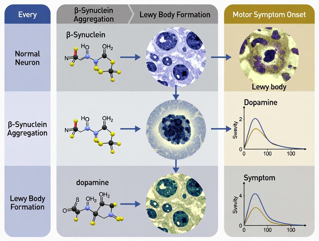
This technical guide examines the temporal progression of Lewy body (LB) pathology, correlating neuroanatomical seeding with the emergence of motor symptoms, specifically gait disturbance and rigidity. Framed within a broader thesis on α-synuclein (α-syn) propagation, we detail the preclinical phases, highlight critical pathological hallmarks, and present standardized experimental protocols for modeling and quantifying this progression in preclinical and human tissue-based research.
The Braak hypothesis, later expanded, posits that Lewy pathology originates in the olfactory bulb and dorsal motor nucleus of the vagus nerve, progressing rostrally through the brainstem to limbic and neocortical regions. Motor symptoms (gait, rigidity, bradykinesia) manifest when pathology reaches the substantia nigra pars compacta (SNc), resulting in significant dopaminergic cell loss. The protracted preclinical phase offers a critical therapeutic window.
Quantitative Correlation: Pathological Burden vs. Clinical Signs
Recent quantitative studies have refined the correlation between α-syn burden, neuronal loss, and motor deficit severity.
Table 1: Correlation Metrics for Key Motor Features in Early Clinical Phase
| Motor Feature | Correlated Pathological Measure | Pearson's r (Range) | Key Brain Region | Typely Threshold for Symptom Onset |
|---|---|---|---|---|
| Gait Disturbance | % Neuronal Loss in SNc | -0.72 to -0.85 | Substantia Nigra, Locus Coeruleus | ~50-60% dopaminergic loss |
| Rigidity | Phosphorylated α-syn Load (LB/cm²) in SNc | 0.65 to 0.78 | Substantia Nigra, Putamen | > 2.5 LB/mm² in SNc |
| Bradykinesia | Dopamine Transporter (DAT) Density in Putamen | -0.80 to -0.90 | Caudate/Putamen | < 30% of age-matched control mean |
| Postural Instability | Neuronal Loss in Locus Coeruleus | -0.60 to -0.75 | Locus Coeruleus, Pedunculopontine Nucleus | Often coincides with >40% LC loss |
Table 2: Preclinical Phase Biomarker Trajectory (Longitudinal Cohort Data)
| Disease Stage (Estimated Years to Diagnosis) | Mean CSF α-syn (pg/ml) | Mean DAT Scan SBR | Mean MDS-UPDRS III Score | Predominant Pathology Location |
|---|---|---|---|---|
| Preclinical (10-15 years) | 1200 ± 150 | 4.5 ± 0.8 | 2 ± 3 | Olfactory Bulb, Dorsal Motor Nucleus X |
| Prodromal (5-10 years) | 900 ± 200 | 3.2 ± 0.7 | 8 ± 5 | Locus Coeruleus, Raphe Nuclei |
| Early Clinical (0-5 years) | 700 ± 250 | 2.1 ± 0.6 | 25 ± 8 | Substantia Nigra, Basal Forebrain |
| Established Disease | 550 ± 300 | 1.5 ± 0.5 | 45 ± 12 | Limbic & Neocortical Regions |
Experimental Protocols
Protocol: Seeding and Propagation of α-Synuclein Pathology in M83 Transgenic Mice
Objective: To model the caudo-rostral progression of LB pathology and assess correlated motor deficits.
- Pre-formed Fibril (PFF) Preparation: Recombinant human α-syn is agitated in PBS at 37°C for 7 days. Fibrillization is confirmed via Thioflavin T assay and transmission electron microscopy.
- Stereotaxic Injection: Anesthetize 3-month-old M83 (hA53T α-syn) mice. Inject 2 µL of sonicated α-syn PFFs (5 µg/µL) or PBS vehicle unilaterally into the striatum (coordinates from Bregma: AP +0.5 mm, ML ±2.0 mm, DV -3.0 mm).
- Longitudinal Motor Phenotyping:
- Gait Analysis (DigiGait): Monthly, record mice walking on a transparent treadmill. Key parameters: stride length variability, stance width, hindlimb swing speed.
- Cylinder Test (Forelimb Use): Assess spontaneous forelimb asymmetry as a proxy for rigidity/bradykinesia.
- Hindlimb Clasping: A measure of axial rigidity, scored from 0 (extended) to 3 (severe clasping).
- Terminal Histopathology: At 1, 3, 6, and 9 months post-injection, perfuse mice. Serial brain sections are stained with:
- Anti-pS129 α-syn: Map LB-like inclusions.
- Anti-Tyrosine Hydroxylase (TH): Quantify nigrostriatal dopaminergic integrity.
- Iba1 & GFAP: Assess neuroinflammation.
- Quantitative Analysis: Stereological counting of TH+ neurons in SNc. pS129 α-syn load quantified via digital pathology (area fraction). Correlate with longitudinal motor scores.
Protocol: Post-Mortem Tissue-Based Correlation in Human Brainstem
Objective: To quantitatively correlate regional LB burden with historical clinical motor scores.
- Tissue Cohort: 50 cases from a brain bank with Lewy pathology (Braak stages 3-6) and detailed longitudinal Unified Parkinson's Disease Rating Scale (UPDRS) records.
- Region-Specific Microdissection: Using a brain matrix, isolate 2mm punches from: Dorsal Motor Nucleus of Vagus (DMV), Locus Coeruleus (LC), Substantia Nigra (SN), and Putamen.
- Biochemical Fractionation: Homogenize each punch. Sequentially extract with:
- High-Salt Buffer: Soluble α-syn.
- Triton-X-100: Membrane-bound.
- SDS/Urea: Insoluble, fibrillar α-syn (pathological).
- ELISA: Perform sandwich ELISA for total and pS129 α-syn in the SDS/Urea fraction for each region.
- Digital Histopathology: Adjacent sections immunostained for pS129 α-syn and NeuN. Whole-slide imaging followed by automated inclusion counting and neuronal density calculation.
- Statistical Correlation: Linear mixed-effects models to correlate regional insoluble pS129 α-syn concentration (pg/µg protein) with ante-mortem UPDRS-III subscores for rigidity and postural instability/gait.
Visualizing Pathways and Workflows
Title: Prion-like α-Synuclein Propagation Pathway
Title: Temporal Correlation of Braak Pathology with Symptom Onset
Title: Preclinical Model Experimental Workflow
The Scientist's Toolkit: Research Reagent Solutions
Table 3: Essential Reagents for Lewy Body Propagation Research
| Reagent / Material | Provider Examples | Function & Application |
|---|---|---|
| Recombinant Human α-Synuclein Protein | rPeptide, Sigma-Aldrich, Abcam | Substrate for generating pre-formed fibrils (PFFs) for seeding experiments in vitro and in vivo. |
| Phospho-S129 α-Synuclein Antibodies (clones MJFR1, EP1536Y) | Abcam, BioLegend, Cell Signaling Tech | Gold-standard for detecting pathological Lewy body-like inclusions in immunohistochemistry and immunoblotting. |
| Tyrosine Hydroxylase (TH) Antibodies | MilliporeSigma, Pel-Freez | Labels dopaminergic neurons for quantifying nigrostriatal degeneration in mouse models and human tissue. |
| α-Synuclein Pre-Formed Fibrils (PFFs), Human | StressMarq, MilliporeSigma | Ready-to-use, characterized seeds for consistent induction of pathology, reducing protocol variability. |
| Protease K | Thermo Fisher, Roche | Used in Paraffin-Embedded Tissue (PET) blot and sequential extraction protocols to isolate proteinase K-resistant, pathological α-syn aggregates. |
| Lumit α-Synuclein Aggregation Immunoassay | Promega | Homogeneous, bioluminescent assay for real-time, high-throughput quantification of α-syn aggregation in cell lysates. |
| Meso Scale Discovery (MSD) α-Synuclein Kits | Meso Scale Diagnostics | Ultrasensitive multiplex immunoassays for quantifying total and phosphorylated α-syn in CSF, plasma, and brain homogenates. |
| NeuN Antibodies (clone D4G4O) | MilliporeSigma, Cell Signaling Tech | Neuronal nuclear marker for quantifying neuronal density in correlation with regional Lewy body counts. |
From Bench to Biomarker: Tools for Tracking Pathology and Motor Decline
This whitepaper provides an in-depth technical guide on advanced neuroimaging methodologies for quantifying nigrostriatal degeneration, a core pathological feature of Lewy body disorders including Parkinson's disease (PD) and Dementia with Lewy bodies (DLB). Within the broader thesis of Lewy body pathology and motor symptom progression, precise in vivo quantification of dopaminergic neuron loss is paramount for staging disease, tracking progression, and evaluating therapeutic efficacy in clinical trials. This document details current MRI and PET tracer techniques, experimental protocols, and data interpretation for the research and drug development community.
Part 1: PET Tracer Imaging of Dopaminergic Integrity
PET imaging utilizes radiolabeled ligands to target specific components of the presynaptic dopaminergic terminal.
Key PET Tracers and Targets
Table 1: Common PET Tracers for Nigrostriatal Imaging
| Tracer | Primary Target | Radiolabel | Key Binding Measure | Clinical Correlation |
|---|---|---|---|---|
| ¹⁸F-FP-CIT | Dopamine Transporter (DAT) | ¹⁸F | Non-displaceable binding potential (BPND) | Strong correlation with UPDRS-III motor scores |
| ¹¹C-CFT | Dopamine Transporter (DAT) | ¹¹C | Striatal Binding Ratio (SBR) | Reductions of 50-70% in early PD vs. controls |
| ¹⁸F-FDOPA | Aromatic L-amino acid decarboxylase (AADC) | ¹⁸F | Influx rate constant (Ki) | ~40-60% reduction in putamen Ki in PD |
| ¹¹C-DTBZ | Vesicular Monoamine Transporter 2 (VMAT2) | ¹¹C | Distribution Volume Ratio (DVR) | Less affected by compensatory changes than DAT |
Experimental Protocol: ¹⁸F-FP-CIT PET Acquisition and Analysis
Protocol Title: Dynamic PET Acquisition for Dopamine Transporter Quantification.
Objective: To measure striatal DAT availability in patients with suspected Lewy body pathology.
Materials & Scanner:
- PET/CT or PET/MR scanner (e.g., Siemens Biograph, GE Discovery).
- ¹⁸F-FP-CIT (approx. 185 MBq dose).
- High-resolution structural T1-weighted MRI for co-registration.
Procedure:
- Patient Preparation: Ensure patient is off medications affecting DAT (e.g., amphetamines, bupropion) for >5 half-lives.
- Transmission Scan: Perform a low-dose CT scan for attenuation correction.
- Tracer Injection: Administer ¹⁸F-FP-CIT intravenously as a bolus.
- Dynamic Acquisition: Initiate a 90-120 minute dynamic emission scan immediately post-injection. Acquire frames as: 6x30s, 4x60s, 5x120s, 5x300s, 2x600s.
- Reconstruction: Reconstruct images using an iterative algorithm (OSEM) with attenuation and scatter correction.
- MRI Co-registration: Co-register the individual's T1 MRI to the mean PET image using rigid-body transformation in SPM or FSL.
- Region of Interest (ROI) Definition: Manually or automatically delineate ROIs for caudate, putamen (anterior/posterior subdivisions), and occipital cortex (reference region) on the co-registered MRI.
- Quantification: Calculate time-activity curves. Compute the Specific Binding Ratio (SBR) or BPND using the simplified reference tissue model (SRTM) with occipital cortex as the reference.
Data Output: Regional BPND or SBR values. A posterior putamen BPND reduction >30% compared to age-matched controls is a typical diagnostic threshold.
Part 2: Advanced MRI Techniques
Quantitative Structural MRI: Nigrosome-1 Imaging
Protocol Title: High-Resolution 3D T2*-Weighted MRI for Nigrosome-1 Detection.
Objective: To visualize the loss of the dorsolateral nigrosome-1 region in the substantia nigra pars compacta (SNc), a hallmark of PD.
Scanner & Sequence: 3T MRI with a 3D multi-echo gradient echo (ME-GRE) or susceptibility-weighted imaging (SWI) sequence.
Parameters (Example):
- TE/TR: 20ms/35ms
- Flip Angle: 15°
- Resolution: 0.5 x 0.5 x 0.5 mm³ isotropic.
- Scan Time: ~8 minutes.
Analysis: Visual assessment of the "swallow tail" appearance on axial slices. Quantitative analysis involves manual or automated segmentation of the SNc and calculation of the nigrosome-1 volume or signal intensity ratio. Loss of the hyperintense nigrosome-1 region has >95% sensitivity and specificity for PD diagnosis in expert settings.
Diffusion MRI: Neurite Orientation Dispersion and Density Imaging (NODDI)
Protocol Title: Multi-Shell Diffusion MRI for Assessing Nigral Microstructure.
Objective: To model and quantify the complex microstructure of the substantia nigra using a multi-compartment biophysical model.
Scanner & Sequence: 3T MRI with a multi-shell diffusion-weighted spin-echo EPI sequence.
Parameters (Example):
- b-values: 0, 700, 2000 s/mm².
- Directions: 30 at b=700, 60 at b=2000.
- Resolution: 2.0 x 2.0 x 2.0 mm³.
- Scan Time: ~12 minutes.
Analysis:
- Preprocess data (denoising, eddy-current/motion correction).
- Fit the NODDI model (using MATLAB toolbox or AMICO) to derive voxel-wise maps of:
- Intracellular Volume Fraction (ICVF): Reflects neurite density.
- Orientation Dispersion Index (ODI): Reflects dendrite complexity.
- Isotropic Volume Fraction (ISOVF): Reflects free water/cerebrospinal fluid contamination.
- Co-register maps to T1 space and extract mean values from a manually segmented substantia nigra ROI.
Table 2: Typical NODDI Findings in Early PD vs. Healthy Controls
| Metric | Healthy Control Mean (SN) | PD Patient Mean (SN) | % Change | P-Value |
|---|---|---|---|---|
| ICVF | 0.52 ± 0.04 | 0.43 ± 0.05 | -17.3% | <0.001 |
| ODI | 0.23 ± 0.03 | 0.27 ± 0.04 | +17.4% | <0.01 |
| ISOVF | 0.18 ± 0.05 | 0.25 ± 0.06 | +38.9% | <0.001 |
Part 3: Multi-Modal Integration and Pathway Analysis
Multi-Modal Assessment of Nigrostriatal Degradation
Multi-Modal Imaging Analysis Pipeline
The Scientist's Toolkit: Research Reagent Solutions
Table 3: Essential Reagents and Materials for Nigrostriatal Degeneration Research
| Item | Function/Application | Example Product/Source |
|---|---|---|
| ¹⁸F-FP-CIT | PET radiopharmaceutical for DAT imaging. Lyophilized kit for radiolabeling. | Radioisotope production facilities (e.g., IBA, CURANOSTUM kit) |
| ¹¹C-Raclopride | PET radiopharmaceutical for D2 receptor imaging (measures post-synaptic changes). | Cyclotron-produced, synthesized on-site. |
| Anti-α-synuclein Antibodies | For post-mortem validation of imaging findings (e.g., LB staining in SN). | Clone 5G4 (Millipore), Phospho-Ser129 (Abcam) |
| Anti-Tyrosine Hydroxylase (TH) Antibodies | Gold-standard immunohistochemical marker for dopaminergic neurons. | Rabbit polyclonal (Pel-Freez), Mouse monoclonal (Sigma) |
| MRI Contrast Agents (for specific protocols) | For blood-brain barrier integrity assessment or vascular-space occupancy. | Gadobutrol (Gadovist), Ferumoxytol (for VASO) |
| 3D Cell Culture/Organoid Kits | For in vitro modeling of nigrostriatal pathways and testing tracer binding. | Human iPSC-derived dopaminergic neuron kits (STEMCELL Tech) |
| Image Analysis Software Licenses | For processing MRI/PET data (segmentation, statistical analysis). | SPM12, FSL, PMOD, AnalyzeDirect, MIM Neuro |
Advanced neuroimaging with MRI and PET tracers provides a powerful, quantitative toolkit for in vivo investigation of nigrostriatal degeneration within Lewy body pathology research. The integration of multi-modal data—from molecular PET targets to microstructural MRI indices—offers a comprehensive pathophysiological profile. This is critical for defining biologically anchored patient strata, identifying progression biomarkers, and objectively measuring outcomes in disease-modifying therapeutic trials.
1. Introduction In the context of Lewy body pathology research, the precise detection and quantification of pathogenic α-synuclein aggregates is a cornerstone for understanding motor symptom progression and staging disease. Seed Amplification Assays (SAAs), particularly real-time quaking-induced conversion (RT-QuIC), have emerged as transformative tools for the ultrasensitive, specific detection of these pathologic seeds in biofluids, offering critical biomarkers for diagnosis and therapeutic development.
2. Core Principles of α-Synuclein SAA SAAs exploit the prion-like seeding capacity of misfolded α-synuclein. A minute quantity of pathogenic seed (from a patient sample) is mixed with an excess of recombinant α-synuclein substrate under conditions that promote templated amplification. This leads to fibrillization, which is monitored in real-time via a fluorescent dye (e.g., Thioflavin T). The time-to-threshold correlates with seed concentration.
3. Key Experimental Protocols
3.1. RT-QuIC Protocol for CSF
- Sample Preparation: Cerebrospinal fluid (CSF) is centrifuged (e.g., 2000 x g, 10 min) to remove cells/debris. Aliquots are stored at ≤ -80°C.
- Reaction Mix:
- Recombinant human wild-type α-synuclein (final conc. 0.1 mg/mL).
- Thioflavin T (final conc. 10 µM).
- Phosphate buffer (e.g., 100 mM sodium phosphate, pH 8.0), 10 µM EDTA.
- NaCl (final conc. 170 mM).
- Procedure:
- Load a black-walled 96-well plate with 85-95 µL of reaction mix per well.
- Add 5-15 µL of CSF sample (typically 2-4 replicates). Include positive (confirmed Lewy body pathology CSF) and negative (healthy control CSF) controls.
- Seal plate, place in a fluorescent plate reader preheated to 42°C.
- Cycle: Incubate with intermittent shaking (e.g., 1 min shake, 14 min rest per cycle).
- Monitor fluorescence (excitation ~450 nm, emission ~480 nm) every 15-45 minutes for 60-150 hours.
- Data Analysis: A sample is considered positive if ≥ 2 replicates cross a predefined fluorescence threshold (typically 5 standard deviations above the mean of negative controls) within the assay time.
3.2. Protocol Adaptations for Other Biofluids
- Saliva/Plasma/Serum: Require more extensive pre-analytical processing (e.g., protease inhibitors, higher dilution, or phosphotungstic acid precipitation) to overcome inhibitors and increase sensitivity.
- Skin/Smell Mucosa Biopsies: Tissue homogenization and sonication are required to extract seeds.
4. Data Presentation
Table 1: Diagnostic Performance of α-Synuclein SAA (RT-QuIC) in CSF
| Condition (Confirmed Diagnosis) | Sensitivity (Range) | Specificity (Range) | Sample Size (Approx.) | Reference Year |
|---|---|---|---|---|
| Isolated REM Sleep Behavior Disorder (iRBD) | 86-95% | 93-100% | 100-1200 | 2021-2023 |
| Parkinson's Disease (PD) | 88-96% | 96-100% | 200-2000 | 2020-2024 |
| Dementia with Lewy Bodies (DLB) | 92-98% | 93-100% | 100-500 | 2021-2023 |
| Multiple System Atrophy (MSA) | 67-95%* | 96-100% | 50-100 | 2022-2024 |
Table 2: SAA Positivity Rates Across Biofluids in PD Cohorts
| Biofluid | Positivity Rate (Range) | Approx. Seed Concentration (Relative to CSF) | Key Technical Notes |
|---|---|---|---|
| Cerebrospinal Fluid (CSF) | 88-96% | Reference (1x) | Standard, most validated matrix. |
| Saliva | 80-90% | 10-100x lower | Requires pre-treatment; submandibular gland fluid shows high promise. |
| Skin Biopsy (Dermal Nerve Fibers) | 90-98% | N/A | High sensitivity; post-mortem and in vivo correlation with pathology. |
| Plasma/Serum | 70-85% | 100-1000x lower | Pre-concentration (e.g., PTA) is essential; higher variability. |
| Olfactory Mucosa | 75-95% | N/A | Invasive collection; direct connection to CNS pathology. |
Note: MSA shows more variable sensitivity, potentially due to structural differences in α-synuclein aggregates (strains).
5. Visualizing the SAA Workflow and Biological Context
Title: RT-QuIC Experimental Workflow
Title: SAA in Lewy Body Pathology & Motor Progression Thesis
6. The Scientist's Toolkit: Key Research Reagent Solutions
Table 3: Essential Materials for α-Synuclein SAA Research
| Item | Function & Critical Notes |
|---|---|
| Recombinant Human α-Synuclein (WT) | High-purity, endotoxin-free monomeric substrate is critical for assay sensitivity and reproducibility. Lyophilized or flash-frozen aliquots recommended. |
| Thioflavin T (ThT) | Fluorescent dye that binds to cross-β-sheet structures of amyloid fibrils. Must be protected from light; stock solutions prepared in buffer. |
| Black-walled 96-well Optical Plate | Plates must be optically clear for bottom reading, have low binding properties, and be compatible with plate sealers. |
| Plate Sealer (Adhesive or Heat Seal) | Prevents evaporation and contamination during multi-day assays. Sealing integrity is paramount. |
| Fluorescent Plate Reader with Temperature Control & Shaking | Requires precise thermal control (≤0.5°C variation) and programmable shaking/rest cycles. Standard equipment for RT-QuIC. |
| Synthetic α-Synuclein Fibrils (Pre-formed) | Used as positive control seeds for assay calibration and optimization. |
| Phosphotungstic Acid (PTA) / Sodium Phosphotungstate | Used for precipitating and concentrating α-synuclein seeds from complex matrices like blood plasma. |
| Protease Inhibitor Cocktails | Essential for processing peripheral biofluids (saliva, plasma) to prevent seed degradation during sample prep. |
| Validated Positive & Negative Control Biofluids | Characterized CSF or tissue homogenates from neuropathologically confirmed cases and controls. Necessary for every assay run. |
This whitepaper details the methodology of digital motor phenotyping for application in clinical trials investigating Lewy body pathology progression. Within the broader thesis on the spatiotemporal spread of alpha-synuclein aggregates and its correlation with motor symptom emergence, this guide provides the technical framework for quantifying gait and movement. The precise, continuous, and objective data generated by these tools are essential for linking pathological burden, as measured by biomarkers, to functional motor decline. This enables sensitive detection of therapeutic efficacy in disease-modifying trials.
Core Technologies & Sensor Platforms
Wearable Sensor Technologies
The foundation of digital phenotyping lies in inertial measurement units (IMUs), which typically integrate tri-axial accelerometers, gyroscopes, and magnetometers.
Table 1: Common Wearable Sensor Specifications for Gait Analysis
| Sensor Type | Primary Metrics | Sample Rate (Typical) | Placement | Clinical Relevance in Lewy Body Trials |
|---|---|---|---|---|
| Tri-axial Accelerometer | Acceleration (m/s²), Jerk, Strike Impact | 50-200 Hz | Lower back (L5), Feet, Wrists | Quantifies gait initiation, step regularity, postural sway. |
| Tri-axial Gyroscope | Angular Velocity (deg/s), Rotation | 50-200 Hz | Shanks, Thighs, Feet | Measures knee flexion/extension, swing phase coordination. |
| Magnetometer | Heading Orientation | 10-50 Hz | Lower back, Feet | Provides context for directionality in turning tasks. |
| Pressure-Sensing Insole | Force (N), Center of Pressure | 100 Hz | Inside Shoes | Detailed stance phase analysis, mediolateral stability. |
| EMG Sensor | Muscle Activation (mV) | 1000-2000 Hz | Tibialis Anterior, Gastrocnemius | Assesses co-contraction, bradykinesia-related activation patterns. |
Key Quantitative Gait Parameters
Raw sensor data is processed into biomechanically meaningful endpoints.
Table 2: Key Digital Gait Parameters and Their Pathophysiological Correlates in Lewy Body Disorders
| Gait Domain | Specific Parameter | Definition | Association with Lewy Body Pathology |
|---|---|---|---|
| Pace | Stride Length (m) | Distance between consecutive heel strikes of the same foot. | Correlates with nigrostriatal dopaminergic deficiency and step scaling. |
| Rhythm | Step Time Variability (CV%) | Coefficient of variation of time between consecutive steps. | Increased variability linked to progressive brainstem and basal ganglia pathology. |
| Asymmetry | Step Time Asymmetry (Abs%) | Absolute percentage difference between left and right step times. | May reflect asymmetric cortical or subcortical Lewy body burden. |
| Postural Control | Stride Velocity (m/s) | Speed of gait, calculated from stride length/time. | Primary endpoint for disease progression and treatment response. |
| Dynamic Stability | Harmonic Ratio (AP/ML) | Ratio of even to odd harmonics of accelerometer signal, indicating smoothness. | Reduced ratio indicates impaired balance control and fall risk. |
| Turning | Turn Duration (s) & Number of Steps | Time and steps required to complete a 180° turn. | Prolonged, multi-step turns are highly specific to Parkinsonism in LBD. |
Experimental Protocols for Clinical Trials
Protocol A: Controlled Clinic-Based Assessment (Standardized Gait Task)
- Objective: To collect a high-fidelity, standardized gait dataset in a controlled environment.
- Equipment: 5 IMUs (lower back, bilateral shanks, bilateral feet), pressure-sensitive walkway.
- Procedure:
- Sensor Calibration: Perform a 3-second static upright calibration, followed by a dynamic calibration (e.g., 10 knee lifts).
- Task Instruction: Walk at a self-selected, comfortable speed along a 20-meter straight hallway.
- Trial Execution: Patient completes 6 continuous walking passes (3 out-and-back cycles). Include an embedded 180-degree turn at each end. Initiate and terminate walking 2 meters before/after the walkway to capture steady-state gait.
- Data Recording: Synchronize all wearable data with the pressure walkway system via a trigger event.
Diagram Title: Clinic-Based Standardized Gait Protocol Workflow
Protocol B: Continuous Free-Living Monitoring (7-Day Protocol)
- Objective: To capture real-world motor fluctuations, activity profiles, and non-linear symptom progression.
- Equipment: 2 IMUs (lower back, dominant wrist) with continuous logging capability (>7-day battery).
- Procedure:
- Sensor Distribution: Fit sensors and verify data streaming to internal storage. Provide waterproof casings.
- Diary & Log: Participants log sleep times, medication ON/OFF states (if applicable), and notable events (falls, freezing).
- Data Collection: Participants wear sensors continuously for 7 consecutive days and nights, removing only for charging (staggered to maintain at least one active sensor).
- Data Upload: Secure, anonymized wireless or physical upload at the end of the monitoring period.
Data Processing & Analytical Workflow
Raw sensor data undergoes a multi-stage pipeline to extract clinical endpoints.
Diagram Title: Sensor Data Processing Pipeline to Endpoints
The Scientist's Toolkit: Research Reagent Solutions
Table 3: Essential Materials for Digital Motor Phenotyping Studies
| Item / Solution | Function / Rationale | Example Vendor/Product |
|---|---|---|
| Research-Grade IMU System | Provides raw, high-frequency data access, precise time synchronization, and open APIs for custom algorithm development. | APDM Opal, DynaPort MoveTest, Shimmer3. |
| Validated Algorithm Library | Software package with peer-reviewed algorithms for gait event detection (IC, FC), turn identification, and posture calculation. | MATLAB Gait and Posture Toolboxes, APDM Mobility Lab algorithms. |
| Standardized Phantoms & Calibrators | Mechanical devices to validate sensor accuracy (e.g., rotation rate, acceleration) and ensure multi-site trial consistency. | Custom motorized jigs, pendulum calibration fixtures. |
| Secure, HIPAA/GCP-Compliant Cloud Platform | For data aggregation from multi-center trials, featuring role-based access, audit trails, and automated preprocessing pipelines. | AWS HealthLake, Google Cloud Healthcare API, Medidata Rave. |
| Digital Biomarker Statistical Package | Specialized software for time-series analysis, handling repeated measures, and calculating intra-individual variability metrics (CV, GVI). | R nparLD package, custom Python scripts using scikit-learn & statsmodels. |
| Synchronization Trigger Device | Generates a simultaneous voltage pulse to all wearable sensors and reference systems (motion capture, EEG) for perfect time alignment. | Biopac STP100C, custom Arduino-based trigger. |
The progression of motor symptoms in Lewy body disorders (LBD), including Parkinson’s disease dementia and dementia with Lewy bodies, is heterogeneous and non-linear. A core challenge in modern neurology is predicting individual symptom trajectories to enable targeted interventions. This whitepaper posits that integrative models, which combine multimodal biomarker data, are essential for deconvoluting this complexity. Such models move beyond single-marker approaches to capture the multifactorial pathogenesis of LBD, where alpha-synuclein pathology, neurodegeneration, and neuroinflammation interact.
Core Biomarker Classes for Integration
The predictive modeling of motor symptom progression relies on quantitative data from several biomarker domains. The table below summarizes key biomarkers, their modalities, and their primary association with Lewy body pathology.
Table 1: Core Biomarker Classes for Lewy Body Symptom Trajectory Prediction
| Biomarker Class | Specific Analytes/Measures | Biofluid/Imaging Modality | Primary Pathological Correlation | Typical Dynamic Range in Early LBD |
|---|---|---|---|---|
| Synucleinopathy | Oligomeric α-synuclein | CSF, Plasma (exosomes) | Presynaptic Lewy body pathology | CSF: 20-50 pg/mL (ELISA) |
| Neuronal Injury | Neurofilament Light Chain (NfL) | CSF, Plasma, Serum | Axonal degeneration & disease severity | CSF: 1000-2500 pg/mL |
| Neuroinflammation | GFAP, YKL-40, IL-6 | CSF, Plasma | Astrogliosis & pro-inflammatory state | CSF GFAP: 8-12 ng/mL |
| Dopaminergic Integrity | DaTscan (Striatal Binding Ratio) | SPECT Imaging | Nigrostriatal terminal loss | Caudate SBR: 1.5-2.5 |
| Functional Network | Resting-state fMRI (network connectivity) | MRI | Cortico-striatal-thalamic dysfunction | Default Mode Network power: -0.5 to +0.5 (z-score) |
Experimental Protocols for Key Biomarker Assays
Protocol: Immunoprecipitation-Mass Spectrometry for CSF α-Synuclein Species
Objective: Quantify pathogenic oligomeric α-synuclein from cerebrospinal fluid (CSF).
- CSF Pre-processing: Centrifuge fresh CSF at 20,000g for 15 minutes at 4°C. Aliquot supernatant.
- Immunoprecipitation: Incubate 500 µL CSF with 5 µg of anti-α-synuclein (MJFR1) antibody conjugated to magnetic beads overnight at 4°C with gentle rotation.
- Washing: Wash beads 3x with PBS-Tween (0.05%).
- Elution: Elute bound proteins using 50 µL of 0.1% formic acid.
- Mass Spectrometry Analysis: Analyze eluate via LC-MS/MS on a Q Exactive HF system. Quantify using parallel reaction monitoring (PRM) targeting unique peptide sequences for α-synuclein. Normalize to a spiked-in heavy isotope-labeled α-synuclein standard.
Protocol: Longitudinal DaTscan SPECT Image Coregistration & Analysis
Objective: Quantify rate of dopaminergic decline over time.
- Image Acquisition: Administer ~185 MBq I-123 Ioflupane. Acquire SPECT images 3-4 hours post-injection using a standardized protocol.
- Preprocessing: Reconstruct images using ordered-subset expectation maximization (OSEM) with attenuation correction.
- Spatial Normalization: Coregister all serial scans from a single patient to their baseline scan using a rigid-body transformation (SPM12 or similar).
- ROI Analysis: Apply the automated "BRASS" or a validated atlas to extract mean counts from bilateral caudate and putamen. Define occipital cortex as reference region.
- Quantification: Calculate Specific Binding Ratio (SBR) = (Target ROI mean counts / Reference ROI mean counts) - 1. Rate of change = (SBRfollow-up - SBRbaseline) / Time (years).
Integrative Modeling Architectures
Predictive models integrate data from Table 1 using various computational architectures.
Table 2: Comparison of Integrative Modeling Approaches
| Model Type | Key Features | Input Data Handling | Suitability for LBD Trajectories | Example Performance (Mean Absolute Error) |
|---|---|---|---|---|
| Linear Mixed-Effects (LME) Model | Handles repeated measures, random intercepts for patients. | Fixed effects for biomarkers, time, and interactions. | High for group-level trend estimation. | UPDRS-III prediction error: ~4.5 points |
| Cox Proportional Hazards with Time-Dependent Covariates | Predicts time-to-event (e.g., need for levodopa). | Biomarker values updated at each visit. | Excellent for clinical milestone prediction. | C-index for "motor complication" event: 0.78 |
| Machine Learning: Random Forest | Non-linear, handles missing data, provides feature importance. | Baseline multimodal data tabulated per subject. | Good for cross-sectional trajectory classification (Slow vs. Fast). | Accuracy for 2-year progression class: 82% |
| Deep Learning: Multi-Modal Neural Network | Learns complex interactions between data types (e.g., image + CSF). | Separate encoder branches for each data modality, fused in latent space. | High potential for personalized, continuous prediction. | RMSE on longitudinal UPDRS-III: 3.8 points |
The Scientist's Toolkit: Research Reagent Solutions
Table 3: Essential Reagents and Materials for Biomarker Research in LBD
| Item | Vendor Examples (Research-Use Only) | Function in Experimental Protocol |
|---|---|---|
| Human α-Synuclein Oligomer-Specific Antibody (e.g., clone Syn-O2) | MilliporeSigma, Abcam | Selective immunoprecipitation/detection of pathogenic oligomeric forms from biofluids. |
| Simoa NF-Light Advantage Kit | Quanterix | Ultrasensitive digital ELISA for quantifying neurofilament light chain in plasma and CSF. |
| Human GFAP ELISA Kit, High Sensitivity | R&D Systems | Quantification of glial fibrillary acidic protein as a marker of astrocytic activation. |
| MagPlex Magnetic Microspheres (Luminex) | Lumipulse G1200 (Fujirebio) | Multiplexed bead-based immunoassay for simultaneous cytokine/chemokine profiling. |
| I-123 Ioflupane (DaTscan) | GE Healthcare | Radiopharmaceutical for binding to dopamine transporters in SPECT imaging. |
| SPM12 Software | Wellcome Centre for Human Neuroimaging | Standard tool for spatial normalization and preprocessing of neuroimaging data. |
Visualizing Pathways and Workflows
From Biomarkers to Trajectory Prediction
Core Pathology Pathway in Lewy Body Disorders
Navigating Complexity: Challenges in Linking Pathology to Clinical Progression
This whitepaper investigates the critical phenomenon in neurodegenerative diseases where the extent of pathological burden (e.g., Lewy body density) does not linearly correlate with the emergence or severity of clinical symptoms. This "mismatch" is central to understanding disease progression in Lewy body disorders, including Parkinson's disease (PD) and dementia with Lewy bodies (DLB). The concepts of symptom thresholds and neural (or cognitive) reserve provide the principal frameworks for explaining this dissociation. For Lewy body pathology, the progression of motor symptoms (bradykinesia, rigidity, tremor) is a key clinical endpoint. Understanding the dynamics between α-synuclein aggregation, neuronal dysfunction, and the system's capacity to compensate is paramount for developing disease-modifying therapies and biomarkers.
Core Concepts: Thresholds and Reserve
The Symptom Threshold Hypothesis
The threshold hypothesis posits that clinical symptoms manifest only when pathological burden exceeds a critical level, overwhelming compensatory mechanisms. This is not a single event but a series of thresholds for different functional systems (motor, cognitive, autonomic).
Neural Reserve
Neural reserve refers to the brain's resilience to pathology, derived from both passive (brain size, synaptic density) and active (network efficiency, cognitive strategies) models. It explains variability in symptom onset among individuals with similar pathological loads.
Quantitative Data in Lewy Body Disorders
The following tables summarize key quantitative findings from recent research illustrating the pathology-symptom mismatch.
Table 1: Postmortem Studies of Lewy Body Pathology vs. Clinical Diagnosis
| Study (Year) | Cohort | Key Finding (Quantitative) | Implication |
|---|---|---|---|
| Postuma et al. (2022) | Prodromal PD | ≥50% loss of striatal dopamine terminals required for motor UPDRS > 6. | Motor threshold requires significant nigrostriatal depletion. |
| Dickson et al. (2021) | DLB/PDD | Neocortical Lewy body count > 5/mm² correlated with dementia, but with high individual variability (R²=0.45). | Cognitive symptoms have a less defined pathological threshold. |
| Surmeier et al. (2023) | PD Brain Bank | 60-70% dopaminergic neuron loss in substantia nigra pars compacta (SNc) at clinical motor diagnosis. | Confirms classic motor threshold; highlights pre-symptomatic phase. |
Table 2: In Vivo Biomarker Correlations with Symptom Severity
| Biomarker | Modality | Correlation with UPDRS-III (r-value) | Correlation with Cognitive Score (MMSE r-value) | Notes |
|---|---|---|---|---|
| Striatal DAT Binding | SPECT | -0.75 to -0.85 | -0.50 | Strong motor correlation, plateaus in advanced disease. |
| Cardiac ¹²³I-MIBG | Scintigraphy | 0.10 (weak) | -0.30 | Poor motor correlation, stronger with autonomic/cognition. |
| CSF α-synuclein | ELISA | -0.40 (moderate) | -0.55 | Moderate correlation, high inter-individual variability. |
Experimental Protocols for Investigating Mismatch
Protocol: Stereological Quantification of Lewy Body Pathology & Neuron Count
Objective: To correlate α-synuclein pathology density with neuronal loss and clinical scores from retrospective histories. Materials: Formalin-fixed paraffin-embedded (FFPE) midbrain and cortical sections, phosphorylated α-synuclein antibodies (e.g., pSer129), stereology workstation. Procedure:
- Sectioning & Staining: Cut serial 40μm sections. Employ immunohistochemistry for pSer129-α-synuclein and Nissl staining on alternating sections.
- Stereological Design: Use systematic random sampling via a fractionator probe. Define SNc boundaries using anatomical landmarks.
- Counting: Using an optical dissector, count (a) α-synuclein-positive Lewy bodies/Lewy neurites and (b) Nissl-stained neurons with visible nucleoli in the SNc.
- Density Calculation: Calculate volumetric density (pathology/mm³, neurons/mm³) using StereoInvestigator or similar software.
- Clinical Correlation: Obtain retrospective UPDRS scores from last clinic visit before death. Perform linear and threshold regression analyses.
Protocol: PET Imaging of Dopaminergic Terminal Integrity and Network Activation
Objective: To measure neural reserve via task-induced fMRI during dopaminergic challenge. Materials: ¹¹C-DTBZ or ¹⁸F-FE-PE2I PET ligand (for VMAT2/DAT), 3T MRI scanner, motor task paradigm (e.g., finger tapping), levodopa. Procedure:
- Baseline Scan: Perform PET scan to quantify striatal binding potential (BPₙᴅ), a measure of terminal integrity.
- fMRI Challenge: In a separate session, conduct fMRI under two conditions: (a) OFF medication (>12hrs withdrawal), (b) ON levodopa (standard dose).
- Motor Paradigm: Use block-design finger-tapping task during fMRI. Measure BOLD signal in motor cortex, supplementary motor area (SMA), and cerebellum.
- Reserve Metric Calculation: Compute "compensatory activation" as the OFF-state hyperactivation in prefrontal-striatal circuits. Calculate "response efficiency" as the normalization (reduction) of this hyperactivation ON medication.
- Correlation: Relate BPₙᴅ from PET to fMRI reserve metrics and clinical UPDRS scores.
Signaling Pathways in Pathology Progression & Compensation
Diagram 1: Threshold Cross via Compensatory Failure
Diagram 2: Basal Ganglia Circuit Dysfunction & Compensation
The Scientist's Toolkit: Research Reagent Solutions
Table 3: Essential Research Reagents for Investigating Mismatch
| Item | Function & Application | Example Product/Specification |
|---|---|---|
| Phospho-Specific α-Synuclein Antibody | Detects pathological, phosphorylated (pSer129) α-synuclein in Lewy bodies and neurites for IHC/IF. | Rabbit monoclonal MJFpS-129 (Abcam), clone EP1536Y. |
| DAT/VMAT2 PET Tracers | In vivo quantification of dopaminergic terminal integrity in rodents and humans. | ¹⁸F-FE-PE2I (DAT), ¹¹C-DTBZ (VMAT2). |
| Pre-formed Fibrils (PFFs) | Recombinant α-synuclein fibrils to seed and propagate pathology in cellular and animal models. | Human α-synuclein PFFs, fluorescently labeled (rPeptide). |
| Stereology Software Suite | Unbiased stereological counting for neuronal and pathological burden quantification. | StereoInvestigator (MBF Bioscience), VIS software. |
| High-Density EEG/fNIRS System | Measures neural network efficiency and compensatory activation non-invasively. | 256-channel EEG systems (EGI, Brain Products). |
| Induced Pluripotent Stem Cells (iPSCs) | Generate patient-specific dopaminergic neurons to model individual reserve capacity. | iPSCs from PD patients with/without rapid progression. |
| Caspase-3/GFAP/IBA1 Antibodies | Markers for apoptosis and glial activation to assess downstream effects of pathology. | Multiplex immunofluorescence antibody panels. |
The pathology-symptom mismatch in Lewy body disorders is governed by dynamic thresholds and variable neural reserve. Advancing therapeutic strategies requires moving beyond static pathological measures to quantify an individual's reserve capacity and proximity to critical thresholds. This necessitates integrated experimental protocols combining molecular pathology, multimodal neuroimaging, and sophisticated computational modeling to predict symptom onset and progression, ultimately guiding targeted, personalized drug development.
This technical guide examines the confounding effects of co-pathologies, specifically Alzheimer's disease (AD) pathology and vascular contributions, within the primary research context of Lewy body pathology and motor symptom progression. The frequent co-occurrence of amyloid-beta plaques, tau neurofibrillary tangles, and cerebral vascular disease in patients diagnosed with Lewy body dementias complicates the accurate assessment of pathological drivers, therapeutic target identification, and clinical trial stratification.
Quantifying Co-pathological Burden
Prevalence data for co-pathologies in Lewy Body Disease (LBD) cohorts, derived from recent clinico-pathological studies, are summarized below.
Table 1: Prevalence of Co-pathologies in Lewy Body Disease (LBD) at Autopsy
| Co-pathology Type | Prevalence in LBD (%) (Range from Recent Studies) | Typical Assessment Method |
|---|---|---|
| Alzheimer's Pathology (Intermediate/High ADNC) | 40-60% | NIA-AA Guidelines (Thal phase, Braak stage, CERAD score) |
| Cerebral Amyloid Angiopathy (CAA) | 30-50% | Modified Vonsattel Criteria |
| Arteriolosclerosis | 50-80% | Vessel Wall Thickness / H&E Staining |
| Macroscopic Infarct(s) | 20-35% | Gross Pathological Examination |
| Microinfarcts | 40-70% | Histology (e.g., H&E, Luxol fast blue) |
| Limbic-predominant Age-related TDP-43 Encephalopathy (LATE) | 20-30% | Phospho-TDP-43 IHC |
ADNC: Alzheimer's Disease Neuropathological Change; IHC: Immunohistochemistry.
Table 2: Impact of Co-pathologies on Clinical Metrics in LBD
| Co-pathology | Association with Faster Cognitive Decline (Hazard Ratio) | Association with Earlier Parkinsonism Onset/Motor Progression | Association with Lower CSF Aβ42 |
|---|---|---|---|
| High AD Tau (Braak ≥IV) | 2.1 [1.5-2.9] | Conflicting Data (Potential for earlier onset) | Strong |
| Moderate-Severe CAA | 1.8 [1.3-2.5] | Significant (p<0.01) for gait impairment | Moderate |
| Presence of ≥2 Microinfarcts | 1.9 [1.4-2.6] | Significant for postural instability progression | Weak/None |
Key Experimental Protocols for Isolating Pathological Contributions
To dissect the specific contributions of each co-pathology, integrated methodologies are required.
Protocol: Multimodal Pathological Staging in Post-Mortem Tissue
Objective: To quantitatively map multiple proteinopathies and vascular pathology within the same tissue specimen.
- Tissue Preparation: Formalin-fixed, paraffin-embedded (FFPE) blocks from neocortical, limbic, and brainstem regions. Serial sections cut at 5-10µm.
- Sequential Immunohistochemistry (IHC) & Staining:
- Cycle 1: Phospho-alpha-synuclein (pSyn#64) IHC for Lewy pathology. Scan slide.
- Cycle 2: Antibody elution (e.g., with glycine buffer, pH 2.0). Phospho-tau (AT8) IHC for AD tau pathology. Scan slide.
- Cycle 3: Elution. Aβ (4G8) IHC for plaques and CAA. Scan slide.
- Cycle 4: Elution. Periodic acid–Schiff (PAS) stain for vascular structures and microinfarcts.
- Image Registration & Analysis: Use digital pathology software (e.g., QuPath, HALO) to align sequential images. Annotate regions of interest (ROIs). Quantify lesion density (per mm²) and distribution for each pathology within the same anatomical footprint.
- Statistical Co-localization Analysis: Perform spatial correlation analyses (e.g., Moran's I) to determine if pathologies cluster independently or synergistically.
Protocol: In Vivo Biomarker Correlation with Post-Mortem Endpoints
Objective: To validate ante-mortem biomarkers against gold-standard pathological assessments.
- Prospective Cohort Selection: Enroll patients with probable DLB/PDD. Obtain standardized ante-mortem biomarkers within 2 years of death:
- PET: [¹⁸F]Flortaucipir (tau), [¹¹C]PIB (amyloid), [¹⁸F]FDG (metabolism).
- MRI: 3T multimodal MRI (structural T1, T2/FLAIR for WMH/infarcts, quantitative susceptibility mapping for microbleeds).
- CSF: Aβ42/40, p-tau181, α-syn RT-QuIC.
- Post-Mortem Validation: Perform comprehensive pathological assessment as per Protocol 3.1. Calculate burden scores for each pathology.
- Correlational Mapping: Use linear mixed models to correlate ante-mortem regional PET signal/MRI metrics with corresponding regional pathological burden scores, controlling for age at death and post-mortem interval.
Protocol: Functional Interplay in Murine Models
Objective: To test mechanistic interactions between α-syn, Aβ, and vascular insult.
- Animal Models: Utilize transgenic mice (e.g., Thy1-hSNCA/APP-PS1) or stereotactic human α-syn pre-formed fibril (PFF) injections into models with amyloidosis.
- Induction of Vascular Insult: Apply a mild, chronic hypoperfusion model (e.g., bilateral common carotid artery stenosis with microcoats) or a microinfarct model (photothrombosis) in one cohort.
- Outcome Measures:
- Motor Phenotyping: Longitudinal assessment using open field, rotarod, and gait analysis (DigiGait).
- Pathology: Terminal brain analysis for α-syn burden (IHC, ELISA), synaptic markers, amyloid load, and neuroinflammation (Iba1, GFAP).
- Vascular Integrity: Measure blood-brain barrier permeability (Evans Blue), capillary density (collagen IV IHC).
- Analysis: Compare outcomes across genotypes/treatments with and without vascular insult using 2-way ANOVA.
Visualizing Pathways and Workflows
Diagram Title: Co-pathology Interactions and Motor Progression
Diagram Title: Sequential Staining and Digital Analysis Protocol
The Scientist's Toolkit: Essential Research Reagents & Materials
Table 3: Key Reagent Solutions for Co-pathology Research
| Item | Function & Application | Example/Format |
|---|---|---|
| Phospho-Specific α-syn Antibodies | Detect pathological Lewy body-associated α-syn (e.g., pSer129). Crucial for accurate LBD pathology quantification. | Rabbit mAb (e.g., pSyn#64, EP1536Y); IHC, WB. |
| AT8 (p-tau) Antibody | Gold standard for detecting Alzheimer's-related phosphorylated tau in NFTs and neurites. | Mouse mAb (clone AT8); IHC, WB. |
| Amyloid-β Antibodies | Differentiate between parenchymal plaques (6E10, 4G8) and cerebrovascular amyloid (CAA). | Mouse mAbs (e.g., 6E10, 4G8); IHC, ELISA. |
| Collagen IV Antibody | Labels basement membranes; essential for quantifying capillary density and vascular integrity. | Rabbit polyclonal; IHC. |
| Multiplex IHC/IF Kits | Enable simultaneous detection of 2+ markers on one slide (e.g., α-syn + GFAP + Aβ). Preserves tissue and reveals spatial relationships. | Commercial kits (e.g., Opal, MACSima). |
| α-syn Pre-formed Fibrils (PFFs) | Induce endogenous α-syn aggregation in cellular and rodent models to study cross-seeding with Aβ/tau. | Recombinant human α-syn PFFs, sonicated. |
| Digital Pathology Software | For whole-slide image analysis, co-localization quantification, and spatial statistics. | QuPath, HALO, Visiopharm. |
| Antibody Elution Buffer | Allows sequential IHC on the same tissue section by stripping antibodies between cycles. | Low pH glycine buffer or commercial eluents. |
| Luminex/Simoa Assay Kits | Ultra-sensitive quantification of proteinopathy biomarkers (α-syn, Aβ, tau) in CSF/biofluids. | Neurology 4-Plex E (N4PE) Kit, Simoa assays. |
This technical whitepaper, framed within ongoing research into Lewy body pathology and motor symptom progression, elucidates the biological underpinnings of phenotypic heterogeneity in Parkinson's disease (PD). It details how the Postural Instability and Gait Difficulty (PIGD) and Tremor-Dominant (TD) subtypes exhibit distinct trajectories of motor decline, largely attributable to differences in underlying neuropathological burden, neurotransmitter system involvement, and neural network dysfunction.
Despite a common neuropathological hallmark—the aggregation of alpha-synuclein into Lewy bodies—PD presents with considerable clinical heterogeneity. The TD subtype is characterized by prominent resting tremor, earlier age of onset, and a generally slower motor progression. In contrast, the PIGD subtype features greater axial impairment (postural instability, gait freezing), more rapid motor decline, and a higher association with cognitive impairment. This divergence suggests that the topographic spread of pathology and its interaction with other neuronal systems differ significantly between subtypes.
Core Pathophysiological Divergence
Topographic Distribution of Lewy Pathology
Current neuropathological and neuroimaging research indicates a more restricted, brainstem-centric pathology in TD, while PIGD is associated with a more diffuse, cortically-predominant pattern.
Table 1: Comparative Pathological & Imaging Features
| Feature | Tremor-Dominant (TD) Subtype | PIGD Subtype |
|---|---|---|
| Lewy Body Topography | Predominantly brainstem (locus coeruleus, substantia nigra pars compacta) | Widespread cortical and limbic involvement |
| Dopaminergic Denervation (PET) | Severe, but focal in posterior putamen | More extensive, affecting anterior and posterior putamen |
| Cholinergic Deficit (PET) | Mild, limited to thalamus | Severe, in basal forebrain and cortex |
| Serotonergic Deficit (PET) | Relatively preserved | Marked loss in raphe nuclei and projections |
| Cortical Thinning (MRI) | Minimal | Significant, in frontal and parietal regions |
Involvement of Non-Dopaminergic Systems
The rapid progression of axial symptoms in PIGD cannot be explained by nigrostriatal dopamine loss alone. Key differentiating systems include:
- Cholinergic Systems: PIGD correlates strongly with degeneration of the pedunculopontine nucleus (PPN, Ch5) and the nucleus basalis of Meynert (NBM, Ch4), critical for gait control and attention.
- Noradrenergic Systems: Locus coeruleus degeneration is more severe in PIGD, impacting posture and arousal.
- Serotonergic Systems: Raphe nucleus dysfunction in PIGD may contribute to imbalance and depression.
- Glutamatergic Cerebello-Thalamo-Cortical Circuit: This pathway is hyperactive in TD, potentially contributing to tremor generation and compensatory mechanisms that slow progression.
Experimental Protocols for Investigating Subtype Heterogeneity
Protocol: Multimodal PET Imaging for System-Specific Degradation
Objective: To quantify and compare dopaminergic, cholinergic, and serotonergic terminal integrity in vivo in TD and PIGD patients.
- Cohort: Recruit 30 TD and 30 PIGD patients (matched for disease duration and age) and 20 healthy controls.
- PET Tracers:
- Dopamine Transporter (DAT): [¹¹C]PE2I or [¹⁸F]FE-PE2I.
- Cholinergic Vesicular Transporter (VAChT): [¹⁸F]FEOBV.
- Serotonin Transporter (SERT): [¹¹C]DASB.
- Image Acquisition: Perform three separate PET scans on a high-resolution scanner (e.g., Siemens HRRT). Co-register all images to individual T1-weighted MRI.
- Analysis: Generate parametric binding potential (BP
ND) images. Define volumes of interest (VOIs: putamen, caudate, thalamus, cortex, raphe nuclei) using automated segmentation (e.g., Freesurfer). Compare VOI BPNDvalues across groups using ANCOVA.
Protocol: Histopathological Staging and Cell Counting
Objective: To map the density and distribution of Lewy bodies and neuronal loss in post-mortem brain tissue.
- Tissue: Obtain fixed hemispheric brain sections from brain banks (e.g., Banner Sun Health Research Institute) from 15 TD and 15 PIGD donors.
- Staining: Perform immunohistochemistry for phosphorylated alpha-synuclein (pSyn#64 antibody) and NeuN (neuronal marker) on serial sections from predefined regions (substantia nigra, locus coeruleus, PPN, NBM, cingulate cortex).
- Quantification: Use stereological counting (Stereo Investigator) to estimate total neuronal count and pSyn-positive inclusions per region. Perform double-label immunofluorescence for pSyn and choline acetyltransferase (ChAT) to assess direct vulnerability of cholinergic neurons.
- Statistical Analysis: Correlate regional pathological load with clinical subtype and last-recorded UPDRS-III subscores.
Signaling Pathways in Phenotype Determination
Title: Pathological Spread Drives Phenotype-Specific Progression Pathways
The Scientist's Toolkit: Key Research Reagent Solutions
| Item | Function in Research | Example Product/Catalog |
|---|---|---|
| Phospho-Synuclein (pSer129) Antibody | Gold-standard for detecting pathological α-synuclein inclusions in IHC/IF. | Clone 64, BioLegend #825701 |
| Choline Acetyltransferase (ChAT) Antibody | Labels cholinergic neurons for assessing vulnerability in PPN/NBM. | MilliporeSigma AB144P |
| VAChT PET Tracer ([¹⁸F]FEOBV) | In vivo quantification of cholinergic terminal density via PET imaging. | Custom synthesis from radiopharmacy cores. |
| DAT PET Tracer ([¹¹C]PE2I) | High-affinity tracer for dopamine transporter density mapping. | ARG Cyclotron Facility |
| Stereology System | Unbiased, quantitative cell counting in histological sections. | Stereo Investigator, MBF Bioscience |
| α-Synuclein PFFs (Pre-formed Fibrils) | To seed and model cell-to-cell propagation of pathology in vitro/in vivo. | rPeptide, AS-55555 |
| Differentiated LUHMES Cells | Human dopaminergic neuron model for studying toxicity and mechanisms. | ATCC, utilized via published differentiation protocol. |
Implications for Therapeutic Development
The phenotypic heterogeneity mandates a precision medicine approach. Disease-modifying therapies targeting α-synuclein may need to be deployed earlier in PIGD, given its rapid progression. Symptomatic therapies for PIGD should prioritize cholinergic and noradrenergic replacement (e.g., cholinesterase inhibitors, α2-adrenergic antagonists). For TD, tremor suppression may focus on modulating cerebellothalamic circuits. Clinical trial design must stratify participants by subtype to detect meaningful therapeutic effects.
The divergence in motor progression between PIGD and TD subtypes stems from fundamental differences in the anatomical spread of Lewy pathology and the consequent differential vulnerability of dopaminergic versus non-dopaminergic neuromodulatory systems. Recognizing PD as a syndrome with distinct biological subtypes is crucial for advancing targeted neuroprotective strategies and personalized patient management.
This technical guide is framed within a broader research thesis investigating the relationship between the progression of Lewy body pathology (LBP) and the emergence and worsening of motor symptoms in Parkinson’s disease (PD) and dementia with Lewy bodies (DLB). The core challenge in developing disease-modifying therapies (DMTs) is the temporal disconnect between pathological progression and clinical manifestation. Traditional clinical rating scales often lack the sensitivity to detect subtle, early pathological changes, leading to prolonged and costly trials with high risk of failure. This whitepaper provides an in-depth analysis of endpoint selection strategies optimized to capture pathological progression, with a specific focus on LBP research.
The Endpoint Selection Framework: Linking Pathology to Clinical Readouts
Endpoint selection must be guided by a clear biological and clinical model. In LBP, the hypothesized sequence involves the spread of alpha-synuclein (α-syn) pathology from brainstem nuclei to limbic and neocortical regions, coupled with progressive neurodegeneration. Endpoints should map directly onto these key pathological processes.
Table 1: Hierarchy of Endpoints for Pathological Progression in LBP Trials
| Endpoint Category | Specific Example(s) | Targeted Pathological Process | Advantages | Limitations |
|---|---|---|---|---|
| Biofluid Biomarker | CSF α-synuclein (total, oligomeric, phosphorylated); Plasma NFL; CSF Aβ42/40 ratio | Neuronal α-syn aggregation & secretion; Axonal degeneration; Co-pathology | Direct molecular readout; Objective; Can be serial; Potential for early signal | Variable pre-analytical protocols; Blood-CSF barrier dynamics; Modest effect sizes in early disease |
| Imaging Biomarker | DaTscan (presynaptic dopamine transporter); α-syn PET (investigational); MRI (volumetry, diffusion) | Nigrostriatal dopaminergic deficit; Aggregated α-syn load; Regional atrophy/connectivity | Anatomical specificity; Objective; Well-validated (DaTscan) | High cost; Availability (PET); α-syn PET ligands still in validation |
| Clinical Composite | MDS-UPDRS Part III (OFF state); Integrated PD Rating Scale | Motor system dysfunction downstream of pathology | Clinically meaningful; Regulatory familiarity | Insensitive to pre-symptomatic change; High placebo response; Subject to symptomatic therapy effects |
| Digital/Sensor-Based | Inertial sensor data (gait, tremor, bradykinesia); Smartwatch-derived activity/sleep metrics | Continuous, real-world motor function & circadian rhythm | High-frequency, objective data; Ecological validity; Sensitive to micro-changes | Data standardization; Analytical pipelines; Validation as primary endpoint ongoing |
| Clinical Endpoint | Time to diagnosis of PD/MCI/DLB; Time to requiring levodopa | Disease state conversion | Unambiguous clinical relevance | Requires very long trials; Late-stage readout |
Key Methodologies for Endpoint Validation
Biofluid Biomarker Assay Protocol: Single-Molecule Array (Simoa) for Neurofilament Light (NFL)
- Objective: Quantify ultra-low levels of plasma NFL, a marker of axonal injury, to track neurodegeneration progression.
- Sample Collection: Blood collected in EDTA tubes, processed to plasma within 2 hours (4°C), aliquoted, and stored at -80°C. Avoid repeated freeze-thaw cycles.
- Assay Principle: Digital ELISA using antibody-coated paramagnetic beads, enzyme-labeled detection antibodies, and fluorogenic substrate in femtoliter wells.
- Procedure:
- Capture: Incubate sample with anti-NFL antibody-coated beads.
- Detection: Add biotinylated detection antibody followed by streptavidin-β-galactosidase conjugate.
- Segmentation: Load bead mixture into Simoa disc to isolate single beads in wells.
- Measurement: Add resorufin β-D-galactopyranoside substrate. Fluorescence from individual wells is counted.
- Analysis: Concentration determined from a standard curve run in parallel.
- Validation: Assess intra- and inter-assay coefficient of variation (<10%), spike-recovery (85-115%), and parallelism of diluted samples.
Imaging Protocol: Dopamine Transporter (DaT) SPECT Quantitative Analysis
- Objective: Quantify striatal dopamine transporter binding as a proxy for nigrostriatal terminal integrity.
- Image Acquisition: Administer I-123 Ioflupane. Perform SPECT imaging 3-6 hours post-injection using a standardized protocol (e.g., specific collimators, acquisition matrix, and orbit).
- Processing Pipeline (Based on Parkinson's Progression Markers Initiative - PPMI):
- Reconstruction: Iterative reconstruction with attenuation correction (CT-based).
- Normalization: Spatially normalize images to a standardized template.
- ROI Definition: Apply pre-defined volumes of interest for left/right caudate, putamen, and occipital cortex (reference region).
- Quantification: Calculate specific binding ratio (SBR) = (Target ROI mean counts / Occipital ROI mean counts) - 1.
- Outcome Measures: Mean striatal SBR, asymmetry index, caudate/putamen ratio.
Visualizing Pathways and Workflows
Title: Pathological Cascade & Endpoint Mapping in LBP
Title: Trial Workflow with Multimodal Endpoints
The Scientist's Toolkit: Research Reagent Solutions
Table 2: Essential Research Reagents for LBP Progression Studies
| Reagent / Material | Supplier Examples | Primary Function in Research |
|---|---|---|
| Phospho-Synuclein (pS129) Antibodies (monoclonal, e.g., EP1536Y, 81A) | Abcam, Cell Signaling Technology, BioLegend | Immunodetection of pathologically relevant phosphorylated α-synuclein in tissue (IHC) and blots. |
| Recombinant Human α-Synuclein Proteins (wild-type, mutant, pre-formed fibrils - PFFs) | rPeptide, Sigma-Aldrich, StressMarq | Seeding assays, in vitro aggregation studies, and in vivo modeling of pathology spread. |
| Single-Molecule Array (Simoa) Neurology Kits (NfL, GFAP, UCH-L1, etc.) | Quanterix | Ultra-sensitive quantification of neurodegenerative biomarkers in blood and CSF. |
| α-Synuclein RT-QuIC Reagents | Custom (in-house) or specialized biotech vendors (e.g., Novandi Chemistry) | Amplification-based seed detection assay for pathological α-syn in CSF, tissue, and other biospecimens with high sensitivity. |
| Dopamine Neuron Markers (TH, DAT Antibodies) | MilliporeSigma, Santa Cruz Biotechnology | Identification and quantification of dopaminergic neurons and terminals in cellular and animal models. |
| PCR Arrays / Panels for Neuroinflammation & Apoptosis | Qiagen, Bio-Rad | Profiling expression changes in pathways associated with pathological progression. |
| Specialized Cell Lines (e.g., SH-SY5Y, LUHMES, iPSC-derived dopaminergic neurons) | ATCC, commercial iPSC repositories (Coriell, Fujifilm CDI) | In vitro modeling of α-syn toxicity, aggregation, and screening of therapeutic compounds. |
| I-123 Ioflupane (DaTscan) | GE Healthcare | Radioactive tracer for in vivo SPECT imaging of presynaptic dopaminergic terminals in clinical and research settings. |
Validation and Translation: Comparing Models, Biomarkers, and Therapeutic Targets
1. Introduction Within the broader thesis on Lewy body (LB) pathology, validating biological models of disease progression is paramount for defining therapeutic windows and clinical trial endpoints. This whitepaper synthesizes evidence from longitudinal cohort studies and biofluid biomarker research to critically appraise current progression models for Lewy body disorders, focusing on the continuum from preclinical synucleinopathy to Parkinson’s disease (PD) and dementia with Lewy bodies (DLB).
2. Current Progression Models: Key Hypotheses Two primary models, supported by longitudinal data, frame current research:
- Braak’s Staging Hypothesis: Proposes caudo-rostral progression of phosphorylated alpha-synuclein (p-α-syn) pathology from the dorsal motor nucleus of the vagus (Stage 1) to the neocortex (Stage 6).
- Body-First vs. Brain-First Model: Posits two distinct entry points for pathology: "Body-first" (originating in the peripheral autonomic nervous system, associated with REM sleep behavior disorder (RBD)) and "Brain-first" (originating in limbic/amygdala regions, associated with early cognitive changes).
3. Quantitative Evidence from Longitudinal Cohorts Key longitudinal cohorts providing validation data include the Parkinson’s Progression Markers Initiative (PPMI), the Tübingen Dementia with Lewy bodies study, and the prospective RBD cohort studies. Core quantitative findings are synthesized below.
Table 1: Key Longitudinal Biofluid Biomarker Trajectories
| Biomarker | Preclinical / Prodromal Phase | Early Clinical Phase | Advanced Disease | Cohort Evidence |
|---|---|---|---|---|
| CSF α-syn | Slight decrease detectable | Significantly decreased vs. controls | Remains low | PPMI, DeNoPa, BioFINDER |
| CSF p-α-syn | Elevated in RBD cohorts | Highly elevated | Plateaus or further increases | Multiple RBD cohorts |
| CSF Aβ42 | May be low in higher-risk subgroups | Decreased in ~60% of PD-MCI/DLB | Consistently low in dementia subgroups | PPMI, DLB cohorts |
| CSF p-tau | Typically normal | Mild elevation in some (cognitive prognosis) | Elevated in DLB vs. PDD | DLB consortium studies |
| Plasma NfL | Within normal range | Slight increase | Steadily increases, correlates with motor/cognitive decline | All major cohorts |
| Seed Amplification Assay (SAA) for α-syn | Positive in >90% of iRBD | Positive in >95% of PD/DLB | Remains positive | PPMI, RBD cohorts |
Table 2: Temporal Sequence of Key Events in Body-First Progression (Model Validation)
| Estimated Years Before Clinical PD/DLB | Event | Supporting Evidence |
|---|---|---|
| -20 to -10 years | Appearance of SAA+ α-syn aggregates in CSF/ tissue; possible olfactory/enteric pathology. | Autopsy studies, RBD cohort biofluids. |
| -10 to -5 years | RBD onset; autonomic dysfunction (constipation, orthostasis); CSF α-syn decrease. | Multiple prospective RBD studies. |
| -5 to 0 years | Subtle dopaminergic PET deficit (caudal putamen); mild cognitive changes. | PPMI-RBD, iRBD neuroimaging studies. |
| Clinical Diagnosis | Motor symptom onset (parkinsonism) leading to PD or DLB diagnosis. | Cohort conversion data. |
| +5 to +10 years | Increasing cognitive decline; rise in plasma NfL; possible tau co-pathology. | Long-term follow-up of incident PD/DLB cohorts. |
4. Experimental Protocols for Key Validation Studies
4.1 Protocol: Longitudinal Biofluid Collection & Analysis in Prodromal Cohorts
- Cohort Definition: Enrichment via idiopathic RBD (iRBD) or genetic risk (e.g., GBA carriers). Baseline assessment includes polysomnography, clinical rating scales (MDS-UPDRS, MoCA), and multimodal imaging.
- Biofluid Collection: CSF via lumbar puncture (LP) and blood plasma collected annually. LP performed in morning after overnight fast. CSF centrifuged, aliquoted, and stored at -80°C within 60 minutes.
- Biomarker Assays:
- ELISA/Simoa: For Aβ42, total/phospho-α-syn, NfL, p-tau. Uses validated commercial kits with internal controls.
- Seed Amplification Assays (SAA): e.g., RT-QuIC. Uses recombinant α-syn substrate, thioflavin T fluorescence. Sample is considered α-syn positive if fluorescence crosses threshold in ≥2 of 4 replicate wells.
- Data Analysis: Linear mixed-effects models to estimate biomarker slopes. Cox regression to evaluate biomarkers as predictors of phenoconversion.
4.2 Protocol: Multimodal Imaging Correlates of Progression
- Imaging Modalities: DAT-SPECT/[18F]FE-PE2I PET (dopaminergic integrity), [18F]FDG PET (metabolic network), and MRI (structural/volumetric).
- Acquisition: Standardized protocols (e.g., PPMI manual) across certified sites.
- Analysis: Voxel-based or ROI-based analysis. For DAT imaging, specific binding ratio (SBR) in putamen and caudate. FDG-PET analyzed using PD-related pattern (PDRP) expression score.
5. Visualization of Pathways and Workflows
Title: Simplified Lewy Body Disease Progression Timeline
Title: Longitudinal Biofluid Study Validation Pipeline
6. The Scientist's Toolkit: Key Research Reagent Solutions
Table 3: Essential Reagents and Materials for Progression Biomarker Studies
| Item / Assay | Function & Role in Validation | Example Vendor/Kit |
|---|---|---|
| Phospho-α-syn (pS129) Antibodies | Detects the primary pathological form of α-syn in LB. Critical for immunohistochemistry and immunoassays. | MJFR, Abcam, BioLegend |
| α-syn RT-QuIC / SAA Kits | Ultrasensitive detection of seeding-competent α-syn aggregates in CSF. Defines biological disease onset. | Novaplex, customized protocols |
| Single Molecule Array (Simoa) Kits | Measures ultra-low concentration biomarkers (e.g., plasma NfL, α-syn) in blood. Enables less invasive tracking. | Quanterix |
| Multiplex ELISA Neurodegeneration Panels | Simultaneous quantification of Aβ42, p-tau, t-tau, α-syn in CSF. Efficient use of precious samples. | Meso Scale Discovery, Luminex |
| Stable Isotope-Labeled Peptide Standards | Internal standards for mass spectrometry-based absolute quantification of biomarkers. Gold-standard precision. | JPT Peptide Technologies, Sigma |
| Aliquot Tubes (DNA/RNA/Protein free) | Prevents biomarker adsorption and ensures sample integrity for longitudinal analysis. | Thermo Scientific, Sarstedt |
| Validated Biofluid Collection Tubes | Standardized tubes (e.g., for plasma EDTA, serum) to minimize pre-analytical variability across sites. | BD P100, Streck cfDNA tubes |
This whitepaper provides an in-depth technical analysis of three core cerebrospinal fluid (CSF) biomarkers—alpha-synuclein seed amplification assay (α-syn SAA), neurofilament light chain (NFL), and phosphorylated tau (p-tau)—for prognosticating Lewy body pathology and motor symptom progression. Framed within the broader thesis of predicting disease trajectory in synucleinopathies, this guide compares the technical performance, prognostic value, and practical implementation of these assays for researchers and drug development professionals.
Lewy body diseases (LBD), including Parkinson's disease (PD) and dementia with Lewy bodies (DLB), are characterized by the accumulation of misfolded α-synuclein. Prognosticating disease progression—particularly motor decline—is critical for clinical trial design and targeted therapeutics. Biomarkers reflecting core pathologies (α-synuclein aggregation, axonal degeneration, and co-pathologies) are essential. CSF provides a direct window into CNS biochemistry, making α-syn SAA (pathology-specific), NFL (neuronal injury), and p-tau (Alzheimer's co-pathology) leading candidate prognostic tools.
Table 1: Core Biomarker Characteristics and Prognostic Performance
| Biomarker | Analytical Method | Biological Correlate | Key Prognostic Association in LBD | Typical CSF Concentration Range | Reported Hazard Ratio (HR) for Motor Progression |
|---|---|---|---|---|---|
| α-syn SAA | Protein misfolding cyclic amplification (PMCA) or real-time quaking-induced conversion (RT-QuIC). | Presence of pathologic, seeding-competent α-synuclein aggregates. | Conversion from prodromal to clinical stage; faster motor decline in SAA+ individuals. | Binary readout (Positive/Negative). | HR: 2.1-3.5 for phenoconversion (REM sleep behavior disorder to PD/DLB). |
| NFL | Single-molecule array (Simoa) or ELISA. | Neuroaxonal injury and degeneration. | Rate of future motor decline (UPDRS-III) and cognitive decline. | ~100-2000 pg/mL (elevated with age/disease). | HR: ~1.8 per SD increase for significant motor progression over 3-5 years. |
| p-tau (p-tau181) | Simoa or ELISA. | Alzheimer's-related tau tangle pathology. | Cognitive prognosis; modest association with motor progression, likely via co-pathology. | ~15-25 pg/mL in PD; elevated in DLB. | HR: ~1.5 for cognitive decline; <1.3 for pure motor progression. |
Table 2: Direct Comparison of Prognostic Utility in PD Cohorts
| Assessment Criteria | α-syn SAA | CSF NFL | CSF p-tau |
|---|---|---|---|
| Specificity for LB Pathology | Very High | Low (general injury marker) | Low (AD pathology marker) |
| Correlation with Baseline Motor Severity | Weak | Moderate | Weak |
| Prediction of Motor Progression Rate | Strong | Strong | Weak to Moderate |
| Prediction of Cognitive Progression | Moderate (for DLB) | Strong | Strong (in DLB/PD-MCI) |
| Assay Standardization Status | Emerging (multi-center validation) | High (well-standardized) | High (well-standardized) |
| Turnaround Time | Days (assay-intensive) | Hours | Hours |
Detailed Experimental Protocols
Protocol 3.1: CSF α-synuclein Seed Amplification Assay (SAA) via RT-QuIC
Principle: Amplification of minute quantities of pathological α-synuclein seeds induces Thioflavin T fluorescence.
Procedure:
- CSF Pre-treatment: Centrifuge CSF at 20,000 x g for 10 min at 4°C. Use supernatant.
- Reaction Plate Setup: In a black 96-well plate with clear bottom, add:
- 98 µL of reaction mix per well: 10 mM Phosphate Buffer (pH 8.0), 170 mM NaCl, 10 µM Thioflavin T, 0.1 mg/mL recombinant human α-synuclein substrate.
- 2 µL of undiluted CSF supernatant (final 1:50 dilution). Run in quadruplicate.
- Seeding & Amplification:
- Seal plate, place in a fluorescent plate reader pre-heated to 42°C.
- Cycle: 1 min of shaking (700 rpm double-orbital), 1 min of rest.
- Measure ThT fluorescence (excitation 450 nm, emission 480 nm) every 45 minutes.
- Analysis:
- Threshold: Mean of negative controls + 5 standard deviations.
- A sample is positive if ≥2 wells show fluorescence exceeding the threshold within 120 hours.
- Report time-to-threshold (lag phase) and maximum fluorescence.
Protocol 3.2: CSF NFL & p-tau181 Quantification via Simoa
Principle: Single-molecule digital ELISA for ultra-sensitive protein quantification.
Procedure (HD-X Analyzer):
- CSF Handling: Aliquot and store CSF at -80°C. Avoid freeze-thaw cycles. Centrifuge before use.
- Reagent Preparation: Reconstitute NFL or p-tau (181) reagent kit (containing capture antibody-coated beads, biotinylated detector antibody, streptavidin-β-galactosidase) as per manufacturer.
- Assay Run:
- Step 1 (Incubation): Mix 20 µL of CSF (diluted 1:4 for NFL, neat for p-tau) with 100 µL of bead solution. Incubate with shaking for 30 min at room temp.
- Step 2 (Wash): Beads are magnetically captured and washed 3x.
- Step 3 (Detection): Incubate with 100 µL of biotinylated detector Ab (30 min), wash, then incubate with 100 µL of streptavidin-β-galactosidase (SβG) enzyme (10 min).
- Step 4 (Signal Generation): Beads are resuspended in resorufin β-D-galactopyranoside substrate. SβG hydrolyzes substrate to generate fluorescent resorufin within femtoliter wells.
- Imaging & Quantification: The analyzer images each well; the ratio of fluorescent to total beads provides average enzymes per bead (AEB). Concentration is calculated from a 4-parameter logistic fit to a standard curve run in parallel.
Visualization of Pathways and Workflows
Diagram Title: Pathophysiology and Biomarker Origins in Lewy Body Disease
Diagram Title: Integrated Prognostic Biomarker Study Workflow
The Scientist's Toolkit: Key Research Reagent Solutions
Table 3: Essential Reagents and Materials for Featured Assays
| Item | Function | Example Vendor/Cat. No. |
|---|---|---|
| Recombinant Human α-synuclein Protein | Substrate for α-syn SAA (RT-QuIC). Purified, monomeric protein is critical for assay sensitivity. | rPeptide, cat. No. S-1001-2 (wild-type). |
| Thioflavin T (ThT) | Fluorescent dye that binds amyloid fibrils; reports amplification in SAA. | Sigma-Aldrich, cat. No. T3516. |
| Anti-NFL Monoclonal Antibodies | Matched pair for capture and detection in NFL immunoassays (Simoa/ELISA). | UmanDiagnostics (clone 47:3/2H3) or Quanterix Simoa NF-Light Kit. |
| Anti-p-tau181 Antibodies | Phospho-epitope specific antibodies for quantifying AD-related tau. | Quanterix Simoa pTau-181 V2 Kit; Fujirebio Lumipulse G pTau181. |
| Simoa Homebrew Assay Kits | Beads, diluents, and detergents for developing custom digital ELISA. | Quanterix, Homebrew Assay Developer Kit. |
| CSF Protein Standard (NFL/p-tau) | Calibrator material traceable to reference methods for assay standardization. | IRMM (International Reference Material) for NFL available. |
| Black 96-well Plate with Clear Bottom | Optical plate for RT-QuIC fluorescence monitoring. | Nunc, cat. No. 265301 or equivalent. |
| CSF Collection Kit (Polypropylene Tubes) | Low-protein binding tubes for standardized CSF collection and storage. | Sarstedt, cat. No. 62.610.018 (polypropylene). |
1. Introduction
Within the context of broader research on Lewy body pathology and its associated motor symptom progression, the validation of experimental models is paramount. Understanding the fidelity with which these models replicate the human condition is critical for elucidating pathogenic mechanisms and for the development of effective therapeutics. This whitepaper provides a technical evaluation of the capacity of predominant animal and cellular models to recapitulate the spatiotemporal progression patterns of Lewy pathology and the correlated emergence of motor deficits, focusing on Parkinson’s disease (PD) and related synucleinopathies.
2. Key Model Systems & Their Pathological Recapitulation
Animal and cellular models are designed to mimic specific aspects of human disease. The following table summarizes their primary features and limitations in recapitulating progression.
Table 1: Comparison of Model Systems for Lewy Pathology Progression
| Model System | Genetic/Induction Method | Key Pathological Features Recapitulated | Motor Phenotype | Progression Fidelity & Major Limitations |
|---|---|---|---|---|
| Transgenic Mouse (α-synuclein) | Overexpression of human SNCA (A53T, A30P) or wild-type under various promoters. | Neuronal α-synuclein aggregation, synaptic dysfunction, neurodegeneration in specific nuclei. | Late-onset, progressive motor deficits (gait, rotarod). | Limited LB-like inclusions; pathology often diffuse, not prion-like; overexpression artifacts. |
| Viral Vector-Mediated (AAV-α-syn) | Stereotaxic injection of AAV expressing human α-synuclein into substantia nigra. | Robust nigrostriatal degeneration, phosphorylated α-syn accumulation, neuroinflammation. | Progressive unilateral motor asymmetry (cylinder test, stepping test). | Acute, localized onset; does not model prodromal or multi-systemic spread from periphery. |
| Preformed Fibril (PFF) Seeding Models | Intrastriatal or intramuscular injection of synthetic α-syn PFFs. | Cell-to-cell transmission, LB-like inclusions spreading through connected circuits (e.g., enteric→CNS). | Progressive motor deficits correlating with nigral pathology. | Best model for prion-like spread; timing and regional vulnerability differ from human. |
| Cellular Models (iPSC-Derived Neurons) | iPSCs from PD patients (SNCA triplication, A53T) or CRISPR-edited lines; exposure to PFFs. | Neuronal α-syn aggregation, phospho-α-syn positivity, mitochondrial dysfunction, synaptic defects. | Not applicable. | Captures human genetic background; lacks circuit-level complexity and systemic factors. |
| Non-Human Primate (MPTP, PFF) | Systemic MPTP administration or intracerebral PFF injection. | Nigrostriatal dopamine neuron loss, parkinsonian motor syndrome. | Bradykinesia, rigidity, tremor (MPTP). | MPTP model lacks Lewy pathology; PFF models show spread but are costly and slow. |
3. Experimental Protocols for Assessing Progression
Protocol 1: Evaluating Spatiotemporal Pathology Spread Using PFF Models
- Objective: To map the time-dependent propagation of α-synuclein pathology in the mouse brain following striatal inoculation.
- Materials: Recombinant mouse α-synuclein preformed fibrils, stereotaxic apparatus, anesthetics, phosphate-buffered saline (PBS), fixation/perfusion system.
- Procedure:
- PFF Preparation: Monomeric α-synuclein is agitated in PBS at 37°C for 7 days. Fibrillization is confirmed by Thioflavin T assay and electron microscopy. Aliquots are stored at -80°C.
- Stereotaxic Surgery: Anesthetize C57BL/6 mice (2-3 months old) and secure in stereotaxic frame. Inject 2 µL of PFF solution (5 µg/µL) unilaterally into the dorsal striatum (coordinates from Bregma: AP +0.2 mm, ML -2.0 mm, DV -2.8 mm) at 0.4 µL/min. Withdraw needle slowly after 5 min.
- Cohort and Sacrifice: Establish cohorts (n=8-10/group) sacrificed at sequential timepoints (1, 3, 6, 9 months post-injection).
- Tissue Processing: Transcardially perfuse with ice-cold PBS followed by 4% paraformaldehyde (PFA). Extract brains, post-fix for 24h, and section coronally (40 µm) using a vibratome.
- Immunohistochemistry: Perform free-floating IHC using antibodies for phospho-α-syn (Ser129, clone 81A), tyrosine hydroxylase (TH), and NeuN. Visualize with DAB or fluorescence.
- Quantitative Analysis: Use stereology (unbiased counting) to quantify TH+ neurons in substantia nigra pars compacta (SNc). Use image analysis software to quantify phospho-α-syn positive inclusions in defined regions (striatum, SNc, amygdala, cortex).
Protocol 2: Longitudinal Motor Phenotyping in Rodent Models
- Objective: To correlate the progression of motor deficits with pathological burden.
- Materials: Rotarod, open field arena, cylinder, adhesive removal test kit, video recording system.
- Procedure:
- Baseline Testing: Habituate and test all animals on behavioral batteries 1 week pre-injection.
- Longitudinal Testing: Conduct tests at monthly intervals post-injection. Test order is randomized, and apparatus is cleaned between animals.
- Tests:
- Rotarod: Measure latency to fall from an accelerating rod (4-40 rpm over 300s). Three trials per session.
- Cylinder Test (for unilateral models): Record forepaw contacts during rearing in a clear cylinder over 5 min. Calculate % impairment contralateral to injection.
- Adhesive Removal Test: Place a small adhesive dot on the distal radial aspect of each forelimb. Record time to contact and remove each dot.
- Data Correlation: Motor performance metrics are plotted against post-injection time and correlated with terminal pathological readouts (e.g., nigral neuron count, striatal α-syn load) using linear regression analysis.
4. Visualization of Pathways and Experimental Logic
Short Title: Experimental Workflow for Validating Disease Progression Models
Short Title: Prion-like Spread Mechanism of α-Synuclein Pathology
5. The Scientist's Toolkit: Research Reagent Solutions
Table 2: Essential Reagents for Lewy Progression Modeling
| Item / Reagent | Function & Application | Key Consideration |
|---|---|---|
| Recombinant α-Synuclein (Monomeric) | Source protein for generating preformed fibrils (PFFs) in-house. Ensures control over fibril preparation. | Purity (>95%), endotoxin level, species-specific sequence (human vs. mouse). |
| AAV-hSYN1-α-synuclein (Human) | For targeted, sustained neuronal overexpression of human α-synuclein in rodent brains. | Serotype (e.g., AAV2/5, AAV2/9 for neurons), titer, promoter specificity (synapsin-1 for pan-neuronal). |
| Phospho-α-Synuclein (pS129) Antibody | Gold-standard IHC/IF marker for pathological, LB-like α-synuclein inclusions. | Clone specificity (e.g., 81A, EP1536Y), validation for species and application (IHC vs. WB). |
| Tyrosine Hydroxylase (TH) Antibody | Marker for dopaminergic neurons. Used to quantify nigrostriatal degeneration. | Host species (e.g., mouse monoclonal vs. rabbit polyclonal) for multiplexing. |
| Isoflurane/Oxygen Vaporizer | For safe, controlled, and reversible anesthesia during survival stereotaxic surgeries. | Precision calibration and scavenging system are required for animal and personnel safety. |
| Stereotaxic Injector & Microsyringe | Enables precise, slow-rate delivery of vectors or PFFs into specific brain nuclei. | NanoFil syringes with 33-34g beveled needles minimize tissue damage and backflow. |
| Vibratome | Produces high-quality, thin (30-50 µm) free-floating brain sections for detailed IHC analysis. | Critical for preserving antigenicity and tissue architecture across large regions. |
| Unbiased Stereology System | Software-microscope setup for accurate, assumption-free counting of neurons (e.g., in SNc). | Requires rigorous user training and standardized sampling protocols for reproducibility. |
6. Conclusion
Current animal and cellular models capture critical facets of Lewy pathology progression, with PFF models offering the most compelling recapitulation of prion-like spread. However, no single model fully replicates the decade-long, multi-systemic progression of human PD from the gut or olfactory bulb to the SNc and cortex. The integration of multi-hit models (combining genetic risk with environmental triggers) and the use of human-derived cellular systems within microfluidic circuits may bridge this gap. For drug development, the choice of model must be explicitly aligned with the specific aspect of progression being targeted, with validation against human biospecimen data remaining the ultimate benchmark.
Within the context of Lewy body pathology research, a central thesis posits that the progression of motor and cognitive symptoms is intrinsically linked to the prion-like, cell-to-cell spread of pathological α-synuclein (α-syn). Target validation in this field is the rigorous process of establishing that a specific biological molecule (e.g., a receptor, enzyme, or oligomeric α-syn species) is causally involved in this spread and that modulating its activity will yield a therapeutic benefit. This whitepaper provides an in-depth technical guide for validating novel targets and therapeutic strategies aimed at halting this pathological cascade.
Core Targets and Mechanisms in α-Synuclein Spread
The following mechanisms represent primary therapeutic nodes for intervention. Quantitative data from recent key studies are summarized in Table 1.
Table 1: Quantitative Efficacy of Therapeutic Strategies in Preclinical Models
| Target/Strategy | Model System | Key Metric | Result (vs. Control) | Citation (Year) |
|---|---|---|---|---|
| LRRK2 Kinase Inhibition | A53T transgenic mouse; PFF-injected rat | % reduction in pS129-α-syn load in striatum | ~40-50% reduction | Bae et al., 2022 |
| CSPα Chaperone Enhancement | AAV-hA53T-SNCA mouse | % reduction in Triton-X insoluble α-syn | ~60% reduction | Sharma et al., 2023 |
| Anti-oligomeric α-syn Antibody | PFF-injected mouse model | % decrease in spread to connected brain regions | ~70% decrease | Nubling et al., 2022 |
| Rab GTPase Modulation | Neuronal cell culture (PFF uptake) | % inhibition of PFF internalization | ~65% inhibition | Fan et al., 2023 |
| NLRP3 Inflammasome Inhibition | Thy1-hSNCA mouse model | % reduction in IL-1β in CSF; motor deficit score improvement | 45% reduction; 30% improvement | Gordon et al., 2023 |
Targeting Pathological α-Syn Species
The primary strategy involves passive immunization with monoclonal antibodies or active vaccination targeting extracellular, oligomeric, or post-translationally modified α-syn. The goal is to block neuron-to-neuron transmission and microglial activation.
Targeting Cellular Uptake and Release Mechanisms
Key mediators include the heparan sulfate proteoglycans (HSPGs), lymphocyte-activation gene 3 (LAG3), and neurexin 1β. Small molecules or biologics that disrupt these interactions inhibit the internalization of pathological seeds.
Targeting Intracellular Clearance Pathways
Enhancing autophagy (via TFEB activators) or the ubiquitin-proteasome system aids in clearing accumulated pathological α-syn, reducing the available seed material for release.
Targeting Neuroinflammation
Inhibiting the NLRP3 inflammasome in microglia or astrocyte reactivity can break the cycle of inflammation-driven neuronal damage and subsequent α-syn release.
Experimental Protocols for Target Validation
A multi-tiered experimental cascade is required for robust validation.
Protocol 3.1: In Vitro Seeding and Spread Assay
Objective: Quantify cell-to-cell transmission of α-syn pathology in a controlled neuronal co-culture system. Methodology:
- Donor Neuron Preparation: Primary wild-type cortical neurons are transfected with human A53T α-syn or treated with pre-formed fibrils (PFFs) (5 µg/mL) on DIV 7.
- Acceptor Neuron Preparation: Primary neurons expressing a fluorescent reporter (e.g., GFP) are plated in a separate vessel.
- Co-culture Establishment: On DIV 14, acceptor neurons are seeded onto a porous (0.4 µm) transwell insert, which is then placed above the donor neuron culture, allowing sharing of media but not cell contact.
- Intervention: The therapeutic candidate (e.g., antibody, small molecule) is added to the shared medium at this time.
- Endpoint Analysis (DIV 28): Acceptor neurons are fixed and immunostained for pathological pS129-α-syn. The number of pS129-positive acceptor neurons per field is quantified via high-content imaging.
Protocol 3.2: In Vivo Pre-Formed Fibril (PFF) Propagation Model
Objective: Assess the ability of a therapeutic to halt the anatomical spread of pathology in vivo. Methodology:
- Stereotactic Injection: 2 µL of sonicated mouse α-syn PFFs (5 µg/µL) or PBS vehicle is unilaterally injected into the striatum of wild-type C57BL/6 mice (coordinates: AP +0.5 mm, ML -2.0 mm, DV -3.0 mm from bregma).
- Therapeutic Administration: Systemic (i.p. or s.c.) or intracerebroventricular treatment begins one week post-PFF injection and continues for 3 months.
- Tissue Processing & Analysis: Mice are perfused at 3-4 months post-injection. Serial brain sections are analyzed via IHC for pS129-α-syn. Pathology load is quantified in connected regions (substantia nigra pars compacta, cortex) relative to the injection site using stereology or automated image analysis (e.g., QuPath software).
Visualizing Key Pathways and Workflows
Diagram 1: α-Synuclein Propagation Cycle & Intervention Points
Diagram 2: Target Validation Experimental Workflow
The Scientist's Toolkit: Research Reagent Solutions
Table 2: Essential Research Reagents for Pathological Spread Studies
| Reagent/Material | Supplier Examples | Function in Experiment |
|---|---|---|
| Recombinant α-Synuclein Pre-Formed Fibrils (PFFs) | StressMarq, rPeptide, in-house preparation | Provide standardized, sonicated pathological seeds for in vitro and in vivo seeding models. |
| Phospho-S129 α-Synuclein Antibody (clone EP1536Y) | Abcam, MilliporeSigma | Gold-standard antibody for detecting pathological, LB-like α-syn aggregates in IHC/ICC. |
| AAV-hSNCA (A53T) Vectors | Vigene, Addgene, in-house packaging | For creating novel in vitro and in vivo models of α-syn overexpression and aggregation. |
| LRRK2 Kinase Inhibitors (e.g., MLi-2, DNL201) | MedChemExpress, Tocris | Pharmacological tools to validate the role of LRRK2 in endolysosomal trafficking and spread. |
| NLRP3 Inhibitors (MCC950, CY-09) | Selleckchem, MedChemExpress | To dissect the role of neuroinflammation in supporting pathological propagation. |
| Proteostat Aggregation Dye | Enzo Life Sciences | A sensitive fluorescent dye for detecting protein aggregates in live or fixed cells. |
| Neuronal Microfluidic Chambers (e.g., XonaChips) | Xona Microfluidics, Emulate | Devices with microgrooves to physically separate somata while allowing axon growth, enabling precise study of axonal transport and trans-synaptic spread. |
Conclusion
The progression of motor symptoms in synucleinopathies is inextricably linked to the predictable, yet complex, spread of Lewy body pathology. Foundational staging systems provide a roadmap, but modern biomarker and digital phenotyping tools are now essential for quantifying this relationship in living patients. Significant challenges remain, including phenotypic heterogeneity and co-pathologies, which complicate clinical correlations. Validated, multimodal biomarker panels that integrate pathological burden with functional measures offer the most promising path forward for patient stratification and evaluating disease-modifying therapies. Future research must prioritize longitudinal studies that bridge neuropathological findings with in vivo biomarkers and detailed clinical data, ultimately enabling interventions at the earliest, most tractable stages of pathological progression.