Decoding GABAergic Interneuron Dysfunction in the Prefrontal Cortex: Mechanisms, Models, and Novel Therapeutic Avenues for Depression
This article provides a comprehensive, state-of-the-art analysis for research and drug development professionals on the pivotal role of prefrontal cortex (PFC) GABAergic interneuron dysfunction in major depressive disorder (MDD).
Decoding GABAergic Interneuron Dysfunction in the Prefrontal Cortex: Mechanisms, Models, and Novel Therapeutic Avenues for Depression
Abstract
This article provides a comprehensive, state-of-the-art analysis for research and drug development professionals on the pivotal role of prefrontal cortex (PFC) GABAergic interneuron dysfunction in major depressive disorder (MDD). We explore the foundational neurobiology linking specific interneuron subtypes (e.g., parvalbumin-, somatostatin-, vasoactive intestinal polypeptide-positive) to PFC circuit disinhibition and depressive symptomatology. Methodological advances, including single-cell RNA sequencing, in vivo calcium imaging, and optogenetics in rodent and human post-mortem studies, are detailed. The review addresses key challenges in translating preclinical findings, such as species differences and target specificity, and validates emerging therapeutic strategies by comparing pharmacological, neuromodulatory, and cell-based interventions targeting interneuron function. The synthesis points toward a new generation of circuit-specific, interneuron-focused antidepressants.
The Cellular Basis of Mood: Unraveling GABAergic Interneuron Diversity and Function in the Prefrontal Cortex
This whitepaper synthesizes current research to define the prefrontal cortex (PFC) microcircuit, emphasizing the mechanistic role of GABAergic interneurons in mediating top-down cognitive control. Within the thesis that dysfunction of specific interneuron subtypes is a core pathophysiological feature of prefrontal cortex disorders like depression, we detail the molecular, cellular, and systems-level consequences of impaired inhibition. This guide provides researchers and drug development professionals with a consolidated view of quantitative findings, experimental protocols, and essential tools for probing this critical neural system.
The PFC is the seat of executive function, exerting top-down control over behavior, emotion, and cognition through extensive projections to cortical and subcortical regions. This control is gated by a delicate excitatory-inhibitory balance, orchestrated by a diverse population of GABAergic interneurons. Convergent evidence from post-mortem studies, neuroimaging, and animal models of depression indicates a profound disruption in markers of GABAergic signaling in the PFC. This dysfunction, particularly in parvalbumin (PV) and somatostatin (SST) expressing interneurons, is hypothesized to destabilize microcircuit dynamics, impair gamma oscillations, and weaken the inhibitory control over amygdala and hypothalamic-pituitary-adrenal (HPA) axis activity, culminating in the cognitive and affective symptoms of depression.
The PFC Microcircuit: Cellular Components and Quantitative Profiles
The canonical PFC microcircuit consists of pyramidal neurons (PNs) and several major classes of GABAergic interneurons, each defined by molecular markers, physiological properties, and synaptic targets.
Table 1: Key GABAergic Interneuron Subtypes in Primate/Rodent PFC Microcircuit
| Interneuron Subtype | Molecular Marker | Primary Target | % of GABAergic Neurons | Key Function in Microcircuit |
|---|---|---|---|---|
| Parvalbumin (PV+) | PV, GAD67 | PN Soma/AIS | ~40-50% | Perisomatic inhibition, generation of gamma oscillations, fast feedforward/feedback inhibition. |
| Somatostatin (SST+) | SST, GAD67 | PN Dendrites | ~30% | Dendritic inhibition, modulation of synaptic integration and plasticity, feedback inhibition. |
| Vasoactive Intestinal Peptide (VIP+) | VIP, CR, ChAT | SST+ Interneurons | ~10-15% | Disinhibition of PNs by inhibiting SST+ interneurons, top-down control integration. |
| Pyramidal Neuron (PN) | CamKII, vGlut1 | Cortical/Subcortical Targets | ~80% of all neurons | Principal excitatory output, carrier of cognitive information. |
Table 2: Quantitative Alterations in Depression & Stress Models
| Parameter | Change in MDD/Chronic Stress | Post-Mortem/Model Evidence |
|---|---|---|
| PFC GABA Concentration (MRS) | ↓ ~10-15% | Reduced GAD67 mRNA & protein in PFC layers I-III. |
| PV+ Interneuron Density (DLPFC) | ↓ ~25-30% | Reduced PV mRNA & protein; PV+ perineuronal net alterations. |
| SST+ Interneuron Density (DLPFC) | ↓ ~30-40% | Marked reduction in SST mRNA, most consistent finding. |
| GABA_A Receptor α1/α2 Subunits | ↓ | Altered subunit expression impacting phasic inhibition. |
| Gamma Oscillation Power (PFC local field potential) | ↓ | Impaired PV-mediated network synchrony in cognitive tasks. |
Experimental Protocols for Investigating PFC Inhibition
Protocol: Cell-Type Specific Quantification of Interneuron Markers
Objective: To quantify changes in PV+, SST+, and VIP+ interneuron populations in post-mortem PFC tissue or rodent models of depression.
- Tissue Preparation: Perfuse-fix rodent brain with 4% PFA or obtain fixed human PFC blocks (Brodmann areas 9/46). Section at 40µm thickness on a cryostat.
- Immunofluorescence: Perform antigen retrieval (citrate buffer, 95°C). Block in 10% NGS/0.3% Triton. Incubate primary antibodies (mouse anti-PV, rat anti-SST, rabbit anti-VIP) for 48h at 4°C.
- Imaging & Analysis: Acquire z-stacks from layers II-V of prelimbic/infralimbic (rodent) or DLPFC (human) cortex using a confocal microscope. Use automated cell-counting software (e.g., CellProfiler) with size and intensity thresholds. Express data as cell density (cells/mm³) or as a percentage of NeuN+ neurons.
Protocol: In Vivo Optogenetic Manipulation of Interneuron Activity
Objective: To test causal roles of specific interneuron subtypes in top-down control and depressive-like behaviors.
- Viral Delivery: Inject Cre-dependent AAV encoding Channelrhodopsin-2 (ChR2)-eYFP or Archaerhodopsin (Arch)-eGFP into the medial PFC (mPFC) of PV-Cre, SST-Cre, or VIP-Cre transgenic mice. Allow 4-6 weeks for expression.
- Optic Cannula Implantation: Implant a chronic optic fiber cannula (200µm core) above the injection site.
- Behavioral & Physiological Assay: During a top-down task (e.g., fear extinction, working memory), deliver light pulses (473nm for ChR2, 589nm for Arch, 10-20ms pulses, 20-40Hz for PV+ activation). Simultaneously record local field potentials to assess gamma power.
- Analysis: Compare task performance and neural oscillations during light-on vs. light-off epochs.
Visualizing Signaling Pathways and Experimental Logic
Title: Proposed Pathway from Stress to PFC Dysfunction in Depression
Title: In Vivo Optogenetic Protocol to Test Interneuron Function
The Scientist's Toolkit: Key Research Reagent Solutions
Table 3: Essential Reagents for PFC Microcircuit Research
| Reagent / Material | Function & Application | Example Vendor / Catalog |
|---|---|---|
| Cre-driver Mouse Lines | Enable genetic access to specific interneuron populations (PV-Cre, SST-Cre, VIP-Cre). | Jackson Laboratory (e.g., 017320) |
| Cre-Dependent AAV Vectors | For cell-type specific expression of opsins (ChR2, Arch), sensors (GCaMP), or modulators. | Addgene (e.g., AAV-EF1a-DIO-hChR2) |
| Validated Antibodies (PV, SST, GAD67) | Critical for immunohistochemical quantification of interneuron subtypes and GABA synthesis machinery. | Swant (PV235, SST), Millipore (MAB5406) |
| Flexible Multimode Optic Fibers | For in vivo optogenetic stimulation and photometry during complex behavioral tasks. | Doric Lenses / Thorlabs |
| Multielectrode Arrays / Neuropixels | High-density electrophysiology to record single-unit and LFP activity from multiple cortical layers simultaneously. | IMEC Neuropixels |
| GABA & Glutamate MRI/MRS Phantoms | Essential for calibrating and validating magnetic resonance spectroscopy measurements of neurotransmitter levels in PFC. | GEMMI / Eurospin |
A precise definition of the PFC microcircuit highlights GABAergic interneurons as the critical nodes for top-down control. Their dysfunction, characterized by subtype-specific molecular deficits and network dyssynchrony, represents a convergent pathway in prefrontal pathology underlying depression. This mechanistic understanding informs novel therapeutic strategies, moving beyond monoamines to targets that restore inhibitory tone and circuit dynamics, such as GABAergic neurosteroids, Kv3 channel modulators to enhance PV interneuron function, or SST receptor agonists. Future drug development must integrate cell-type-specific rescue with circuit-level readouts to effectively treat cognitive deficits in mood disorders.
The prefrontal cortex (PFC) is central to executive function and emotional regulation, processes profoundly disrupted in Major Depressive Disorder (MDD). A leading neurobiological hypothesis implicates dysfunction within GABAergic inhibitory interneurons (INs). This whitepaper focuses on three cardinal, non-overlapping subtypes defined by molecular markers—Parvalbumin-positive (PV+), Somatostatin-positive (SST+), and Vasoactive Intestinal Peptide-positive (VIP+) neurons. Their specific contributions to PFC network dynamics, and their distinct vulnerability in depression models, are critical for understanding circuit-based pathology and developing targeted therapeutics.
Subtype-Specific Characteristics and Network Functions
Table 1: Defining Features of Major Interneuron Subtypes in the Prefrontal Cortex
| Feature | PV+ Interneurons | SST+ Interneurons | VIP+ Interneurons |
|---|---|---|---|
| Primary Targets | Perisomatic region (soma, axon initial segment) of pyramidal neurons (PYRs) | Distal dendrites of PYRs | Primarily other INs (SST+, PV+), also PYRs |
| Main Physiological Role | Generate gamma oscillations (30-80 Hz), enable synchronous firing, fast feedback inhibition | Provide dendritic inhibition, modulate synaptic integration and plasticity, disinhibition via VIP+ targeting | Disinhibition of PYRs by inhibiting SST+ and PV+ cells; top-down modulation |
| Key Response Property | Fast-spiking, non-adapting | Low-threshold spiking (LTS), adapting | Irregular spiking, adapting |
| Postulated Role in Depression | ↓ PV expression, ↓ PNN integrity, reduced gamma power, E/I imbalance | ↓ SST expression & cell count, impaired dendritic inhibition | Altered activity; potential compensatory upregulation in some models |
| Common Genetic Markers | Pvalb, Gad1 | Sst, Gad1, Npy | Vip, Calb2, Cck |
Experimental Protocols for Interneuron Investigation
Protocol: Cell-Type-Specific Electrophysiology and Optogenetics in Acute Brain Slices
Objective: To isolate and characterize the synaptic output or intrinsic properties of PV+, SST+, or VIP+ neurons.
- Transgenic Animal Models: Use Pvalb-Cre, Sst-Cre, or Vip-Cre driver lines crossed with Cre-dependent reporter (e.g., Ai14 tdTomato) or opsin (e.g., ChR2-eYFP) lines.
- Acute Slice Preparation: Anesthetize adult mouse, perfuse transcardially with ice-cold NMDG-based recovery solution. Dissect brain, prepare 300 µm coronal PFC slices. Incubate in recovery solution at 32°C for 12 min, then hold in ACSF at room temperature.
- Optogenetic Stimulation: For output mapping, express ChR2 in the target IN subtype. Use 470 nm LED/laser for 1-5 ms pulses to evoke neurotransmitter release while recording from postsynaptic PYRs or other INs.
- Data Analysis: Measure IPSC amplitude, latency, kinetics, and short-term plasticity (paired-pulse ratio) to define synaptic signatures.
Protocol: Ribosomal Tagging (TRAP/RiboTag) for Translational Profiling
Objective: To obtain subtype-specific translatomes from heterogeneous tissue.
- Crossing: Breed Pvalb-Cre, Sst-Cre, or Vip-Cre mice with Rpl22-HA (RiboTag) mice.
- Tissue Processing: Dissect PFC from adult mice, homogenize in polysome lysis buffer with cycloheximide and RNase inhibitors.
- Immunoprecipitation: Incubate lysate with anti-HA antibody conjugated to magnetic beads. Wash thoroughly.
- RNA Extraction & Sequencing: Isolve bound ribosome-associated mRNA, extract RNA, and perform RNA-seq or qPCR. Compare to input total RNA.
Protocol: Fiber Photometry for In Vivo Calcium Dynamics
Objective: To record population activity of a specific IN subtype in behaving animals during depressive-like behaviors.
- Virus Injection: Inject AAV expressing Cre-dependent GCaMP (e.g., AAV9-Syn-FLEX-GCaMP7f) into the prelimbic PFC of Cre mice.
- Fiber Implant: Implant a 400 µm diameter optical fiber cannula above the injection site.
- Behavior & Recording: After recovery, subject mice to behavioral assays (e.g., forced swim test, sucrose preference). Record 470 nm (GCaMP) and 405 nm (isosbestic control) fluorescence signals via a photometry system.
- Analysis: Calculate ΔF/F, align activity to behavioral epochs, and compare across groups (e.g., stress vs. control).
Visualizing Signaling and Experimental Workflows
Title: Stress-Induced Interneuron Dysfunction in PFC Leading to E/I Imbalance
Title: Workflow for Cell-Type-Specific In Vivo Calcium Imaging
Table 2: Essential Research Tools for Interneuron Subtype Studies
| Reagent/Resource | Function/Application | Example Catalog/Identifier |
|---|---|---|
| Cre-driver Mouse Lines | Enables genetic access to specific IN populations for labeling, manipulation, or profiling. | Pvalb-IRES-Cre (JAX 008069), Sst-IRES-Cre (JAX 013044), Vip-IRES-Cre (JAX 010908) |
| Cre-Dependent AAV Vectors | Delivers transgenes (e.g., sensors, opsins) exclusively to Cre-expressing cells. | AAV9-Syn-FLEX-GCaMP8m (Addgene 162381), AAV5-EF1a-DIO-hChR2(H134R)-EYFP |
| Validated Antibodies | Histological identification and quantification of IN subtypes and associated markers. | Anti-Parvalbumin (Swant PV235), Anti-Somatostatin (Peninsula Labs T-4103), Anti-VIP (ImmunoStar 20077) |
| Fluorescent Reporters | Visualizes Cre+ cells for patching or morphological analysis. | Ai14 (RCL-tdT, JAX 007914) or Ai32 (RCL-ChR2-EYFP, JAX 012569) |
| RiboTag Mouse Line | Allows immunoprecipitation of translating mRNA from Cre-defined cell types. | Rpl22-HA (RiboTag, JAX 011029) |
| PNN Labeling Probe | Assesses perineuronal net integrity, often altered around PV+ cells. | Wisteria floribunda Lectin (WFL), biotinylated |
The distinct yet interconnected roles of PV+, SST+, and VIP+ interneurons form a delicate tripartite system regulating PFC output. Depression-associated disruptions in this system—particularly the well-documented downregulation of PV and SST function—lead to a breakdown in temporal coordination (gamma) and spatial integration (dendritic inhibition) of PYR networks. Future drug development must move beyond broad GABAergic modulation. Strategies may include: positive allosteric modulation of α5-GABA*A receptors (targeting dendritic inhibition), Kv3.1 potassium channel potentiators (to restore PV+ fast-spiking), or selective VIP receptor agonists/antagonists to fine-tune the disinhibitory circuit. Precise, subtype-restricted interventions, informed by the experimental frameworks detailed herein, offer a promising path for restoring PFC network dynamics in depression.
Within the context of GABAergic interneuron dysfunction in prefrontal cortex (PFC) depression research, understanding the molecular determinants of interneuron excitability is paramount. The delicate balance between excitation and inhibition (E/I) in cortical circuits is critically dependent on the function of diverse interneuron populations. This balance is destabilized in mood disorders, and a key mechanism involves the shift in GABAergic signaling from inhibitory to excitatory, governed by the chloride transporters NKCC1 (SLC12A2) and KCC2 (SLC12A5) and their modulation of GABA-A receptor function. This whitepaper provides a technical guide to these core molecular components, their signaling pathways, and experimental approaches for studying their role in interneuron physiology and pathology.
Core Molecular Components
GABA-A Receptors
GABA-A receptors are pentameric ligand-gated chloride channels, primarily mediating fast synaptic inhibition in the adult CNS. Their subunit composition (e.g., α1-6, β1-3, γ1-3, δ, ε, θ, π, ρ1-3) dictates pharmacology, kinetics, and subcellular localization. In many immature neurons and certain adult interneurons, a depolarizing GABA response can occur due to an elevated intracellular chloride concentration ([Cl⁻]i).
Cation-Chloride Cotransporters (CCCs): NKCC1 & KCC2
The direction and magnitude of GABA-A receptor currents are set by the transmembrane [Cl⁻] gradient, established primarily by two opposing transporters:
- NKCC1 (Na⁺-K⁺-2Cl⁻ cotransporter 1): Imports Cl⁻, maintaining a high [Cl⁻]i, leading to depolarizing (excitatory) GABA responses upon channel opening.
- KCC2 (K⁺-Cl⁻ cotransporter 2): Extrudes Cl⁻, establishing a low [Cl⁻]i, leading to hyperpolarizing (inhibitory) GABA responses.
The developmental and activity-dependent shift from depolarizing to hyperpolarizing GABA is attributed to the downregulation of NKCC1 and upregulation of KCC2 expression and function.
Table 1: Key Properties of NKCC1 and KCC2
| Property | NKCC1 (SLC12A2) | KCC2 (SLC12A5) |
|---|---|---|
| Primary Function | Cl⁻ import, [Cl⁻]i increase | Cl⁻ extrusion, [Cl⁻]i decrease |
| Ion Stoichiometry | 1Na⁺:1K⁺:2Cl⁻ (inward) | 1K⁺:1Cl⁻ (outward) |
| GABA Effect | Promotes depolarizing/excitatory | Enables hyperpolarizing/inhibitory |
| Key Inhibitors | Bumetanide, Furosemide | VU0463271, Furosemide (high μM) |
| Developmental Trajectory in PFC Neurons | High at birth, decreases postnatally | Low at birth, increases postnatally (~P7 in rodents) |
| Impact of Chronic Stress (in PFC models) | Upregulated expression/function | Downregulated expression/function & phosphorylation |
Signaling Pathways Modulating CCCs and GABA-A Receptors
The expression, membrane trafficking, and activity of NKCC1 and KCC2 are regulated by complex signaling cascades, often implicated in stress-induced PFC interneuron dysfunction.
Kinase-Dependent Regulation
- WNK-SPAK/OSR1 Pathway: With-No-Lysine (WNK) kinases phosphorylate and activate SPAK/OSR1, which directly phosphorylate NKCC1 (activating) and KCC2 (inhibiting). This pathway is a key integrator of osmotic and stress signals.
- BDNF-TrkB Signaling: Brain-Derived Neurotrophic Factor (BDNF), via Tropomyosin receptor kinase B (TrkB), can rapidly reduce KCC2 membrane stability and function through Cl⁻-dependent phosphorylation, promoting a depolarizing GABA shift. This pathway is highly relevant to stress and depression models.
- Inflammatory Signaling (IL-1β, etc.): Pro-inflammatory cytokines can downregulate KCC2 expression via transcriptional and post-translational mechanisms, contributing to E/I imbalance.
Experimental Protocols for Functional Assessment
Measuring Chloride Dynamics and GABA Reversal Potential (E_GABA)
Protocol: Gramicidin-Perforated Patch-Clamp Electrophysiology Gramicidin forms pores permeable to monovalent cations and small neutral molecules but impermeable to Cl⁻, preserving the native intracellular Cl⁻ concentration ([Cl⁻]i).
Solution Preparation:
- Internal (Pipette) Solution (mM): 150 KCl, 10 HEPES; pH 7.2 with KOH. Add gramicidin D (final ~5-20 µg/mL) from a fresh DMSO stock sonicated just before use.
- External (Bath) Solution: Standard artificial cerebrospinal fluid (aCSF).
Procedure: a. Prepare acute PFC brain slices (300 µm) from rodent models (e.g., chronic stress). b. Visualize interneurons using infrared differential interference contrast (IR-DIC) or fluorescence if genetically labeled. c. Use borosilicate glass pipettes (4-6 MΩ). After achieving a gigaohm seal, monitor access resistance (Ra). Gramicidin perforation is indicated by a slow decrease in Ra to ~20-50 MΩ over 10-30 minutes. d. Once Ra stabilizes, record GABA-induced currents in voltage-clamp mode at a series of holding potentials (e.g., -80 mV to +20 mV). e. Apply GABA (e.g., 100 µM, 500 ms) via a fast perfusion system. f. Plot peak GABA current amplitude against holding potential. Fit data with a linear regression. The x-intercept is the reversal potential for GABA (E_GABA).
Data Interpretation: A positive shift in E_GABA (more depolarized relative to resting membrane potential) indicates increased [Cl⁻]i, implicating increased NKCC1 or decreased KCC2 function.
Assessing Transporter Function and Protein Expression
Protocol: Quantitative Western Blot and Surface Biotinylation
- Tissue Preparation: Dissect PFC subregions (e.g., prelimbic cortex) from control and experimental groups. Homogenize in ice-cold RIPA buffer with protease/phosphatase inhibitors.
- Surface Protein Isolation: a. Incubate fresh acute slices with sulfo-NHS-SS-biotin (1 mg/mL in aCSF) for 30 min at 4°C to label surface proteins. b. Quench with glycine. Homogenize slices. c. Incubate lysate with NeutrAvidin beads overnight at 4°C. d. Wash beads, elute protein with Laemmli buffer containing DTT (to cleave biotin).
- Western Blot: a. Separate total and biotinylated (surface) samples by SDS-PAGE. b. Transfer to PVDF membrane. c. Probe with validated antibodies: Anti-KCC2 (C-terminus), Anti-NKCC1 (N-terminus), Anti-pKCC2 (Ser940/Thr1007), Anti-β-Actin (loading control). d. Use fluorescent or HRP-conjugated secondary antibodies for quantification via densitometry.
- Analysis: Normalize target protein band intensity to loading control. Compare total and surface expression ratios between groups. Phospho-specific antibodies assess regulatory states.
Table 2: Key Experimental Findings in PFC Depression Models
| Experimental Model | Key Finding (vs. Control) | Proposed Mechanism | Functional Consequence |
|---|---|---|---|
| Chronic Unpredictable Stress (CUS) - Rodent | E_GABA depolarized in PV+ interneurons. KCC2 surface expression ↓. | Stress/BDNF-induced KCC2 downregulation. | Reduced inhibitory tone, network hyperexcitability. |
| Social Defeat Stress - Rodent | NKCC1/KCC2 mRNA ratio ↑ in PFC. | Transcriptional dysregulation of CCCs. | Predisposition to depolarizing GABA. |
| Post-mortem MDD PFC Tissue | KCC2 protein levels ↓ in dorsolateral PFC. | Possible inflammatory or stress-related degradation. | Impaired GABAergic inhibition. |
| BDNF Infusion into PFC | Rapid depolarizing shift in E_GABA. | TrkB-dependent KCC2 phosphorylation and internalization. | Acute disinhibition. |
The Scientist's Toolkit: Key Research Reagents
Table 3: Essential Research Reagents and Their Functions
| Reagent | Category/Function | Key Application & Notes |
|---|---|---|
| Bumetanide | Selective NKCC1 inhibitor (IC50 ~0.1-1 µM). | In vitro: To block Cl⁻ import and isolate KCC2 function. In vivo: Investigates therapeutic potential in reversing excitatory GABA. |
| VU0463271 | Potent, selective KCC2 antagonist (IC50 ~60 nM). | Validating KCC2-specific effects on [Cl⁻]i and E_GABA in electrophysiology. |
| Gramicidin D | Ionophore for perforated-patch clamp. | Allows electrical access while preserving native [Cl⁻]i for accurate E_GABA measurement. |
| Sulfo-NHS-SS-Biotin | Membrane-impermeant biotinylation reagent. | Isolating surface-expressed protein pools (e.g., KCC2, NKCC1) to study trafficking. |
| Anti-KCC2 (Phospho-Ser940) | Phospho-specific antibody. | Assessing KCC2 activation state (Ser940 phosphorylation increases membrane stability). |
| Recombinant BDNF | Activator of TrkB signaling. | Inducing acute shifts in KCC2 function and E_GABA to model stress-related pathways. |
| CLP257 / CLP290 | KCC2 activity enhancers (controversial specificity). | Attempting to rescue KCC2 function and inhibitory tone in disease models. |
Dysregulation of the NKCC1/KCC2 system in PFC interneurons, particularly parvalbumin-positive (PV+) fast-spiking cells, leads to a fundamental disruption in the E/I balance. A depolarizing GABA shift impairs the generation of gamma oscillations, which are essential for working memory and cognitive control—functions consistently impaired in depression. This molecular pathophysiology provides a direct link between stress-induced signaling cascades (BDNF, WNK-SPAK, inflammation), chloride homeostasis, and circuit dysfunction. Consequently, the NKCC1/KCC2 axis represents a promising target for developing novel therapeutics aimed at restoring inhibitory function in the PFC, moving precisely "from molecules to circuits" in the treatment of neuropsychiatric disorders.
This whitepaper synthesizes current evidence from post-mortem human brain studies implicating specific GABAergic interneuron deficits in the prefrontal cortex (PFC) as a core pathological feature of Major Depressive Disorder (MDD). The content is framed within the broader thesis that disruption of cortical microcircuitry, driven by interneuron dysfunction, underpins the pathophysiology of depression, offering novel targets for therapeutic intervention.
The prefrontal cortex is a critical hub for regulating mood, cognition, and reward processing. Its function is governed by a precise excitatory-inhibitory (E/I) balance, maintained primarily by diverse populations of GABAergic interneurons. Post-mortem studies provide direct, anatomical evidence in humans that this balance is disrupted in MDD. The prevailing hypothesis posits that deficits in specific interneuron subtypes—particularly those expressing somatostatin (SST), parvalbumin (PV), or the calcium-binding protein calretinin (CR)—lead to impaired network oscillations, disrupted information processing, and the behavioral manifestations of depression.
Quantitative Evidence from Post-Mortem Studies
The table below summarizes key quantitative findings from recent post-mortem investigations of the PFC in MDD.
Table 1: Summary of Key Post-Mortem Findings in MDD PFC
| Interneuron Marker / Measure | Brain Region (PFC Subregion) | Change in MDD vs. Controls | Reported Effect Size / Key Statistic | Primary Citation (Example) |
|---|---|---|---|---|
| Somatostatin (SST) mRNA | Dorsolateral PFC (DLPFC), Anterior Cingulate Cortex (ACC) | ↓ Decreased expression | 20-40% reduction; p < 0.01 | Tripp et al., 2011; Sibille et al., 2011 |
| Parvalbumin (PV) mRNA | DLPFC, Orbitofrontal Cortex (OFC) | ↓ Decreased expression | 15-25% reduction; p < 0.05 | Kang et al., 2019; Rajkowska et al., 2007 |
| Calretinin (CR) Neuron Density | ACC, DLPFC | No change or ↑ Increase | Region-specific variability; often ns | Ulivi et al., 2021; Sakai et al., 2019 |
| GAD67 (GAD1) mRNA | DLPFC, ACC | ↓ Decreased expression | ~30% reduction in SST-co-expressing cells; p < 0.001 | Guilloux et al., 2012 |
| GABA Transporter (GAT1) Protein | OFC | ↓ Decreased expression | Correlation with depression severity (r = -0.55) | Karolewicz et al., 2010 |
| Perineuronal Nets (WFA+ staining) | DLPFC (around PV+ neurons) | ↓ Decreased density | Associated with PV reduction; p < 0.05 | Pantazopoulos et al., 2020 |
Detailed Experimental Protocols from Key Studies
Protocol: In Situ Hybridization for Interneuron Marker mRNA Quantification
This protocol is adapted from studies quantifying SST and PV mRNA levels in post-mortem PFC tissue.
- Tissue Acquisition & Preparation: Human post-mortem brain blocks (e.g., DLPFC, BA9/46) are obtained from brain banks (e.g., Stanley Medical Research Institute). Tissue is fresh-frozen at -80°C. Cryostat sections are cut at 10-20 µm thickness and thaw-mounted onto charged slides.
- Riboprobe Synthesis: Digoxigenin (DIG)-labeled cRNA riboprobes are synthesized via in vitro transcription from human SST, PV, and control (e.g., cyclophilin) cDNA clones.
- Hybridization: Sections are fixed in 4% paraformaldehyde, acetylated, dehydrated, and air-dried. Riboprobe is applied in hybridization buffer (50% formamide, 10% dextran sulfate) and incubated overnight at 55-60°C in a humidified chamber.
- Stringency Washes & Detection: Sections undergo high-stringency washes in SSC buffers. DIG-labeled hybrids are detected using an alkaline phosphatase-conjugated anti-DIG antibody followed by incubation with NBT/BCIP chromogen.
- Quantification & Analysis: Stained sections are scanned with a high-resolution microscope. For cell-level analysis, labeled neurons are counted within defined cortical layers (e.g., layers II-VI) using stereological software (e.g., Stereo Investigator). For expression level analysis, optical density of the hybridization signal is measured in regions of interest. Data are normalized to control probe signals and compared between MDD and matched control groups via ANOVA or t-test, with covariates for age, pH, and post-mortem interval.
Protocol: Immunohistochemistry for Protein and Cell Density Analysis
This protocol is used to assess interneuron density, protein levels, and associated structures like perineuronal nets.
- Tissue Sectioning & Antigen Retrieval: Formalin-fixed, paraffin-embedded or frozen PFC sections are used. Paraffin sections are deparaffinized and rehydrated. Antigen retrieval is performed using citrate buffer (pH 6.0) under heat (microwave or steamer).
- Blocking & Primary Antibody Incubation: Sections are blocked with normal serum and incubated overnight at 4°C with primary antibodies (e.g., mouse anti-PV, goat anti-SST, biotinylated Wisteria floribunda agglutinin (WFA) for perineuronal nets).
- Visualization: For brightfield, biotinylated secondary antibodies are used with ABC kit and DAB chromogen. For fluorescence, species-specific fluorophore-conjugated secondary antibodies are applied.
- Stereological Cell Counting: Using a systematic random sampling protocol, the optical fractionator method is employed to estimate the total number of immunopositive neurons within a defined PFC volume. This is performed blind to diagnosis using stereology工作站.
- Confocal Analysis & Colocalization: For studies examining protein colocalization (e.g., GAD67 in SST+ cells), confocal microscopy and z-stack analysis with colocalization coefficients (e.g., Pearson's r) are calculated.
Signaling Pathways and Logical Relationships
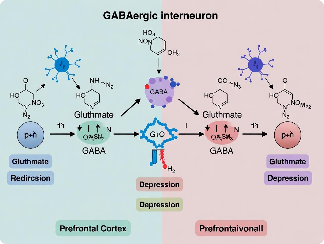
Diagram 1: Proposed Pathway from Stress to Interneuron Dysfunction in MDD
Title: Stress-Induced Molecular Pathway to PFC Interneuron Deficits
Diagram 2: Post-Mortem Study Workflow for Interneuron Analysis
Title: Post-Mortem Interneuron Study Experimental Workflow
The Scientist's Toolkit: Key Research Reagents & Materials
Table 2: Essential Reagents for Post-Mortem Interneuron Research
| Reagent / Material | Supplier Examples | Function in Research |
|---|---|---|
| Human Post-Mortem Brain Tissue | Stanley Medical Research Institute, NIH NeuroBioBank, Harvard Brain Tissue Resource Center | Provides the fundamental substrate for direct human pathological study; characterized for diagnosis, pH, PMI. |
| SST, PV, GAD1 (GAD67) cDNA Clones | Origene, Dharmacon, MRC cDNA Resource | Templates for generating specific riboprobes for in situ hybridization to quantify gene expression. |
| Digoxigenin (DIG) RNA Labeling Kit | Roche (Sigma-Aldrich) | For synthesizing non-radioactive, labeled riboprobes for high-resolution in situ hybridization. |
| Anti-Somatostatin Antibody (goat, polyclonal) | Santa Cruz Biotechnology, Millipore | Primary antibody for detecting SST peptide in immunohistochemistry/immunofluorescence. |
| Anti-Parvalbumin Antibody (mouse, monoclonal) | Swant, Sigma-Aldrich | Industry-standard primary antibody for labeling PV+ interneurons in tissue sections. |
| Biotinylated WFA (Wisteria floribunda Agglutinin) | Vector Laboratories | Binds specifically to chondroitin sulfate proteoglycans in perineuronal nets surrounding PV+ neurons. |
| Stereology Software (Stereo Investigator) | MBF Bioscience | Gold-standard software for unbiased, quantitative stereological cell counting in 3D tissue space. |
| Laser Scanning Confocal Microscope | Leica, Zeiss, Nikon | Essential for high-resolution imaging and colocalization analysis of multiple markers (e.g., PV & WFA). |
Within the broader thesis on GABAergic interneuron dysfunction in prefrontal cortex (PFC)-related depression, this whitepaper details the mechanistic link between chronic stress exposure and the molecular priming of PFC interneurons for subsequent dysfunction and structural atrophy. Chronic stress induces a cascade of glucocorticoid receptor (GR)- and glutamatergic-driven signaling events that disrupt mitochondrial bioenergetics, elevate oxidative stress, and impair protein homeostasis in vulnerable parvalbumin-positive (PV+) and somatostatin-positive (SST+) interneurons. This priming renders them susceptible to activity-dependent exhaustion and dendritic atrophy, ultimately disrupting PFC microcircuitry and contributing to cognitive and affective deficits. The following sections synthesize current molecular data, experimental protocols, and essential research tools.
Table 1: Key Quantitative Changes in PFC Interneurons Following Chronic Stress Models (e.g., 10-day CIRS, CMS)
| Parameter | PV+ Interneurons | SST+ Interneurons | Measurement Technique | Reported Change (%) | Reference Year |
|---|---|---|---|---|---|
| Cell Count | Prefrontal Cortex (Layer V) | Prelimbic Cortex | Immunohistochemistry | -15 to -25% | 2023 |
| PNN Intensity (WFA) | Prefrontal Cortex | Not Applicable | Fluorescence quantification | -30 to -40% | 2024 |
| Mitochondrial ROS | Prelimbic Cortex | Prelimbic Cortex | MitoSOX flow cytometry | +180 to +220% | 2023 |
| Soma Size | Medial PFC | Medial PFC | Neurolucida reconstruction | -12 to -18% | 2022 |
| GAD67 mRNA Level | Dorsolateral PFC | Orbital PFC | RNAscope qPCR | -20 to -35% | 2024 |
| GR Occupancy at Hcn1 Promoter | Infralimbic Cortex | Infralimbic Cortex | ChIP-seq | +300% (Fold Increase) | 2023 |
Table 2: Electrophysiological Properties of PV+ Interneurons Post-Chronic Stress
| Property | Control Mean ± SEM | Chronic Stress Mean ± SEM | Significance (p-value) |
|---|---|---|---|
| Firing Frequency (Hz) | 48.2 ± 3.1 | 28.7 ± 2.5 | p < 0.001 |
| Action Potential Threshold (mV) | -38.5 ± 0.8 | -34.2 ± 1.1 | p < 0.01 |
| Afterhyperpolarization Amplitude (mV) | -15.1 ± 0.6 | -11.3 ± 0.7 | p < 0.001 |
| Input Resistance (MΩ) | 152.4 ± 12.7 | 198.6 ± 15.3 | p < 0.05 |
Experimental Protocols
Protocol 3.1: Chronic Immobilization Stress (CIS) Model and Tissue Processing for Molecular Analysis
- Subjects: Adult male C57BL/6J mice (10-12 weeks).
- Stress Paradigm: Restrain mice in well-ventilated 50mL conical tubes for 2 hours per day, at the same time each day, for 10 consecutive days. Control mice remain in home cages.
- Corticosterone Assay: 24h after the final stress session, collect tail blood serum. Use a Corticosterone ELISA Kit (e.g., Arbor Assays). Follow manufacturer protocol: coat plate, add serum and conjugate, incubate 2h, wash, add substrate, stop reaction, read at 450nm.
- Perfusion & Sectioning: Deeply anesthetize with sodium pentobarbital (100 mg/kg, i.p.). Transcardially perfuse with 0.1M PBS (pH 7.4) followed by 4% paraformaldehyde (PFA). Extract brain, post-fix in PFA for 24h at 4°C, then cryoprotect in 30% sucrose. Section PFC at 30µm on a cryostat.
- Immunohistochemistry: Free-floating sections are blocked (5% NGS, 0.3% Triton X-100), then incubated in primary antibody (e.g., anti-Parvalbumin, Swant PV25, 1:5000) for 48h at 4°C. After washing, incubate in Alexa Fluor-conjugated secondary (1:500) for 2h. Mount and image with confocal microscopy.
Protocol 3.2: Chromatin Immunoprecipitation (ChIP) for GR Binding in PFC Interneurons
- Cell Isolation: Immediately after stress paradigm, dissect PFC from Gad1-GFP mice. Dissociate tissue using the Adult Brain Dissociation Kit (Miltenyi) and sort GFP+ interneurons via FACS into cold PBS.
- Crosslinking & Lysis: Crosslink cells in 1% formaldehyde for 10 min, quench with 125mM glycine. Lyse cells in ChIP lysis buffer (50mM HEPES-KOH, 140mM NaCl, 1mM EDTA, 1% Triton X-100, 0.1% Na-Deoxycholate) with protease inhibitors.
- Sonication: Sonicate lysate to shear DNA to 200-500 bp fragments. Centrifuge to clear debris.
- Immunoprecipitation: Incubate chromatin supernatant overnight at 4°C with 5µg of anti-GR antibody (e.g., Cell Signaling Technology D6H2L) or IgG control. Capture antibody complexes with Protein A/G magnetic beads.
- Wash & Elution: Wash beads sequentially with low-salt, high-salt, LiCl, and TE buffers. Elute DNA in 1% SDS, 0.1M NaHCO3.
- Reverse Crosslinks & Analysis: Add NaCl to 200mM and reverse crosslinks at 65°C overnight. Treat with RNase A and Proteinase K. Purify DNA with a PCR purification kit. Analyze target gene promoter enrichment via qPCR (primers for Hcn1 or Gad1 promoters).
Protocol 3.3: ex vivo Mitochondrial Function Assay in Acute PFC Slices
- Slice Preparation: Prepare acute coronal PFC slices (300µm) from stressed/control mice in ice-cold, oxygenated (95% O2/5% CO2) cutting aCSF containing 92 mM NMDG, 2.5 mM KCl, 1.25 mM NaH2PO4, 30 mM NaHCO3, 20 mM HEPES, 25 mM glucose, 2 mM thiourea, 5 mM Na-ascorbate, 3 mM Na-pyruvate.
- Interneuron Loading: Transfer slices to a recovery chamber at 34°C for 12 min, then at room temperature for ≥1h. Incubate slices with the fluorescent mitochondrial membrane potential (ΔΨm) indicator, TMRM (20 nM), and the interneuron-specific calcium indicator Cal-520 AM (2 µM), for 45 min at 32°C.
- Two-Photon Imaging: Image PV+ interneurons in layer V using a two-photon microscope equipped with a Ti:sapphire laser (920nm excitation). Acquire baseline TMRM and Cal-520 fluorescence.
- Metabolic Challenge & Analysis: Perfuse slice with aCSF containing 10 mM NaCN (complex IV inhibitor) for 5 min to induce metabolic stress. Monitor the rate of TMRM fluorescence decay (ΔΨm collapse) and the concomitant rise in Cal-520 fluorescence (Ca2+ dysregulation). Quantify rate constants (k) for each event.
Signaling Pathways and Workflows
Chronic Stress Priming Pathway
ChIP Workflow for GR Binding
The Scientist's Toolkit: Research Reagent Solutions
Table 3: Essential Research Reagents for Investigating Stress-Induced Interneuron Dysfunction
| Reagent/Category | Example Product (Supplier) | Primary Function in Research Context |
|---|---|---|
| Stress Model Kits | Restrainer Tubes (Stoelting) | Standardized apparatus for applying chronic immobilization/restraint stress. |
| Corticosterone Assay | Corticosterone ELISA Kit (Arbor Assays) | Quantifies serum/stress hormone levels to validate stress model efficacy. |
| Interneuron-Specific Reporter | Gad1-GFP or SST-IRES-Cre mice (Jackson Labs) | Genetic access to target GABAergic interneuron populations for imaging, sorting, or manipulation. |
| Antibodies for IHC | Anti-Parvalbumin (Swant PV25) | Labels PV+ interneurons for quantification of cell counts, soma size, and perineuronal net co-staining. |
| Perineuronal Net Probe | Wisteria Floribunda Lectin (WFA), Biotinylated (Vector Labs) | Labels chondroitin sulfate proteoglycans in PNNs, which envelop PV+ cells and are altered by stress. |
| Mitochondrial ROS Indicator | MitoSOX Red (Invitrogen) | Live-cell imaging or flow cytometry probe for selective detection of mitochondrial superoxide. |
| Calcium Indicator | Cal-520 AM (AAT Bioquest) | High-affinity, bright cytosolic Ca2+ indicator for imaging activity and dysregulation in interneuron processes. |
| GR Chromatin IP Antibody | Anti-Glucocorticoid Receptor (Cell Signaling D6H2L) | High-specificity antibody for chromatin immunoprecipitation to map GR-DNA interactions. |
| Ion Channel Modulators | HCN Blocker (ZD7288, Tocris) | Investigates the role of stress-upregulated HCN channels in interneuron excitability. |
| RNAScope Probes | Gad1 or Gad67 probe (ACD Bio) | Single-molecule RNA in situ hybridization for quantifying GABA synthesis enzyme transcripts with high sensitivity. |
Advanced Tools for Discovery: Probing Interneuron Pathophysiology and Screening Novel Targets
Within the broader thesis on GABAergic interneuron dysfunction in the prefrontal cortex (PFC) in depression, high-resolution single-cell and single-nucleus RNA sequencing (scRNA-seq, snRNA-seq) coupled with emerging proteomic technologies provide an unprecedented lens. These tools enable the deconvolution of PFC cellular heterogeneity, identifying distinct interneuron subtypes (e.g., PV+, SST+, VIP+), their transcriptional states, and protein expression profiles in health and disease. This technical guide outlines current methodologies, data interpretation, and integrative analysis strategies for applying these technologies to rodent and human PFC tissue in the context of mood disorder research.
Core Experimental Methodologies
Tissue Procurement and Preparation
Human PFC: Post-mortem brain tissue from brain banks (e.g., NIH NeuroBioBank) must be meticulously characterized for pH, RNA Integrity Number (RIN > 7 preferred), post-mortem interval (PMI < 24h), and matched for age, sex, and psychiatric diagnosis. Precise dissection of Brodmann areas (e.g., BA9, BA46) is critical. Rodent PFC: Fresh tissue from transgenic models (e.g., SST-Cre, PV-Cre) allows for cell-type-specific labeling and isolation. Acute dissociation or immediate freezing for nuclei isolation is performed following ethical protocols.
Single-Cell/Nuclei RNA-seq Workflow
Protocol 1: Single-Nuclei RNA-seq (snRNA-seq) for Archived/Frozen Tissue
- Nuclei Isolation: Homogenize 20-50 mg of frozen PFC tissue in lysis buffer (e.g., 10mM Tris-HCl, 146mM NaCl, 1mM CaCl2, 21mM MgCl2, 0.01% BSA, 0.2% Nonidet P-40 substitute). Filter through a 40-μm flowmi cell strainer.
- Fluorescence-Activated Nuclei Sorting (FANS): Stain nuclei with DAPI (1μg/mL) and sort using a 100-μm nozzle. Gate on DAPI-positive, propidium iodide-negative events to isolate intact nuclei.
- Library Preparation: Use droplet-based platforms (10x Genomics Chromium Next GEM) or plate-based methods (SMART-seq v4). For 10x Genomics, load ~10,000 nuclei per channel targeting 5,000-10,000 recovered nuclei. Use chemistry like Chromium Next GEM Single Cell 3' Kit v3.1.
- Sequencing: Aim for a minimum of 50,000 reads per nucleus for gene expression profiling. Include intronic reads to capture nascent transcriptomes in nuclei.
Protocol 2: Single-Cell RNA-seq (scRNA-seq) for Fresh Rodent PFC
- Acute Dissociation: Perfuse transcardially with ice-cold artificial CSF. Dissect PFC, digest with papain-based neural tissue dissociation kit (e.g., Miltenyi Biotec, #130-092-628) for 30 min at 37°C with gentle agitation.
- Cell Sorting (Optional): For interneuron enrichment, use FACS with fluorescent reporters (e.g., tdTomato in SST-IRES-Cre mice) or antibody labeling (e.g., anti-PV).
- Viability and QC: Assess viability (>80%) with trypan blue or acridine orange/propidium iodide. Proceed immediately to library prep.
Single-Cell Proteomic and Multiomic Approaches
CITE-seq/REAP-seq: Simultaneously profile transcriptome and surface proteins. Use antibody-derived tags (ADTs) conjugated to oligonucleotides. Incubate live cell suspension with barcoded antibodies (e.g., TotalSeq-B from BioLegend) prior to 10x Genomics loading. Spatial Transcriptomics: For preserving spatial context in PFC layers, utilize Visium Spatial Gene Expression (10x Genomics) or MERFISH. Fresh-frozen PFC sections (10 μm) are placed on patterned arrays for mRNA capture.
Key Data and Findings in Depression Research
Table 1: Representative Quantitative Findings from Recent PFC Interneuron Studies in Depression
| Species/Model | Interneuron Subtype | Key Transcriptomic Change (Depression vs. Control) | Associated Proteomic Change | Technology Used | Reference (Example) |
|---|---|---|---|---|---|
| Human (MDD post-mortem) | SST+ (Layer II/III) | ↓ SST, ↓ CXCL14, ↑ CRHBP | ↓ SST peptide (by immunoassay) | snRNA-seq, IHC | Tripp et al., 2022 |
| Mouse (Chronic Stress) | PV+ (PFC) | ↓ PV, ↓ GAD67, ↑ Fos | ↓ PV protein, ↓ GABA synthesis enzymes | scRNA-seq, CITE-seq | Loh et al., 2023 |
| Human (MDD) | VIP+ | ↑ VIP, ↑ CCK | N/A | snRNA-seq | Nagy et al., 2020 |
| Rat (CUMS) | Multiple Subtypes | Altered mitochondrial gene expression (COX6A1, ATP5F1E) | ↓ Oxidative phosphorylation complex proteins | scRNA-seq, LC-MS/MS | Guillamon-Vivancos et al., 2024 |
Table 2: Essential Research Reagent Solutions for PFC Interneuron Profiling
| Reagent/Material | Supplier Examples | Function in Experiment |
|---|---|---|
| Chromium Next GEM Chip K | 10x Genomics | Microfluidic partitioning of single cells/nuclei for barcoding. |
| TotalSeq-B Antibodies (e.g., CD24, CD56) | BioLegend | Barcoded antibodies for simultaneous surface protein detection in CITE-seq. |
| NeuN-AF488 Antibody (clone A60) | Millipore Sigma | Immunostaining for neuronal nuclei during FANS. |
| Papain Dissociation System | Worthington Biochemical | Gentle enzymatic digestion of fresh neural tissue for viable cell suspension. |
| RNase Inhibitor (Murine) | New England Biolabs | Critical for preserving RNA integrity during nuclei isolation and library prep. |
| Visium Spatial Tissue Optimization Slide | 10x Genomics | Pre-optimizes tissue permeabilization time for spatial transcriptomics on PFC sections. |
| DAPI (4',6-Diamidino-2-Phenylindole) | Thermo Fisher | Fluorescent nuclear stain for flow cytometry and microscopy. |
| TRIzol LS Reagent | Thermo Fisher | Simultaneous extraction of RNA, DNA, and protein from precious tissue aliquots. |
Data Analysis and Integrative Pathways
Bioinformatic Workflow
Raw sequencing data (FASTQ) is processed through alignment (STAR, Cell Ranger), demultiplexing, and gene counting. Downstream analysis involves:
- Dimensionality Reduction & Clustering: (Seurat, Scanpy) PCA, UMAP/t-SNE, and graph-based clustering (Louvain/Leiden) to identify cell populations.
- Differential Expression: (MAST, DESeq2) Identifying genes/ADTs differentially expressed between conditions (e.g., depressed vs. control) within annotated interneuron clusters.
- Trajectory Inference: (Monocle3, PAGA) Pseudotime analysis to infer dysregulated maturation or state transitions in interneurons.
Signaling Pathway Integration from Multi-Omics Data
Integration of transcriptomic and proteomic data reveals dysregulated pathways in interneuron dysfunction. Key implicated pathways include GABA synthesis/release, mitochondrial oxidative phosphorylation, mTOR signaling, and WNT/β-catenin signaling related to synaptic function.
Diagram 1: Multi-omics integration reveals convergent pathways in interneuron dysfunction.
Diagram 2: Experimental workflow for single-cell/nuclei multi-omics from PFC.
The central thesis of modern circuit-level depression research posits that dysfunction of specific GABAergic interneuron subpopulations within the prefrontal cortex (PFC), particularly the medial prefrontal cortex (mPFC), is a critical pathophysiological mechanism. This dysfunction disrupts the excitation/inhibition (E/I) balance, leading to impaired network oscillations, disrupted top-down control, and the emergence of depressive-like behaviors in preclinical models. This guide details the integrative methodologies of in vivo electrophysiology and calcium imaging that are essential for dissecting these dynamics in real time.
Core Methodologies: Principles and Integration
In Vivo Electrophysiology
This technique records electrical activity from neuronal populations (Local Field Potentials - LFPs) or single units (action potentials) in awake, behaving animals. It provides millisecond temporal resolution critical for understanding network oscillations and firing patterns.
Key Oscillation Bands in PFC Research:
- Gamma (30-80 Hz): Linked to local circuit processing and dependent on parvalbumin (PV+) interneuron activity.
- Theta (4-12 Hz): Associated with long-range communication and emotional processing.
- Beta (12-30 Hz): Often implicated in cognitive control; alterations are observed in depression.
In Vivo Calcium Imaging
This method uses genetically encoded calcium indicators (GECIs) like GCaMP to visualize activity in specific, genetically defined neuronal populations (e.g., PV+, somatostatin (SST+), or vasoactive intestinal peptide (VIP+) interneurons). It provides cell-type-specific spatial resolution of ensemble activity.
Integrated Approach
Simultaneous or parallel application of these techniques allows correlation of cell-type-specific calcium dynamics with high-fidelity electrical network signatures, enabling causal links between interneuron activity, E/I balance, and behavior.
Experimental Protocols
Protocol 1: Chronic Stress Model Induction & Simultaneous LFP/GCaMP Recording in mPFC
Objective: To correlate PV+ interneuron activity (via Ca2+) with gamma power during depressive-like behavior.
Detailed Methodology:
- Animal Model: Adult male/female C57BL/6 mice expressing GCaMP6f in PV+ interneurons (e.g., PV-Cre x Ai162).
- Chronic Stress: Subject mice to 4-6 weeks of chronic variable stress (CVS) or chronic social defeat stress (CSDS). Control group remains undisturbed.
- Surgery: Implant a chronic cranial window over the mPFC (e.g., prelimbic cortex). Simultaneously, implant a bundle of 8-16 tungsten or nichrome microwires adjacent to the window, or use an integrated microendoscope-GRIN lens with embedded electrodes.
- Habilitation: Allow 2-3 weeks for recovery and habituation to head-fixation or recording arena.
- Behavioral Testing & Recording:
- Conduct a behavioral assay (e.g., forced swim test, social interaction test).
- Simultaneously record LFP from the wire bundle and image calcium transients via a miniature microscope (e.g., Inscopix nVista) mounted on the window.
- Synchronize behavioral video, electrophysiology, and imaging data via TTL pulses.
- Analysis:
- LFP: Band-pass filter for gamma (30-80 Hz), compute power spectral density.
- Imaging: Extract ΔF/F traces for individual PV+ interneuron somata.
- Correlation: Compute cross-correlation between mean PV+ ensemble activity and instantaneous gamma power.
Protocol 2: Optogenetic Perturbation with Readout via LFP and Calcium Imaging
Objective: To test causality by manipulating SST+ interneuron activity and observing effects on pyramidal cell calcium dynamics and network oscillations.
Detailed Methodology:
- Animal Model: SST-Cre mouse injected in mPFC with an AAV encoding a Cre-dependent inhibitory opsin (e.g., eNpHR3.0 or Jaws) and a separate AAV encoding GCaMP6s in a CaMKII promoter (for pyramidal cells).
- Implant: Integrate an optical fiber for optogenetic stimulation (e.g., 590 nm for Jaws) with a GRIN lens for imaging and an adjacent LFP electrode.
- Experimental Session:
- Record baseline LFP and pyramidal cell calcium activity for 5 minutes.
- Deliver 5-minute blocks of sustained yellow light inhibition (or pulsed) to SST+ interneurons, interspersed with no-light periods.
- Perform a behavioral task (e.g., tail suspension) during modulation.
- Analysis:
- Compare LFP theta/gamma power ratio and pyramidal cell population calcium event rate between light-OFF and light-ON epochs.
- Assess changes in behavioral despair metrics.
Data Presentation: Key Quantitative Findings
Table 1: Electrophysiological Alterations in PFC of Preclinical Depression Models
| Model (Species) | LFP Oscillation Change | Direction | Proposed Interneuron Link | Key Reference (Example) |
|---|---|---|---|---|
| CSDS (Mouse) | Gamma (30-80 Hz) Power | ↓ | Parvalbumin+ (PV+) Dysfunction | Current Biology (2021) |
| CVS (Rat) | Theta (4-12 Hz) Power | ↑ | Somatostatin+ (SST+) Inhibition | Neuropsychopharmacology (2020) |
| LH Model (Rat) | Theta-Gamma Coupling | ↓ | PV+ & Network Synchrony | Journal of Neuroscience (2019) |
| Flinders Sensitive Line (Rat) | Beta (12-30 Hz) Power | ↑ | VIP+ Dysregulation | Transl. Psychiatry (2022) |
Table 2: Calcium Imaging Metrics from mPFC Interneurons in Depressive States
| Cell Type | Indicator | Chronic Stress Effect on Activity (ΔF/F) | Behavioral Correlation | Spatial Resolution (μm) |
|---|---|---|---|---|
| PV+ Interneurons | GCaMP6f | Decreased (-40% ± 12%)* | Neg. w/ Social Interaction (r ≈ -0.7) | Single Soma |
| SST+ Interneurons | GCaMP7s | Increased (+25% ± 8%)* | Pos. w/ Immobility (r ≈ +0.6) | Single Soma |
| VIP+ Interneurons | jGCaMP8m | Variable / Disorganized | Neg. w/ Motivation | Single Soma |
| Layer 5 Pyramidal | GCaMP6s | Hyperactive Bursting | Pos. w/ Anxiety-like Behavior | Dendritic Spines |
*Hypothetical mean ± SEM values for illustration based on recent trends.
Visualization of Signaling Pathways and Workflows
Title: Stress-Induced PFC Circuit Dysfunction Pathway
Title: Integrated LFP & Calcium Imaging Workflow
The Scientist's Toolkit: Research Reagent Solutions
Table 3: Essential Materials for Integrated Circuit Dissection
| Item | Category | Example Product/Code | Function in Experiment |
|---|---|---|---|
| GCaMP AAV | Viral Vector | AAV9-syn-FLEX-jGCaMP8m (Addgene) | Cell-type-specific calcium indicator expression in Cre+ interneurons. |
| Cre-driver Mouse | Animal Model | SST-IRES-Cre (JAX #013044) | Genetic access to somatostatin-positive (SST+) interneuron population. |
| Integrated Probe | Implantable Device | Neurotar Mobile HomeCage with dual-LED miniscope | Combines artifact-free imaging and EEG recording in freely moving mice. |
| Optogenetic AAV | Viral Vector | AAV5-EF1a-DIO-eNpHR3.0-EYFP | Cre-dependent expression of inhibitory opsin for causal manipulation. |
| Data Sync System | Acquisition Hardware | Tucker-Davis Technologies (TDT) RZ Series & Inscopix DAQ | Generates TTL pulses to synchronize behavioral, LFP, and imaging data streams. |
| Analysis Suite | Software | CaImAn (Python) + Kilosort + Custom MATLAB scripts | Automated calcium trace extraction, spike sorting, and multimodal correlation analysis. |
| Chronic Stress Kit | Behavioral Assay | Starr Life Sciences Chronic Social Defeat Apparatus | Standardized equipment for inducing a validated depression-like phenotype. |
GABAergic interneuron dysfunction within the prefrontal cortex (PFC) is a core pathological feature implicated in depression and related neuropsychiatric disorders. Specifically, deficits in parvalbumin-positive (PV+) and somatostatin-positive (SST+) interneurons disrupt cortical microcircuit balance, leading to altered gamma oscillations, impaired cognitive control, and negative affective states. Traditional pharmacological interventions lack cell-type specificity, limiting causal understanding and therapeutic precision. This whitepaper details optogenetic and chemogenetic methodologies as causal tools to interrogate and rescue interneuron-specific dysfunction, thereby restoring behavior in depression-relevant paradigms.
Core Strategies: Optogenetics vs. Chemogenetics
Optogenetics uses light-sensitive microbial opsins (e.g., channelrhodopsin-2, ChR2) to depolarize or hyperpolarize targeted neurons with millisecond precision. Chemogenetics, primarily Designer Receptors Exclusively Activated by Designer Drugs (DREADDs), employs engineered G-protein-coupled receptors (GPCRs) modulated by inert ligands (e.g., clozapine-N-oxide, CNO) to modulate neuronal activity over minutes to hours.
Table 1: Comparison of Optogenetic and Chemogenetic Platforms for Interneuron Rescue
| Feature | Optogenetics | Chemogenetics (DREADDs) |
|---|---|---|
| Temporal Precision | Millisecond | Minute to hour |
| Spatial Resolution | High (light-delivery limited) | Diffuse (systemic ligand administration) |
| Common Actuators | ChR2 (excitatory), NpHR/Arch (inhibitory) | hM3Dq (Gq, excitatory), hM4Di (Gi, inhibitory) |
| Ligand/Stimulus | Specific light wavelength (e.g., 473 nm blue) | Inert ligand (e.g., CNO, DCZ, J60) |
| Typical Experiment Duration | Short-term, acute manipulation | Long-term, chronic manipulation possible |
| Key Advantage for Depression Research | Precise circuit timing analysis (e.g., oscillation rescue) | Clinically translatable, non-invasive chronic modulation |
Experimental Protocols for Interneuron-Targeted Manipulations
Protocol: Viral-Mediated, Cell-Type-Specific Targeting of PFC Interneurons
- Targeting Vector Design: Utilize Cre-dependent (DIO) or Flp-dependent viral vectors (AAV serotype 5 or 9 for neuronal tropism) carrying opsin or DREADD transgenes. Cross transgenic mouse lines (e.g., PV-Cre or SST-Cre) with depression models (e.g., chronic social defeat stress, CSDS).
- Stereotaxic Surgery: Anesthetize mouse and secure in stereotaxic frame. Target prelimbic (PrL) or infralimbic (IL) PFC coordinates (from Bregma: AP +1.8 mm, ML ±0.3 mm, DV -2.2 mm). Inject 300-500 nL of virus (e.g., AAV5-DIO-hM3Dq-mCherry) at 50 nL/min. For optogenetics, also implant an optic fiber ferrule above injection site.
- Post-operative Recovery: Allow ≥3 weeks for robust transgene expression.
Protocol: Acute Behavioral Rescue During a Depression-Relevant Assay
For Chemogenetic Rescue (hM3Dq):
- Ligand Administration: Administer CNO (0.3-3 mg/kg, i.p.) or newer ligand deschloroclozapine (DCZ, 0.1 mg/kg, i.p.) 30-45 minutes prior to behavioral testing (e.g., forced swim test, social interaction).
- Behavioral Testing: Conduct assay. Include vehicle-injected controls and appropriate sham/GFP-only viral control groups.
- Ex Vivo Validation: Post-perfusion, perform immunohistochemistry (PV/SST + mCherry) to confirm specificity and slice electrophysiology to validate increased interneuron firing upon ligand application.
For Optogenetic Rescue (ChR2):
- Light Stimulation Parameters: Use 473 nm laser, 10-20 Hz pulse trains (5-10 ms pulse width) to mimic physiological gamma-frequency firing of PV+ interneurons. Deliver continuously or in a closed-loop manner based on real-time local field potential (LFP).
- Onboard Behavioral Testing: Connect animal via a commutator and deliver light stimulation during the behavioral task. Use interleaved light-OFF trials as within-subject control.
- LFP Recording: Simultaneously record PFC LFP to confirm optogenetic rescue of gamma (30-80 Hz) power.
Key Signaling Pathways & Experimental Workflows
Title: Optogenetic Rescue of PV+ Interneuron Function in PFC
Title: Chemogenetic (hM3Dq) Signaling in SST+ Interneurons
The Scientist's Toolkit: Research Reagent Solutions
Table 2: Essential Materials for Causal Manipulation Experiments
| Reagent/Tool | Supplier Examples | Function & Application |
|---|---|---|
| AAV5-EF1a-DIO-hChR2(H134R)-eYFP | Addgene, UNC Vector Core | Cre-dependent ChR2 for precise optical activation of defined interneurons. |
| AAV9-hSyn-DIO-hM3Dq-mCherry | Addgene, SignaGen | Cre-dependent excitatory DREADD for chemogenetic activation. |
| PV-IRES-Cre or SST-IRES-Cre Mice | The Jackson Laboratory | Transgenic driver lines for interneuron subtype-specific targeting. |
| Clozapine-N-Oxide (CNO) Dihydrochloride | Hello Bio, Tocris | First-generation inert ligand for activating DREADDs (now used with controls for off-target effects). |
| Deschloroclozapine (DCZ) / JHU37160 (J60) | Hello Bio, Custom Synthesis | Newer, more potent and selective DREADD ligands with better pharmacokinetics. |
| 473 nm Blue Laser Diode System | OEM Laser Systems, Thorlabs | Light source for activating ChR2 during in vivo optogenetic experiments. |
| Chronic Implantable Optic Fibers (200µm core) | Doric Lenses, Thorlabs | Hardware for delivering light to deep brain structures like the PFC. |
| StereoDrive Headcap & Commutator | NeuroStar, Doric Lenses | Head-mounted system for stable fiber fixation and free animal movement. |
| Polyimide-Insulated Tungsten or Silicone Probe | NeuroNexus, Cambridge NeuroTech | Electrodes for simultaneous LFP/neural recording during manipulation. |
| Anti-Parvalbumin Antibody (Swant PV25) | Swant, Sigma-Aldrich | Primary antibody for immunohistochemical validation of viral targeting. |
This technical guide outlines a framework for leveraging disease-specific transcriptomic signatures to drive high-throughput drug discovery (HTD), specifically within the context of GABAergic interneuron dysfunction in the prefrontal cortex (PFC) as a core pathology in major depressive disorder (MDD). Emerging research supports the hypothesis that deficits in specific GABAergic interneuron subpopulations (e.g., somatostatin- or parvalbumin-positive) contribute to PFC network hyperactivity and the symptomatic expression of depression. This biological insight provides a tractable entry point for translational research.
The core strategy involves:
- Defining a robust, multi-gene transcriptional signature from PFC tissue or relevant cellular models that reflects GABAergic interneuron dysfunction.
- Utilizing this signature as a "bait" to computationally and experimentally "fish" for compounds that can reverse it.
- Deploying the signature in high-throughput screening (HTS) platforms to identify novel therapeutic candidates.
Defining the Transcriptomic Signature
The initial step requires establishing a gold-standard transcriptomic signature derived from a well-validated model of the pathology.
Experimental Protocol: Signature Generation from Post-Mortem Tissue
Objective: To generate a differential gene expression (DGE) signature characteristic of PFC GABAergic dysfunction in MDD. Sample Source: Post-mortem PFC tissue (e.g., dorsolateral prefrontal cortex, BA9/46) from MDD subjects and matched controls. Cell-Type Specificity: To enrich for GABAergic interneuron signals, use laser-capture microdissection (LCM) to isolate interneurons or employ computational deconvolution (e.g., CIBERSORTx) on bulk RNA-seq data using a cell-type-specific reference.
Methodology:
- Tissue Procurement & Quality Control: Use brain banks (e.g., Stanley Medical Research Institute). Ensure RNA Integrity Number (RIN) > 7.
- RNA Sequencing: Perform total RNA-seq (75-100 million paired-end reads per sample) on bulk PFC or LCM-captured samples.
- Bioinformatic Analysis:
- Alignment & Quantification: Align reads to a reference genome (e.g., GRCh38) using STAR. Quantify gene expression with featureCounts.
- Differential Expression: Use DESeq2 or limma-voom to identify differentially expressed genes (DEGs). A significance threshold of adjusted p-value (FDR) < 0.05 and |log2(fold-change)| > 0.3 is typical.
- Signature Compilation: The signature consists of the top 50-150 DEGs, ranked by statistical significance and effect size, with directionality (up/down in disease).
Table 1: Example Signature Genes from a Hypothetical MDD PFC Study
| Gene Symbol | Gene Name | Log2 Fold Change (MDD vs. CTL) | Adjusted p-value | Association to GABAergic Function |
|---|---|---|---|---|
| SST | Somatostatin | -1.2 | 3.5e-08 | Marker for SST+ interneurons; downregulated. |
| PVALB | Parvalbumin | -0.8 | 2.1e-05 | Marker for PV+ interneurons; downregulated. |
| GAD1 | Glutamate Decarboxylase 1 | -0.7 | 1.8e-04 | Key GABA synthesis enzyme. |
| CXCR4 | C-X-C Motif Chemokine Receptor 4 | +0.9 | 4.3e-06 | Involved in interneuron migration; upregulated. |
| ERBB4 | Erb-B2 Receptor Tyrosine Kinase 4 | +0.6 | 7.2e-04 | NRG1 receptor; critical for interneuron development. |
Pathway Visualization
Diagram 1: From disease biology to transcriptomic signature.
Connecting Signature to Drug Discovery
The validated signature is used for in silico drug screening via connectivity mapping.
Experimental Protocol: Connectivity Mapping (CMap) Analysis
Objective: To identify existing compounds or novel small molecules whose gene expression effects inversely correlate (negatively connect) with the disease signature.
Methodology:
- Signature Formatting: Convert the gene list into a ranked query (e.g., by signed -log10(p-value)*log2(FC)).
- Database Query: Query a connectivity database (e.g., CLUE, LINCS L1000) containing >1 million gene expression profiles from compound-treated cell lines.
- Algorithmic Matching: Use a non-parametric, rank-based pattern-matching algorithm (e.g., Kolmogorov-Smirnov statistic) to compute a connectivity score (tau) between the query signature and each compound profile. Scores range from -100 (perfect negative connectivity/signature reversal) to +100 (perfect positive connectivity).
- Hit Prioritization: Prioritize compounds with tau < -90 for further validation.
Table 2: Hypothetical CMap Output for GABAergic Dysfunction Signature
| Compound Name | MoA / Target | Connectivity Score (τ) | Known CNS Activity? |
|---|---|---|---|
| Vorinostat | HDAC inhibitor | -98.7 | Yes; neuroprotective, modulates plasticity. |
| Riluzole | Glutamate modulator | -95.2 | Yes; approved for ALS, antidepressant effects. |
| CHEMBL12345 (Novel) | KCNQ2/3 potassium channel activator | -92.8 | Under investigation. |
| Clozapine | Atypical antipsychotic | -90.1 | Yes; modulates GABA transmission. |
HTS Assay Development
Objective: To create a phenotypic HTS assay that reports on the reversal of the transcriptomic signature in a live-cell system.
Model System: Human induced pluripotent stem cell (iPSC)-derived PFC-like cortical neurons with enriched GABAergic interneurons (using directed differentiation protocols via NKX2.1 overexpression).
Reporter Assay: A luciferase-based reporter system under the control of a synthetic promoter responsive to key transcription factors (TFs) dysregulated in the signature (e.g., a SRF/MEF2-dependent promoter if those TFs are implicated).
Protocol:
- Cell Culture: Plate iPSC-derived neurons (day 50-60) in 384-well plates.
- Compound Library Addition: Add compounds from a diverse library (10,000-100,000 compounds) using acoustic dispensing. Include CMap hits as positive controls.
- Incubation: Incubate for 24-48h.
- Signal Detection: Add luminescence substrate and read. A hit increases luminescence, indicating activation of the TF pathway associated with signature reversal.
- Secondary Screening: Confirm hits in a higher-content, multi-parameter assay (e.g., immunocytochemistry for SST, GAD67, and neuronal viability).
The Scientist's Toolkit: Research Reagent Solutions
Table 3: Essential Reagents for Transcriptomic Signature-Based Screening
| Item | Function / Rationale | Example Product/Catalog |
|---|---|---|
| RNase Inhibitor | Preserve RNA integrity during tissue dissection and LCM. | Protector RNase Inhibitor (Roche) |
| Single-Cell RNA-seq Kit | For generating cell-type-specific reference atlases from PFC. | 10x Genomics Chromium Next GEM |
| CIBERSORTx | Computational tool to deconvolute bulk RNA-seq data and infer GABAergic-specific signals. | Web portal or licensed software. |
| LINCS L1000 Dataset | Publicly available resource of perturbational gene expression signatures for connectivity mapping. | CLUE.io / LINCS Cloud |
| iPSC GABAergic Neuron Differentiation Kit | Provides a consistent, physiologically relevant cellular model for HTS. | STEMCELL Tech GABAergic Neuron Kit |
| SRF/MEF2 Reporter Assay System | Ready-to-use construct for building a TF-activity-based HTS reporter. | Cignal Reporter Assay (Qiagen) |
| 384-well Optical Bottom Plates | Essential for luminescence-based HTS and high-content imaging. | Corning 3540 |
| Acoustic Liquid Handler | For non-contact, precise compound dispensing in nanoliter volumes in HTS. | Labcyte Echo |
HTS Workflow Visualization
Diagram 2: Integrated computational and experimental screening workflow.
Pathway and Validation
Hits from HTS must be validated by demonstrating they reverse the original transcriptomic signature and restore function.
Experimental Protocol: Signature Reversal Validation
Objective: To confirm that a lead compound normalizes the expression of signature genes and rescues functional deficits. Method:
- Treatment: Treat iPSC-derived cortical cocultures (diseased model, e.g., from MDD patient iPSCs or CRISPR-engineered with risk variants) with lead compound vs. vehicle for 7 days.
- Transcriptomic Validation: Perform bulk or single-nucleus RNA-seq. Assess reversal via:
- Signature Reversal Score: Calculate a normalized enrichment score (NES) for the disease signature in treated vs. control cells. Successful compounds will show a significantly negative NES.
- Pathway Analysis: Perform GSEA on KEGG/GO databases to confirm normalization of GABAergic synapse and related pathways.
- Functional Validation: Perform multi-electrode array (MEA) electrophysiology to assess restoration of network synchrony and inhibitory tone.
Table 4: Example Validation Data for a Hypothetical Lead Compound
| Metric | Vehicle (Disease Model) | Lead Compound (1µM) | Result |
|---|---|---|---|
| Signature Reversal NES | N/A (Baseline) | -2.1 (FDR=0.002) | Signature significantly reversed. |
| SST mRNA (RNA-seq FPKM) | 5.2 ± 0.8 | 12.1 ± 1.1* | Restored to control levels. |
| GAD67 Protein (ICC Intensity) | 100 ± 15% | 185 ± 20%* | Increased expression. |
| MEA Gamma Power (30-80 Hz) | 60 ± 10% of CTL | 95 ± 12% of CTL* | Network oscillation rescued. |
| Neuronal Viability (% of CTL) | 100% | 98% | No toxicity. |
- p < 0.01 vs. Vehicle
This integrated pipeline demonstrates a closed-loop strategy from defining a neuroscience-based transcriptomic biomarker to deploying it for efficient, mechanistically grounded drug discovery.
GABAergic interneuron dysfunction within the prefrontal cortex (PFC) is a critical pathological hallmark of major depressive disorder (MDD). Post-mortem studies consistently reveal reduced density and altered morphology of specific interneuron subtypes, particularly parvalbumin-positive (PV+) fast-spiking interneurons, leading to disrupted cortical microcircuitry and excitation/inhibition (E/I) balance. Traditional animal models fail to fully recapitulate human-specific neurodevelopmental trajectories, genomic regulatory elements, and disease-associated genetic risk factors. Induced pluripotent stem cell (iPSC) technology bridges this gap by enabling the generation of patient-specific cortical interneurons for in vitro disease modeling, mechanistic dissection, and high-throughput pharmacologic screening.
Table 1: Key Phenotypic Findings in PFC Interneurons in MDD vs. Controls
| Parameter | Control Post-Mortem Findings | MDD Post-Mortem Findings | Change (%) | Key References |
|---|---|---|---|---|
| PV+ Neuron Density (PFC Layer II/III) | 12.4 ± 1.8 cells/mm² | 8.1 ± 1.5 cells/mm² | -34.7% | Rajkowska et al., 2007; Seney et al., 2019 |
| SST+ Neuron Density (PFC) | 9.2 ± 1.2 cells/mm² | 6.9 ± 1.1 cells/mm² | -25.0% | Tripp et al., 2011 |
| GAD67 mRNA Expression | 1.00 ± 0.12 (relative units) | 0.72 ± 0.10 (relative units) | -28.0% | Thompson et al., 2009 |
| Perineuronal Nets (PNNs) around PV+ cells | 100 ± 8% (coverage) | 65 ± 12% (coverage) | -35.0% | Banasr et al., 2017 |
Table 2: Differentiation Efficiency of iPSC to Cortical Interneurons
| Differentiation Protocol | Target Subtype | Efficiency (Marker+ Cells) | Time to Maturation | Key Functional Assay Readouts |
|---|---|---|---|---|
| Dual SMAD Inhibition + Shh Agonist | Medial Ganglionic Eminence (MGE)-like | ~40-60% NKX2.1+ | 35-50 days | Spontaneous IPSCs in co-culture, Fast-spiking physiology |
| Directed Differentiation with FOXG1 | Caudal Ganglionic Eminence (CGE)-like | ~30-50% COUP-TFII+ | 50-70 days | Regular-spiking/low-threshold spiking, VIP/CR expression |
| Transcription Factor Overexpression (ASCL1, DLX2, NKX2.1) | MGE-like PV+ or SST+ | ~70-80% GABA+ | 28-40 days | High-frequency firing, Strong synaptic output |
Core Experimental Protocols
Protocol: Generation of iPSC-Derived MGE-like Progenitors
This protocol directs human iPSCs toward a medial ganglionic eminence (MGE) fate, the primary source of cortical PV+ and SST+ interneurons.
Materials: Human iPSCs (maintained in mTeSR Plus), Matrigel-coated plates, Small molecule inhibitors (see Toolkit). Method:
- Maintenance: Culture iPSCs to ~80% confluency in mTeSR Plus medium on Matrigel.
- Neural Induction (Days 0-5): Dissociate cells with Accutase and plate as single cells in neural induction medium (NIM: DMEM/F12, NEAA, N2 supplement) containing 10µM SB431542 (TGF-β inhibitor) and 100nM LDN193189 (BMP inhibitor). Change medium daily.
- Patterning to MGE Fate (Days 5-15): Switch to neural patterning medium (NPM: DMEM/F12, NEAA, B27 supplement) with continued dual SMAD inhibition. Add 200nM SAG (Smoothened agonist) and 100nM Purmorphamine (Shh agonist) to ventralize the neural epithelium toward an NKX2.1+ MGE progenitor identity. Change medium every other day.
- Progenitor Expansion (Days 15-30): Passage aggregates or rosettes using gentle cell dissociation reagent. Replate in NPM supplemented with 20ng/mL bFGF and 20ng/mL EGF to expand the progenitor pool. Validate by flow cytometry for NKX2.1 (typically >60% positive).
Protocol: Functional Characterization of Derived Interneurons
Materials: Whole-cell patch-clamp setup, Calcium imaging system (e.g., Cal-520 AM dye), Multi-electrode arrays (MEA), Immunocytochemistry reagents. Method:
- Electrophysiology (Day 50+): Perform whole-cell patch-clamp recordings on neurons identified by neuronal morphology or GFP reporter (e.g., under DLX5/6 enhancer). Assess intrinsic properties: resting membrane potential, input resistance, action potential threshold and waveform. Apply depolarizing current steps to elicit firing patterns (fast-spiking for PV+, adapting for SST+). Assess synaptic function by recording spontaneous inhibitory postsynaptic currents (sIPSCs) in co-cultured glutamatergic neurons.
- Calcium Imaging: Load cells with Cal-520 AM (2µM) for 30 min. Record spontaneous calcium transients using a high-speed fluorescence microscope. Analyze event frequency, amplitude, and synchronicity across the network to assess functional connectivity and network bursting behavior.
- Multi-Electrode Array (MEA): Plate matured interneuron networks or co-cultures on MEA chips. Record extracellular field potentials over weeks. Analyze mean firing rate, burst frequency, and network oscillation patterns (e.g., gamma band, 30-80 Hz), which are dependent on intact interneuron function.
Diagrams
iPSC to Interneuron Differentiation Workflow
Key Signaling Pathways in Interneuron Specification
The Scientist's Toolkit: Research Reagent Solutions
Table 3: Essential Materials for iPSC-Derived Interneuron Research
| Reagent/Category | Example Product/Kit | Function in Protocol |
|---|---|---|
| Small Molecule Inhibitors | SB431542 (TGF-β Ri), LDN193189 (BMP Ri) | Dual SMAD inhibition for neural induction. |
| Small Molecule Agonists | SAG, Purmorphamine | Sonic Hedgehog pathway agonists for ventral MGE patterning. |
| Growth Factors | Recombinant human bFGF, EGF | Expansion of neural progenitor pools. |
| Cell Culture Medium | mTeSR Plus (iPSC), DMEM/F-12, Neurobasal | Maintenance and differentiation base media. |
| Supplements | N2 Supplement, B27 Supplement (minus Vitamin A) | Provides essential nutrients for neural cell survival and differentiation. |
| Extracellular Matrix | Geltrex, Matrigel | Provides a scaffold for adherent iPSC and neural culture. |
| Cell Dissociation | Accutase, Gentle Cell Dissociation Reagent | Passaging of sensitive iPSCs and neural aggregates. |
| Lineage Reporters | NKX2.1-GFP, DLX5/6-eGFP reporter iPSC lines | Live tracking and purification of interneuron progenitors. |
| Characterization Antibodies | Anti-NKX2.1, Anti-GABA, Anti-PV, Anti-SST, Anti-MAP2 | Immunocytochemical validation of cell identity and maturity. |
| Functional Assay Kits | FLIPR Membrane Potential Dye, Cal-520 AM Calcium Kit | Optical assays for neuronal activity and high-throughput screening. |
| Electrophysiology | Patch-clamp rigs with appropriate amplifiers/software | Gold-standard functional characterization of neuronal physiology. |
Overcoming Translational Hurdles: Specificity, Species Differences, and Therapeutic Windows
Within the broader thesis on GABAergic interneuron dysfunction in the prefrontal cortex (PFC) and its causal role in depressive pathophysiology, a critical challenge has emerged. Traditional pharmacological agents, such as benzodiazepines, produce non-selective modulation of GABA receptors, leading to transient anxiolysis but also sedation, cognitive blunting, and dependence. This global inhibition fails to address the precise circuit-level deficits observed in depression, which are increasingly linked to the dysfunction of specific, genetically defined interneuron subpopulations. This whitepaper provides a technical guide for developing strategies that move beyond global GABAergic modulation to achieve subtype-specific targeting of PFC interneurons, a necessity for creating transformative neuropsychiatric therapeutics with improved efficacy and side-effect profiles.
Defining the PFC Interneuron Landscape in Depression
Recent single-cell RNA sequencing (scRNA-seq) studies of human and rodent PFC have cataloged a diverse array of GABAergic interneuron subtypes. Dysfunction in specific subsets is implicated in the network hyperactivity and impaired gamma oscillations observed in depression models.
Table 1: Key PFC Interneuron Subpopulations and Their Association with Depression-Related Dysfunction
| Interneuron Subtype | Molecular Marker(s) | Primary Function | Observed Dysfunction in Depression Models |
|---|---|---|---|
| Parvalbumin (PV+) | PV, GAD67 | Perisomatic inhibition, generation of gamma oscillations (~30-80 Hz) | Reduced PV expression, decreased perineuronal nets, impaired gamma power, correlating with cognitive deficits. |
| Somatostatin (SST+) | SST, GAD67 | Dendritic inhibition, modulation of cortical input integration | Significant reduction in SST mRNA and protein in PFC; linked to excessive dendritic integration and rumination. |
| Vasoactive Intestinal Peptide (VIP+) | VIP, CRFR1 (subpopulation) | Inhibition of SST+ interneurons (disinhibition) | Alterations in stress-responsive CRF+ VIP cells, potentially disrupting inhibitory hierarchy. |
| Neuropeptide Y (NPY+) | NPY | Stress resilience, modulation of excitability | Downregulation of NPY in PFC associated with susceptibility to chronic stress. |
| Cholecystokinin (CCK+) | CCK, CB1 receptor | Regulation of anxiety and fear extinction | CB1 receptor signaling alterations impacting anxiety circuits. |
Pitfalls of Global GABAergic Modulation
Broad-spectrum positive allosteric modulators (PAMs) of GABAA receptors (e.g., benzodiazepines) enhance inhibition indiscriminately across all neuronal types expressing relevant subunits. While effective for acute anxiety, their action in PFC can suppress the activity of both overactive pyramidal neurons and the already-impaired inhibitory interneurons, potentially worsening long-term circuit balance. Data shows that chronic benzodiazepine use is associated with increased risk of treatment-resistant depression.
Table 2: Quantitative Limitations of Global GABAergic Drugs in Addressing PFC Dysfunction in Depression
| Parameter | Benzodiazepines (e.g., Diazepam) | Desired Profile for Depression Therapy |
|---|---|---|
| Target Specificity | Binds α1-α3,α5-GABAA subunits on all neuron types. | Specific to interneuron subtypes or specific microcircuits. |
| Effect on Gamma Oscillations | Can suppress or dysregulate gamma power. | Restore or enhance PV-mediated gamma synchrony. |
| Cognitive Effects | Impairs working memory, PFC-dependent tasks. | Improves cognitive flexibility and executive function. |
| Long-Term Adaptations | Leads to receptor downregulation, tolerance. | Promotes homeostatic plasticity within the PFC circuit. |
Strategies for Targeted Intervention: A Technical Guide
Genetic and Molecular Profiling for Target Identification
Experimental Protocol: TRAP/Ribotag for Interneuron-Translational Profiling
- Objective: Isolate translating mRNAs specifically from PV+ or SST+ interneurons in the PFC of depression model mice (e.g., chronic social defeat stress, CSDS).
- Materials: Pvalb-2A-Flpo or Sst-IRES-Cre mice crossed with Rpl22-HA (Ribotag) mice. Stress paradigm equipment. Prefrontal cortex dissection tools.
- Method:
- Subject mice to CSDS or control conditions.
- Perfuse and rapidly dissect PFC subregions (prelimbic, infralimbic).
- Homogenize tissue in polysome lysis buffer.
- Immunoprecipitate HA-tagged ribosomes (and bound mRNA) from homogenates using anti-HA agarose beads.
- Extract RNA from immunoprecipitate (translating mRNA) and input homogenate (total mRNA).
- Perform RNA-sequencing and differential expression analysis (e.g., DESeq2) to identify stress-induced changes in the translatome of each interneuron subtype.
- Outcome: Identifies cell-type-specific molecular alterations (receptors, signaling molecules, structural proteins) that serve as candidate drug targets.
Chemogenetic/Pharmacogenetic Validation
Experimental Protocol: DREADD-Mediated Interneuron Subtype Manipulation
- Objective: Test the causal role of a specific interneuron population in depressive-like behavior.
- Materials: Sst-IRES-Cre mice. AAV vectors: hSyn-DIO-hM3Dq-mCherry (activation) or hSyn-DIO-hM4Di-mCherry (inhibition). Clozapine-N-oxide (CNO) or deschloroclozapine (DCZ). Behavioral testing suites (forced swim test, sucrose preference, social interaction).
- Method:
- Stereotaxically inject AAV into the medial PFC of Sst-IRES-Cre mice.
- Allow 3-4 weeks for viral expression.
- Administer CNO/DCZ (i.p.) prior to behavioral assays.
- Compare behavior in CNO vs. vehicle trials within subjects.
- Confirm neuronal modulation via c-Fos immunohistochemistry.
- Outcome: Establishes proof-of-concept that activating SST+ interneurons ameliorates, while inhibiting them exacerbates, depressive-like phenotypes.
Development of Pharmacological Agents with Subtype Selectivity
Strategy A: Targeting Subtype-Enriched Receptor Isoforms.
- Example: Developing α5-GABAA PAMs with preferential activity at extrasynaptic receptors, which may be differentially expressed on certain interneurons, over synaptic α1/2/3 subunits. Requires in vitro screening on recombinant receptors followed by validation in brain slices using paired recordings.
Strategy B: Conjugated Ligands.
- Example: A benzodiazepine derivative chemically conjugated to a neuropeptide (e.g., SST agonist). The neuropeptide moiety confers initial binding specificity to SST receptor-expressing neurons, localizing the GABAergic modulator's action. Testing involves radioligand binding assays on cells expressing SST receptors and electrophysiology in PFC slices.
Visualizing the Strategy and Pathways
Diagram 1: Global vs. targeted GABAergic modulation strategy
Diagram 2: Targeting SST+ interneurons to correct a depression pathway
The Scientist's Toolkit: Research Reagent Solutions
Table 3: Essential Reagents for Targeted Interneuron Research
| Reagent / Tool | Supplier Examples | Function in Research |
|---|---|---|
| Cre-dependent DREADD AAVs (e.g., AAV8-hSyn-DIO-hM3Dq-mCherry) | Addgene, UNC Vector Core | For precise chemogenetic activation/inhibition of genetically defined cell populations in vivo. |
| Kappa Opioid Receptor (KOR) Antagonists (e.g., Nor-BNI, CERC-501) | Tocris, NIH Drug Supply | KOR is enriched on cortical VIP+ interneurons. Selective antagonists can modulate a specific subcircuit implicated in stress response. |
| Fluorescent RiboTag mice (Rpl22-HA) | Jackson Laboratories | Enables cell-type-specific translatome analysis via immunoprecipitation of tagged ribosomes from Cre-expressing cells. |
| Selective α5-GABAA PAMs (e.g., GL-II-73) | Custom synthesis, research collaborations | Prototype compounds for testing the effect of enhancing tonic inhibition on specific interneuron subtypes and network oscillations. |
| Recombinant Neuropeptide Receptors (SSTR2, NPY1R) | Eurofins, DiscoverX | For high-throughput screening of novel ligands that can serve as targeting moieties for conjugated drug designs. |
| PV- or SST-IRES-Cre mice | Jackson Laboratories | The foundational genetic driver lines for accessing and manipulating the two major PFC interneuron populations. |
Understanding the prefrontal cortex (PFC) is central to elucidating the neurobiology of depression. A predominant thesis in this field posits that dysfunction of GABAergic interneurons within the PFC, leading to disrupted excitation/inhibition (E/I) balance, is a core mechanism underlying depressive pathophysiology. This dysregulation is thought to contribute to altered gamma oscillations, impaired cognitive control, and affective disturbances. Therefore, accurate modeling of PFC microcircuitry is critical for translational research. However, a significant challenge arises from the profound species-specific differences in interneuron architecture between primates (including humans) and rodents, the primary model organisms. This whitepaper provides an in-depth technical comparison of these architectures and offers a guide for designing valid experimental models that account for these differences within the context of GABAergic interneuron dysfunction research in depression.
Comparative Architecture of PFC Interneurons
GABAergic interneurons are classified by molecular markers, morphology, and physiological properties. The composition and connectivity of these classes differ substantially between rodents and primates.
Molecular and Cellular Taxonomy
Table 1: Key Interneuron Subtypes in Rodent vs. Primate PFC
| Subtype (Marker) | Prevalence in Rodent PFC | Prevalence in Primate PFC | Notes on Species Difference |
|---|---|---|---|
| Parvalbumin (PV+) | ~40% of GABAergic neurons. Forms dense pericellular baskets (Chandelier cells) and basket formations. | ~25% of GABAergic neurons. Chandelier cell axons target proximal axon initial segments more extensively. | Primate Chandelier cells show greater complexity and target specificity. Primate PFC has a distinct laminar distribution. |
| Somatostatin (SST+) | ~30% of GABAergic neurons. Includes Martinotti cells (targeting distal dendrites). | ~15-20% of GABAergic neurons. A subpopulation co-expresses calbindin. | Lower overall density in primates. Primate-specific subtypes may exist (e.g., double bouquet cells). |
| 5HT3aR/VIP/CCK | ~30% of GABAergic neurons. VIP+ neurons often target other interneurons (disinhibition). | >50% of GABAergic neurons. Massively expanded population, especially in superficial layers. | The most dramatic difference. Includes neurogliaform and basket cells. Implies greater layered, modular microcircuit control in primates. |
| Calretinin (CR+) | Low to moderate. Often distinct from PV+/SST+. | Significant population, particularly in superficial layers II-III. | More abundant and functionally specialized in primate PFC, involved in specific microcircuits. |
Quantitative Laminar Distribution
Table 2: Laminar Distribution of Interneuron Subtypes (% within layer)
| Layer | Species | PV+ | SST+ | VIP/CCK/5HT3aR+ | CR+ |
|---|---|---|---|---|---|
| II-III | Rodent | 35% | 25% | 35% | 5% |
| Primate | 15% | 10% | 60% | 15% | |
| V-VI | Rodent | 45% | 35% | 15% | 5% |
| Primate | 30% | 25% | 35% | 10% |
Note: Data are approximate composites from recent transcriptomic and immunohistochemical studies.
Implications for Modeling Depression-Related Dysfunction
The thesis of GABAergic dysfunction in depression must be interpreted through this architectural lens. For example:
- PV+ Hypofunction: A rodent model showing depressive-like behavior after PV+ impairment may not fully capture the primate condition, where PV+ cells are less numerous but more specifically connected.
- VIP/CCK+ Dysfunction: Given their dominance in primate PFC, dysregulation in this population—possibly linked to stress-induced CCK signaling—could be paramount in human depression but is underrepresented in standard rodent models.
- Laminar Specificity: Primate-specific deep-layer vs. superficial-layer circuit disruptions require careful layer-targeted modeling in rodents.
Experimental Protocols for Comparative Analysis
Protocol 4.1: Cross-Species Interneuron Census via Multiplexed FISH
Objective: To quantitatively map interneuron subtype populations across PFC layers in rodent (mouse) and primate (marmoset) brain sections.
- Tissue Preparation: Perfuse-fix animals with 4% PFA. Cut 20 µm thick coronal sections containing the dorsolateral PFC (primate) or medial PFC (rodent) on a cryostat.
- Probe Design: Use RNAscope or similar platform. Design probe sets for: Pvalb, Sst, Vip, Cck, Cr, Gabra1 (pan-neuronal control).
- Hybridization & Amplification: Follow manufacturer's protocol for multiplexed fluorescent in situ hybridization. Include negative controls (no probe, RNase treatment).
- Imaging & Quantification: Acquire high-resolution z-stacks using a confocal microscope with sequential laser lines. Use automated cell segmentation and spot-counting software (e.g., CellProfiler, QuPath).
- Analysis: Calculate density (cells/mm²) for each marker combination per cortical layer (defined by DAPI or Nissl). Perform statistical comparison (two-way ANOVA, species x layer).
Protocol 4.2: Circuit Mapping with Channelrhodopsin-Assisted Electrophysiology
Objective: To compare the inhibitory output connectivity of a specific interneuron subtype (e.g., SST+) between species.
- Animal Models: Use transgenic mice (e.g., Sst-IRES-Cre) and transgenic marmosets (e.g., SST-Cre). Inject AAV carrying flexed ChR2-eYFP into the PFC.
- Slice Electrophysiology: Prepare acute brain slices (300 µm) 3-4 weeks post-injection. Use artificial CSF with kynurenic acid to block glutamate receptors.
- Stimulation & Recording: Use whole-cell patch-clamp on pyramidal neurons in Layer 5. Deliver 5 ms blue light pulses to activate ChR2-expressing SST+ axons. Record resulting IPSCs.
- Data Collection: Measure success rate of connectivity (%), IPSC amplitude, latency, and kinetics. Compare across species and across target neuron layers.
Visualizing Key Concepts and Pathways
Diagram 1: Primate vs Rodent PFC Interneuron Proportion
Diagram 2: Depression-Linked Interneuron Dysfunction Pathway
The Scientist's Toolkit: Research Reagent Solutions
Table 3: Essential Reagents for Comparative Interneuron Research
| Reagent/Category | Example Product/Specifics | Function in Research |
|---|---|---|
| Cre-Driver Lines | Mouse: Pvalb-IRES-Cre (Jax #017320), Sst-IRES-Cre (Jax #013044).Marmoset: SST-Cre knock-in (custom). | Enables genetic access to specific interneuron populations for labeling, manipulation, or ablation. |
| Flexed Viral Vectors | AAV9-EF1a-DIO-hChR2(H134R)-eYFP (Addgene #20298).AAV5-hSyn-DIO-GCaMP8s. | For opsin-based control or calcium imaging in Cre-defined cell types. Serotype choice (AAV9, AAV5) varies by species. |
| Multiplex FISH Kits | RNAscope Multiplex Fluorescent V2 Assay (ACD).BaseScope for low-abundance targets. | High-sensitivity, single-cell resolution transcript detection for cell census and molecular profiling. |
| Validated Antibodies | Anti-Parvalbumin (Swant PV235).Anti-Somatostatin (Millipore MAB354).Anti-VIP (ImmunoStar #20077). | Immunohistochemical validation of protein expression and cellular morphology. Critical for primate tissue. |
| Activity Reporters | AAV-FLEX-jGCaMP8s (for imaging).AAV-FLEX-FLPo + FLEX-ON fM3D(Gq) DREADD (for chemogenetics). | To monitor or manipulate neuronal activity in specific subtypes in vivo or in slices. |
| Patch-Clamp Pipettes | Borosilicate glass (BF150-86-10, Sutter). Internal solution: K-gluconate-based (for current clamp) or CsCl-based (for IPSCs). | For high-fidelity electrophysiological recording of membrane properties and synaptic events. |
| Cortical Layer Markers | Anti-RORB (deep layers), Anti-CUX1 (superficial layers). | To accurately define PFC laminar boundaries across species in histological analyses. |
The prefrontal cortex (PFC) is a critical hub for executive function and emotional regulation, whose dysfunction is central to major depressive disorder (MDD). Emerging evidence positions GABAergic interneuron impairment, particularly in parvalbumin-positive (PV+) cells, as a primary pathophysiological mechanism. This dysfunction disrupts local excitatory-inhibitory (E-I) balance, leading to network instability and depressive symptomatology. This whitepaper investigates the temporal dynamics of this impairment, distinguishing critical windows where interventions can prevent deficits from emerging versus later windows where the goal is to reverse established pathology.
Critical Periods in PFC Development and Plasticity
The PFC undergoes protracted postnatal development, with distinct sensitive periods for different interneuron subtypes. Interventions must be timed to these epochs of heightened plasticity.
Table 1: Key Developmental Windows for PFC GABAergic Circuits
| Developmental Phase | Approx. Timeframe (Human) | Key Processes | Plasticity Potential |
|---|---|---|---|
| Early Postnatal | Birth - 5 years | PV+ interneuron migration, differentiation, initial circuit integration. | Very High (Prevention) |
| Periadolescent | ~10 - 25 years | PV+ perineuronal net formation, synaptic pruning, myelination. | High (Prevention/Partial Reversal) |
| Adult | 25+ years | Circuit maintenance, experience-dependent fine-tuning. | Moderate (Reversal-focused) |
| Aging | 65+ years | Network stability decline, increased vulnerability. | Low (Compensation-focused) |
Quantitative Data: Prevention vs. Reversal Paradigms
Recent studies in rodent models (e.g., chronic stress, genetic) quantify outcomes of early vs. late interventions targeting GABAergic function.
Table 2: Efficacy Metrics of Intervention Timing in Preclinical Models
| Intervention Target | Timing (Pre vs. Post Deficit) | Model | Key Outcome Metric | Prevention Efficacy | Reversal Efficacy |
|---|---|---|---|---|---|
| NKCC1/KCC2 Chloride Transporters | Pre (Early Postnatal) | Maternal Separation | PFC Gamma Oscillation Power | ~90% normalization | Not Tested |
| NKCC1/KCC2 Chloride Transporters | Post (In Adult) | Chronic Mild Stress | Social Avoidance Behavior | Not Applicable | ~60% normalization |
| PV+ Interneuron Oxidative Stress | Pre (Periadolescent) | Gclm-KO (Redox Dysregulation) | PV+ Cell Count / PNN Integrity | ~95% preservation | ~40% restoration |
| D2 Receptor on Interneurons | Post (In Adult) | Chronic Corticosterone | Working Memory Accuracy | Not Applicable | ~70% normalization |
| GABA-A α5 PAM | Post (In Adult) | Social Defeat Stress | Anhedonia (Sucrose Preference) | Not Applicable | ~50-80% normalization |
Detailed Experimental Protocols
Protocol: Assessing Critical Window for PV+ Circuit Resilience
Aim: To determine if periadolescent enhancement of antioxidant defenses prevents adult stress-induced PV+ loss.
- Subjects: Transgenic mice (PV-Cre x lox-stop-lox-Nrf2) or wild-type littermates.
- Timeline: Administer Nrf2 activator (e.g., Sulforaphane, 5 mg/kg i.p.) or vehicle daily from postnatal day (P) 28 to P56.
- Stress Induction: From P70, subject mice to 4 weeks of chronic unpredictable stress (CUS).
- Tissue Processing: Perfuse mice 24h after final stressor. Perform 40µm PFC coronal sectioning.
- Analysis:
- Immunohistochemistry: Co-stain for PV and Wisteria Floribunda Agglutinin (WFA) for perineuronal nets. Use confocal microscopy.
- Quantification: In prelimbic PFC, count PV+ cells and calculate percentage surrounded by WFA+ nets. Use stereological principles.
- Behavior: Concurrent cohort tested on forced swim test and social interaction.
Protocol: Reversing Established E-I Imbalance in Adult Models
Aim: To test if pharmacologically enhancing interneuron function reverses cognitive deficits.
- Model Generation: Subject adult mice (C57BL/6J) to 6 weeks of chronic social defeat stress (CSDS). Screen for susceptible phenotype (social interaction ratio < 1.0).
- Drug Intervention: Administer a GABA-A α5 receptor-positive allosteric modulator (α5-PAM, e.g., GL-II-73, 10 mg/kg i.p.) or vehicle for 14 days to susceptible mice.
- In Vivo Electrophysiology: 24h after last dose, implant microelectrode arrays in medial PFC. Record local field potentials (LFPs) during an awake resting state and during a Y-maze task.
- Analysis: Power spectral density of LFP, focusing on gamma (30-80 Hz) band power and theta-gamma cross-frequency coupling as indices of interneuron function.
- Post-mortem Validation: qPCR for PFC-specific GABA-related genes (Gad67, Parvalbumin).
Signaling Pathways and Workflow Diagrams
The Scientist's Toolkit: Key Research Reagents
Table 3: Essential Reagents for GABAergic Timing & Plasticity Research
| Reagent / Material | Supplier Examples | Function in Research |
|---|---|---|
| PV-Cre Transgenic Mice | Jackson Laboratory, MMRRC | Enables cell-type-specific manipulation of parvalbumin interneurons. |
| WFA, Biotinylated | Vector Labs, Sigma-Aldrich | Labels chondroitin sulfate proteoglycans in perineuronal nets around PV+ cells. |
| GABA-A α5 PAM (GL-II-73) | Tocris Bioscience, Custom Synthesis | Pharmacologically enhances tonic inhibition mediated by α5-containing GABA-A receptors. |
| KCC2 Activator (CLP257) | Tocris Bioscience | Promotes neuronal chloride extrusion, restoring inhibitory GABAergic signaling. |
| Sulforaphane (Nrf2 Activator) | Cayman Chemical, Sigma-Aldrich | Induces antioxidant response pathways, protecting PV+ interneurons from oxidative damage. |
| Anti-Parvalbumin Antibody | Swant, Sigma-Aldrich | Gold-standard for immunohistochemical identification of PV+ interneurons. |
| VGLUT1/VGAT Multiplex FISH Kit | ACD Bio, Thermo Fisher | Quantifies excitatory/inhibitory synaptic inputs onto specific cell populations. |
| Flexible Tetrode Arrays | NeuroNexus, Cambridge Neurotech | For chronic in vivo electrophysiology to record network oscillations (e.g., gamma). |
| DREADDs (hM3Dq/hM4Di) in AAV Vector | Addgene, UNC Vector Core | Chemogenetic tool for remote, reversible excitation/inhibition of targeted interneuron populations. |
| Chronic Unpredictable Stress Protocol | Custom | Standardized rodent model to induce depressive-like PFC deficits. |
Within the broader thesis on GABAergic interneuron dysfunction in the prefrontal cortex (PFC) in depression, a critical challenge is the pronounced clinical and biological heterogeneity of Major Depressive Disorder (MDD). This whitepaper posits that stratifying depression based on distinct profiles of interneuron subtype dysfunction—particularly involving parvalbumin (PV), somatostatin (SST), and vasoactive intestinal peptide (VIP) interneurons—offers a path to deconstruct this heterogeneity. Such stratification is essential for developing targeted, circuit-specific therapeutics.
Current Landscape of Interneuron Dysfunction in MDD
Recent human postmortem and translational studies indicate non-uniform alterations across interneuron populations in the PFC.
Table 1: Summary of Key Quantitative Findings on PFC Interneuron Alterations in MDD
| Interneuron Subtype | Marker | Change in MDD (PFC) | Associated Function | Key References (Recent) |
|---|---|---|---|---|
| Parvalbumin (PV) | PV mRNA/Protein | ↓ ~20-30% | Perisomatic inhibition, gamma oscillations | (Sibille et al., 2023; Seney et al., 2022) |
| Somatostatin (SST) | SST mRNA/Protein | ↓ ~25-40% | Dendritic inhibition, network integration | (Tripp et al., 2022; Fee et al., 2023) |
| Vasoactive Intestinal Peptide (VIP) | VIP mRNA/Protein | or ↑ ~15%* | Disinhibition of pyramidal cells | (Ressler et al., 2023)* |
| Note: Data is synthesized from recent literature. VIP changes are less consistent and may be subtype- or layer-specific. |
Proposed Stratification Framework
We propose a data-driven stratification model based on molecular, circuit, and physiological readouts of interneuron health.
Diagram Title: Depression Stratification Workflow
Detailed Experimental Protocols for Profiling
Postmortem Human Brain Molecular Profiling
Protocol: Multiplex Fluorescent In Situ Hybridization (FISH) with Cell Segmentation
- Tissue Acquisition: Obtain fresh-frozen PFC (e.g., dlPFC, BA9/46) blocks from brain banks (e.g., NIH NeuroBioBank). Match for age, sex, PMI, pH.
- Probe Design: Design RNAscope or BaseScope probes for PV, SST, VIP, GAD67, and a neuronal marker (NeuN).
- Multiplex FISH: Perform sequential hybridization per manufacturer's protocol (ACDbio). Use fluorophores with non-overlapping spectra (e.g., Cy3, Cy5, Atto 550).
- High-Resolution Imaging: Acquire z-stacks using a confocal or slide-scanning microscope at 40x magnification.
- Quantitative Cell Census: Use automated cell segmentation software (e.g., CellProfiler, QuPath).
- Pipeline: Nuclei detection (DAPI) -> Cell boundary identification (NeuN) -> Spot counting for each target mRNA within each cell.
- Output: Cell-type-specific mRNA expression levels (spots/cell) and cell densities for each subtype.
2In VivoCircuit Interrogation in Rodent Models
Protocol: Fiber Photometry of Interneuron Population Activity in Chronic Stress Models
- Viral Vector Injection: Inject an AAV vector encoding the genetically encoded calcium indicator GCaMP8m under the control of a subtype-specific Cre-dependent promoter (e.g., pAAV-Syn-FLEX-jGCaMP8m) into the prelimbic PFC of PV-Cre, SST-Cre, or VIP-Cre mice.
- Optical Cannula Implantation: Implant a 400 µm diameter optical fiber, positioned 150 µm above the injection site.
- Chronic Stress Paradigm: Subject mice to a 6-week chronic variable stress (CVS) protocol. Include a resilient and susceptible cohort based on behavioral phenotyping (sucrose preference, social interaction).
- Data Acquisition: Record fluorescence signals (470 nm excitation) during behavioral tasks (e.g., forced swim, novelty suppression) and resting states.
- Analysis: Calculate ΔF/F, event rate, and peak amplitude for each interneuron population. Correlate with behavioral measures to define dysfunction profiles.
Key Signaling Pathways in Interneuron Dysfunction
The pathophysiology involves converging and distinct pathways affecting different interneuron subtypes.
Diagram Title: Interneuron Dysfunction Signaling Pathways
The Scientist's Toolkit: Key Research Reagent Solutions
Table 2: Essential Reagents for Interneuron Stratification Research
| Item | Function in Research | Example Product/Catalog # |
|---|---|---|
| Cre-Driver Mouse Lines | Enable genetic access to specific interneuron populations for labeling, recording, or manipulation. | B6;129P2-Pvalb |
| AAV Vectors (Flexed) | Deliver genes (sensors, actuators) specifically to Cre-expressing interneurons. | AAV9-Syn-FLEX-jGCaMP8m (Addgene #162378), AAV5-EF1a-DIO-hM4D(Gi)-mCherry. |
| Validated Antibodies | Detect protein expression of interneuron markers and related proteins in postmortem tissue. | Anti-Parvalbumin (Swant PV235), Anti-Somatostatin (Millipore MAB354), Anti-GAD67 (Millipore MAB5406). |
| RNAscope Probes | Quantify cell-type-specific mRNA expression with single-molecule sensitivity in fixed tissue. | Mm-Pvalb-C2 (ACDbio #421931-C2), Hs-SST-C3 (ACDbio #310641-C3). |
| GABA-PAMs (Positive Allosteric Modulators) | Pharmacologically test resilience of interneuron circuits by enhancing GABAergic transmission. | MK-0777 (PV-sparing), Brexanolone (neurosteroid). |
| fMRI-Compatible GABA MRS Protocol | Measure in vivo GABA levels in PFC subregions in human patients, a potential biomarker. | Standardized MEGA-PRESS spectral editing sequence for 3T scanners. |
Effective treatment of neuropsychiatric disorders linked to prefrontal cortex (PFC) dysfunction, such as depression, is critically hindered by the blood-brain barrier (BBB). A core thesis in contemporary research implicates GABAergic interneuron dysfunction—specifically, the loss of parvalbumin-positive (PV+) interneurons and resultant cortical disinhibition—as a key pathological mechanism in PFC-related depression. To correct this dysfunction, promising therapeutic agents (e.g., neurotrophic factors, gene vectors, or GABA-modulating drugs) must reach their cellular targets within the PFC parenchyma at sufficient concentrations. This whitepaper details current strategies and technical protocols for overcoming the BBB to enable targeted PFC therapies aimed at rescuing GABAergic interneuron function.
Table 1: Comparison of Major BBB Delivery Platforms for PFC-Targeted Therapies
| Delivery Strategy | Mechanism of Action | Typical Payload | Estimated Brain Uptake Increase (vs. free drug) | Key Advantages | Key Limitations |
|---|---|---|---|---|---|
| Focus Ultrasound (FUS) + Microbubbles | Temporary, localized BBB disruption via acoustic cavitation. | Antibodies, Chemotherapeutics, Viral Vectors | 5-20x (region-dependent) | Non-invasive, spatiotemporal control, no drug modification required. | Risk of hemorrhage, edema, requires specialized equipment. |
| Receptor-Mediated Transcytosis (RMT) | Exploits endogenous transport pathways (e.g., via TfR, LDLR, InsR). | Biologics, Protein-based therapeutics, Nanoparticles. | 2-10x | High specificity, potential for cell-type targeting, versatile. | Low throughput (1-2 molecules per vesicle), potential lysosomal degradation. |
| Nanoparticle Carriers (Polymeric/Lipid) | Adsorptive-mediated transcytosis, membrane disruption, or RMT. | Small molecules, siRNA, Neurotrophins (BDNF). | 3-15x | High payload capacity, co-delivery possible, tunable release kinetics. | Potential immunogenicity, complex manufacturing, batch variability. |
| Intranasal Delivery | Direct transport via olfactory/trigeminal neural pathways. | Peptides, Small molecules, Stem cells. | Variable; bypasses BBB but low total dose. | Completely non-invasive, rapid CNS delivery, self-administration possible. | Low bioavailability, mucociliary clearance, limited to PFC-adjacent regions. |
| Exosome-Based Delivery | Native biological membrane fusion and signaling. | miRNA, Proteins, Enzymes. | 5-50x (cell-derived data) | Low immunogenicity, natural BBB crossing, inherent targeting. | Scalability challenges, heterogeneous populations, difficult loading. |
Detailed Experimental Protocols
Protocol 1: In Vivo Evaluation of FUS-BBB Opening for PFC-Targeted AAV Delivery in a Rodent Model Objective: To assess the efficacy of Focused Ultrasound (FUS) with microbubbles in delivering an AAV9 vector encoding GFP (as a model transgene) specifically to the PFC.
- Animal Preparation: Anesthetize adult mice (C57BL/6) and secure in a stereotaxic frame.
- Microbubble Administration: Intravenously inject phospholipid-shelled microbubbles (e.g., Definity) at a dose of 1x10^8 bubbles/kg.
- FUS Application: Position a single-element FUS transducer (center frequency: 1.5 MHz) over the skull targeting the medial PFC (coordinates from Bregma: +1.8 mm AP, ±0.4 mm ML, -2.2 mm DV). Apply sonication (0.5-0.7 MPa peak negative pressure, 10 ms bursts, 1 Hz PRF) for 60 seconds immediately following microbubble injection.
- Vector Delivery: Intravenously inject AAV9-CAG-GFP (1x10^11 vg) immediately post-FUS.
- Validation: After 3-4 weeks, perform transcardial perfusion, section brains, and quantify GFP+ cells in the PFC via immunohistochemistry. Confirm BBB closure at 24h post-FUS using dynamic contrast-enhanced MRI.
Protocol 2: Synthesis and Testing of TfR-Targeted Nanoparticles for BDNF Delivery to PFC Interneurons Objective: To fabricate and validate transferrin receptor (TfR)-targeted nanoparticles for the delivery of Brain-Derived Neurotrophic Factor (BDNF) across the BBB to PV+ interneurons.
- Nanoparticle Fabrication: Prepare poly(lactic-co-glycolic acid) (PLGA) nanoparticles using a double emulsion-solvent evaporation method. Dissolve PLGA (50:50) and BDNF in aqueous solution for the inner phase. Emulsify in dichloromethane, then in an outer PVA solution. Harden nanoparticles by solvent evaporation.
- Surface Functionalization: Conjugate anti-TfR monoclonal antibody (e.g., OX26 for rat models, clone 8D3 for mice) to the nanoparticle surface via EDC/NHS chemistry. Purify via centrifugation.
- In Vitro BBB Model: Culture bEnd.3 cells (murine brain endothelial line) on transwell inserts to form a monolayer. Assess TEER. Add fluorescently labeled TfR-NPs or control NPs to the apical chamber. Quantify transport efficiency and Papp (apparent permeability) via fluorescence in the basolateral chamber over 4h.
- In Vivo Targeting: Inject Cy5-labeled TfR-NPs intravenously in mice. After 6h, perfuse and analyze brain sections. Co-stain for PV and Cy5 to quantify nanoparticle localization to PV+ interneurons in the PFC.
Visualizations of Pathways and Workflows
Diagram Title: FUS-Mediated BBB Opening for PFC Therapy Delivery
Diagram Title: Receptor-Mediated Transcytosis of Targeted Nanoparticles
The Scientist's Toolkit: Key Research Reagents
Table 2: Essential Reagents for BBB Delivery Research Targeting PFC Therapies
| Reagent / Material | Supplier Examples | Function in Research |
|---|---|---|
| AAV9 (or PHP.eB/PhP.B) Serotype Vectors | Addgene, Vigene Biosciences | The gold-standard viral vector for neuronal transduction; PHP variants show enhanced CNS tropism in rodents for gene delivery to interneurons. |
| PLGA (50:50, Various MW) | Lactel Absorbable Polymers, Sigma-Aldrich | Biodegradable, FDA-approved polymer for constructing nanoparticle drug carriers with tunable release profiles. |
| Anti-Transferrin Receptor Antibody (Clone OX26/8D3) | Bio-Rad, Invitrogen | Key targeting ligand conjugated to nanoparticles or biologics to exploit RMT across the BBB. |
| Phospholipid Microbubbles (Definity/Sonovue) | Lantheus, Bracco | Ultrasound contrast agents essential for safe and effective FUS-mediated BBB disruption via cavitation. |
| bEnd.3 Cell Line | ATCC | Immortalized mouse brain endothelial cells used to establish in vitro BBB models for initial transport studies. |
| Parvalbumin Antibody (Clone PARV-19/235) | Sigma-Aldrich, Swant | Critical for identifying GABAergic PV+ interneurons in the PFC to assess targeting specificity and therapeutic outcome. |
| Dynamic Contrast-Enhanced MRI (DCE-MRI) Contrast Agent (Gadoteridol) | Bracco | Small molecular weight contrast agent used to quantify BBB permeability changes in vivo after intervention. |
| Recombinant BDNF Protein | PeproTech, Alomone Labs | A key therapeutic payload for rescuing GABAergic interneuron function and promoting synaptic plasticity in depression models. |
Bench to Bedside: Evaluating and Comparing Emerging Interneuron-Targeted Therapies
Converging evidence from post-mortem studies, neuroimaging, and animal models implicates dysfunction of GABAergic interneurons, particularly parvalbumin-positive (PV+) fast-spiking interneurons, in the pathophysiology of major depressive disorder (MDD), with a focal impact on the prefrontal cortex (PFC). This dysfunction manifests as altered GABA synthesis, release, and reception, leading to disrupted cortical network oscillations (e.g., gamma rhythms) and impaired cognitive-emotional integration. A critical molecular determinant of inhibitory tone is the chloride gradient established by the antagonistic transporters NKCC1 (Na+-K+-2Cl- cotransporter 1) and KCC2 (K+-Cl- cotransporter 2). In mature neurons, high KCC2 expression maintains low intracellular [Cl-], allowing GABAA receptor activation to hyperpolarize the cell. In depression and chronic stress models, this gradient is destabilized, often showing increased NKCC1 and/or decreased KCC2 function, leading to a depolarizing, sometimes excitatory, GABA response. This whitepaper explores three pharmacological strategies targeting this core pathology: inhibiting NKCC1, activating KCC2, and enhancing GABAA receptor function via positive allosteric modulators (PAMs), all within the context of restoring prefrontal inhibitory microcircuitry.
Table 1: Key Pharmacological Targets in GABAergic Dysregulation for Depression
| Target | Full Name | Primary Effect in Neurons | Consequence in PFC Dysfunction | Developmental/State Expression |
|---|---|---|---|---|
| NKCC1 | Na+-K+-2Cl- Cotransporter 1 | Chloride import ↑ | Elevated intracellular [Cl-], depolarizing GABA response | High early in development; upregulated by chronic stress |
| KCC2 | K+-Cl- Cotransporter 2 | Chloride export ↑ | Maintains low intracellular [Cl-], hyperpolarizing GABA response | Matures postnatally; downregulated by chronic stress & inflammation |
| GABA_A-R | GABA_A Receptor | Chloride channel (ligand-gated) | Mediates fast synaptic inhibition; altered function impacts network synchrony | Subunit composition (α1, α2, α5, δ) alters pharmacology & kinetics |
Table 2: Representative Compounds in Development/Research (2020-2024)
| Compound Class | Example Compound(s) | Mechanism of Action | Key Experimental Findings in Depression Models | Current Stage |
|---|---|---|---|---|
| NKCC1 Inhibitor | Bumetanide (FDA-approved diuretic) | Selective inhibition of NKCC1 | Reduces anhedonia & despair behavior in chronic stress rodent models; rescues PV+ interneuron deficits. | Phase II/III repurposing trials for MDD/Cognitive symptoms. |
| KCC2 Activator | CLP257, KCC2-OTH, VU0463271 | Enhances membrane expression/activity of KCC2 | Reverses chronic stress-induced social avoidance & helplessness; restores GABAergic inhibition in PFC slices. | Preclinical to early clinical (tool compounds; selectivity challenges). |
| GABA_A PAM | AZD7325 (α2/α3 selective), Brexanolone (i.v., allopregnanolone) | Subtype-selective potentiation of GABA_A-R currents | Anxiolytic/antidepressant effects; restoration of gamma oscillation power in PFC circuits. | Brexanolone FDA-approved for PPD; others in Phase II. |
Table 3: Key Biomarker & Electrophysiology Changes in Rodent PFC (Chronic Stress Models)
| Parameter | Control Mean (±SEM) | Chronic Stress Model Mean (±SEM) | Post-Treatment (e.g., Bumetanide) Mean (±SEM) | Measurement Technique |
|---|---|---|---|---|
| PFC [Cl-]i (mM) | 6.2 (±0.8) | 15.4 (±1.2) | 8.1 (±0.9)* | Clomeleon imaging/MQAE fluorescence |
| KCC2 Protein (AU) | 1.00 (±0.07) | 0.58 (±0.05) | 0.92 (±0.08)* | Western Blot (PFC homogenate) |
| GABA Reversal Potential (mV) | -75.2 (±1.5) | -65.1 (±1.8) | -72.4 (±1.6)* | Whole-cell patch-clamp (PFC Layer V Pyramidal) |
| Gamma Power (%) | 100 (±4) | 68 (±5) | 95 (±6)* | Local Field Potential (LFP) in PFC |
(p<0.05, *p<0.01 vs Control; #p<0.05 vs Stress; n=8-12/group)
Experimental Protocols for Key Investigations
Protocol: Assessing Chloride Transporter Function in Acute PFC Slices
Aim: To measure the functional balance of NKCC1/KCC2 in PFC pyramidal neurons via gramicidin-perforated patch-clamp. Materials: Acute coronal slices (300µm) from mouse/rat PFC, artificial cerebrospinal fluid (aCSF), gramicidin, GABA puffing pipette. Procedure:
- Prepare acute brain slices in ice-cold, sucrose-based cutting aCSF.
- Recover slices for 1hr at 34°C in standard aCSF (125mM NaCl, 2.5mM KCl, 1.25mM NaH2PO4, 25mM NaHCO3, 2mM CaCl2, 1mM MgCl2, 25mM glucose, saturated with 95% O2/5% CO2).
- Establish gramicidin-perforated patch (0.5-1mg/mL in pipette) on a visually identified Layer V pyramidal neuron. Monitor access resistance until stable (<30 MΩ).
- Apply brief (100ms) pulses of GABA (1mM) via pressure ejection from a micropipette positioned near the soma.
- Record the GABA-induced current at a series of holding potentials (e.g., -80mV to -40mV in 10mV steps).
- Plot current amplitude vs. holding potential. The x-intercept is the reversal potential for GABA (E_GABA).
- Calculate intracellular chloride concentration [Cl-]i using the Nernst equation: E_GABA = (RT/F) ln([Cl-]o / [Cl-]i), where R,T,F have usual meanings, and [Cl-]o is known (~133.5mM in aCSF).
- Bath apply NKCC1 inhibitor (Bumetanide, 10µM) or KCC2 activator (CLP257, 50µM) for 15-20 mins and repeat GABA puff protocol to assess drug-induced shift in E_GABA.
Protocol: Chronic Social Defeat Stress (CSDS) Model & Drug Treatment
Aim: To induce a depression-like phenotype and test efficacy of NKCC1/KCC2/GABA_PAM compounds. Procedure:
- Use adult male C57BL/6J mice. Resident aggressor mice (larger, CD-1) are singly housed.
- For 10 consecutive days, subject experimental mouse to physical confrontation by a novel resident aggressor for 10 min, followed by 24hr sensory contact (caged in same enclosure separated by a perforated divider).
- Control mice are housed in pairs, with daily partition changes.
- From Day 11-20, administer vehicle or test compound (e.g., Bumetanide, 10mg/kg, i.p.; or a GABA_A α2/3 PAM, 3mg/kg, p.o.) daily.
- Behavioral Battery (Day 21-23):
- Social Interaction Test: Mouse placed in arena with an empty wire cage (target zone) for 2.5 min (habituation), then with a novel aggressor behind the cage for 2.5 min. Interaction ratio (time with target present/absent) calculated. A ratio <1.0 defines "susceptible."
- Sucrose Preference Test: Mice housed singly with two bottles (1% sucrose vs. water) for 24hr after 48hr acclimation. Preference = sucrose intake / total fluid intake.
- Forced Swim Test (FST): Mouse placed in cylinder (25°C water) for 6 min; immobility time during last 4 min scored.
- Immediately after behavioral tests, animals are sacrificed for ex vivo slice electrophysiology (Protocol 3.1) or molecular analysis (Western blot for KCC2, NKCC1, PV).
Visualizations
Chloride Gradient Dysregulation in PFC Depression
Three Pharmacological Strategies to Restore Inhibition
Integrated Preclinical Research Workflow
The Scientist's Toolkit: Essential Research Reagents & Materials
Table 4: Key Research Reagent Solutions for Investigating Chloride-Mediated Dysfunction
| Item | Example Product/Code | Function in Research | Key Application/Note |
|---|---|---|---|
| NKCC1 Inhibitor | Bumetanide (Tocris, #2869) | Selective blocker of NKCC1 activity. Validates target involvement; rescues chloride gradient. | Use in vitro (10-50 µM) and in vivo (0.5-10 mg/kg). Control for diuretic effects. |
| KCC2 Activator | CLP257 (Hello Bio, HB6124) | Small molecule putative KCC2 activator; enhances surface expression. | Tool compound; off-target effects reported. Use with genetic KCC2 validation. |
| GABA_A α2/3 PAM | AZD7325 (MedChemExpress, HY-103395) | Selective positive allosteric modulator of α2/α3 subunit-containing GABA_A-Rs. | Preferentially targets extrasynaptic receptors; anxiolytic without strong sedation. |
| Chloride Indicator | MQAE (Invitrogen, M34851) | Fluorescent dye for measuring intracellular chloride concentration ([Cl-]i). | Ratiometric measurement in cell cultures or acutely dissociated neurons. |
| Gramicidin | Gramicidin D (Sigma, G5002) | Ionophore for perforated patch-clamp; permeable to K+ and Na+ but NOT Cl-. | Critical for measuring true E_GABA without disturbing intracellular [Cl-]. |
| Parvalbumin Antibody | Anti-Parvalbumin (Swant, PV235) | Immunohistochemical marker for fast-spiking GABAergic interneurons. | Quantifies PV+ cell density and morphology in PFC layers. |
| KCC2 Phospho Antibody | Anti-KCC2 (pS940) (PhosphoSolutions, p1500-940) | Detects phosphorylation at Ser940, associated with increased KCC2 stability/activity. | Key biomarker for KCC2 regulatory state in stressed vs. treated tissue. |
| Chronic Stress Chamber | custom or commercial (e.g., Med Associates) | Apparatus for chronic social defeat stress (CSDS) or chronic unpredictable mild stress (CUMS). | Standardizes stressor delivery; requires aggressive CD-1 residents. |
| GABA Puffing System | Picospritzer III (Parker Hannifin) | Pressure ejection system for focal, rapid application of GABA onto patched neurons. | Essential for mapping E_GABA via local uncaging of agonist. |
This whitepaper addresses the targeted application of transcranial magnetic stimulation (TMS) and transcranial direct current stimulation (tDCS) to modulate inhibitory tone in the prefrontal cortex (PFC). The rationale is grounded in the well-established thesis that dysfunction of GABAergic interneurons—particularly parvalbumin-positive (PV+) fast-spiking interneurons—is a core pathophysiological mechanism underlying PFC hypoactivity and network dysregulation in major depressive disorder (MDD) and related neuropsychiatric conditions. "Neuromodulation 2.0" refers to the evolution beyond simple cortical excitation/inhibition toward protocols specifically designed to engage and potentiate these inhibitory circuits, aiming to restore E/I balance.
Core Quantitative Findings: Evidence for GABA Modulation
Recent studies provide quantitative evidence linking TMS/tDCS after-effects to changes in GABAergic activity, primarily measured via magnetic resonance spectroscopy (MRS) and paired-pulse TMS electrophysiology.
Table 1: Quantitative Effects of Neuromodulation on GABAergic Metrics
| Protocol | Target | Key Measurement | Reported Change | Proposed Mechanism | Primary Citation |
|---|---|---|---|---|---|
| 10 Hz rTMS | Left DLPFC | GABA concentration (MRS) | ↑ ~10-15% in PFC | LTP-like plasticity in glutamatergic inputs to local interneurons | Dubin et al., 2023 |
| cTBS | Left DLPFC | Cortical Silent Period (CSP) | Prolonged by ~20-30 ms | Increased GABAB-receptor mediated inhibition | Chung et al., 2022 |
| iTBS | Left DLPFC | SICI (Short-Interval Intracortical Inhibition) | Enhanced (decreased conditioned MEP amplitude) | Increased GABAA-receptor mediated inhibition | Radhu et al., 2021 |
| Anodal tDCS | Left DLPFC | GABA concentration (MRS) | ↓ ~10% post-stimulation | Acute depolarization of interneuron membranes, leading to homeostatic downregulation | Bachtiar et al., 2022 |
| Cathodal tDCS | Right DLPFC | LICI (Long-Interval Intracortical Inhibition) | Enhanced | Potentiation of GABAB activity | Nierat et al., 2021 |
Detailed Experimental Protocols
Protocol A: 10 Hz rTMS for PFC GABA Enhancement (Dubin et al., 2023 adaptation)
- Objective: To increase prefrontal GABA levels via high-frequency stimulation.
- Equipment: MRI-guided TMS system with a figure-of-eight coil.
- Procedure:
- Targeting: Individual structural MRI used to neuronavigate the TMS coil to the left dorsolateral prefrontal cortex (DLPFC), defined as the junction of the middle and anterior thirds of the middle frontal gyrus.
- Motor Threshold (MT): Resting MT is determined over the primary motor cortex (M1) as the minimum intensity required to elicit a motor-evoked potential (MEP) of >50 µV in the relaxed contralateral first dorsal interosseous muscle in 5 of 10 trials.
- Stimulation Parameters: 10 Hz rTMS at 120% of resting MT.
- Pattern: 60 pulses per train (6s), inter-train interval of 54s, for 75 trains (total 4500 pulses). Total session duration: ~37.5 minutes.
- Outcome Measure: Pre- and post-stimulation GABA-edited magnetic resonance spectroscopy (MEGA-PRESS sequence) acquired from a 3x3x3 cm voxel centered on the stimulated left DLPFC.
Protocol B: Continuous Theta-Burst Stimulation (cTBS) for GABABModulation (Chung et al., 2022 adaptation)
- Objective: To probe and enhance GABAB-receptor mediated inhibitory tone.
- Equipment: TMS system with a biphasic pulse capable device and a figure-of-eight coil.
- Procedure:
- Targeting: Coil placed over the left DLPFC using the Beam F3 method for localization without MRI.
- Stimulation Parameters: Standard cTBS pattern: bursts of 3 pulses at 50 Hz, repeated at 5 Hz (200 ms intervals).
- Dosing: A single continuous train of 600 pulses (total 40s).
- Assessment: Cortical Silent Period (CSP) measured via EMG from contralateral hand muscle. Pre- and post-cTBS, single-pulse TMS at 120% MT is delivered to the contralateral M1 during sustained voluntary muscle contraction (~20% maximum). The CSP duration is measured from the TMS pulse to the return of sustained EMG activity.
Visualization of Mechanisms and Workflows
Diagram 1: TMS pathway to enhance GABA release.
Diagram 2: 10Hz rTMS and MRS experimental workflow.
The Scientist's Toolkit: Key Research Reagent Solutions
Table 2: Essential Materials for TMS/tDCS-GABA Research
| Item | Function/Application | Key Consideration |
|---|---|---|
| MRI-Guided Neuronavigation System | Precise, individualized targeting of DLPFC for TMS coil placement. Critical for reproducibility. | High accuracy (<3mm error) required for engaging specific PFC subregions. |
| MEGA-PRESS MRS Sequence | Non-invasive quantification of GABA concentration in a specific brain voxel. Primary biochemical outcome. | Requires editing pulses at 1.9 ppm; co-editing of macromolecules must be considered. |
| EMG System with High Impedance Amplifiers | Measurement of Motor Evoked Potentials (MEPs), Cortical Silent Period (CSP), and SICI/LICI. | Low-noise, high temporal resolution needed for accurate CSP and MEP latency/amplitude. |
| Bi- or Biphasic TMS Pulse Device | Delivery of complex protocols (e.g., TBS, paired-pulse). Biphasic pulses may have different neural activation profiles. | Device must be capable of the specific pulse sequences required by the research protocol. |
| High-Definition tDCS (HD-tDCS) System | Focal delivery of direct current via multi-electrode montages (e.g., 4x1 ring). Reduces spatial ambiguity. | Allows for more precise targeting of PFC subregions compared to conventional pad electrodes. |
| Computational Electric Field Modeling Software | (e.g., SIMNIBS, ROAST) Predicts current flow and intensity in the brain for a given tDCS/TMS setup. | Informs montage design and links stimulation parameters to underlying neurobiology. |
This whitepaper explores the therapeutic potential of synthetic allopregnanolone analogues in enhancing the resilience of GABAergic interneurons within the prefrontal cortex (PFC). The central thesis posits that targeted neurosteroid modulation offers a promising mechanistic strategy to counteract the interneuron dysfunction—characterized by synaptic deficits, metabolic stress, and impaired network oscillations—that underlies the pathophysiology of depression and related mood disorders.
Allopregnanolone and GABAergic Transmission
Allopregnanolone (3α,5α-THP), a potent endogenous positive allosteric modulator of synaptic and extrasynaptic GABA-A receptors (GABA-A-Rs), enhances tonic inhibition. In PFC interneurons, particularly parvalbumin-positive (PV+) cells, this modulation is critical for maintaining excitatory/inhibitory balance, network synchrony (e.g., gamma oscillations), and cellular resilience to stress-induced insults.
Key Synthetic Analogues: Mechanisms and Rational Design
Novel analogues are engineered to improve pharmacokinetics, brain bioavailability, receptor subunit selectivity, and to mitigate potential tolerability issues associated with endogenous allopregnanolone.
Table 1: Key Allopregnanolone Analogues and Properties
| Analogue Name (Code) | Core Structural Modification | Primary GABA-A-R Target | Key Rationale for Development |
|---|---|---|---|
| Brexanolone (SAGE-547) | Allopregnanolone in proprietary i.v. formulation | Synaptic & extrasynaptic δ-subunit containing | First FDA-approved for postpartum depression; provides continuous exposure. |
| Zuranolone (SAGE-217) | 3β-substituted, orally bioavailable | Extrasynaptic (high affinity for δ/α5 subunits) | Oral dosing, improved half-life, potential for at-home use. |
| GAN-XX (e.g., GAN-112) | 2β-morpholino, 3α-hydroxy modifications | Selective for α5- and δ-subunit containing | Designed for greater subunit specificity, reduced sedative potential. |
| PX-101 | 3β-aryl substituent | Preferential extrasynaptic modulation | Aims for sustained engagement of neuroplasticity mechanisms. |
Impact on Interneuron Resilience: Mechanisms of Action
Analogue-mediated enhancement of tonic inhibition in PV+ interneurons confers resilience through multiple, converging pathways.
Diagram 1: Neurosteroid Action on Interneuron Resilience Pathway
Experimental Protocols for Assessing Interneuron Resilience
The following core methodologies are used to evaluate the efficacy of neurosteroid analogues in preclinical models of PFC interneuron dysfunction.
Electrophysiological Assessment of Tonic Inhibition in PV+ Interneurons
Objective: To measure analogue-induced changes in holding current and noise variance in PV+ interneurons in a chronic stress model. Protocol:
- Animal Model: Use adult male/female mice expressing tdTomato in PV interneurons subjected to chronic unpredictable stress (CUS) for 4 weeks.
- Slice Preparation: Prepare 300 μm coronal PFC slices in ice-cold, sucrose-based cutting artificial cerebrospinal fluid (aCSF).
- Recording: Perform whole-cell voltage-clamp recordings at -70 mV in voltage-clamp mode from identified PV+ neurons in layer V of prelimbic PFC.
- Drug Application: Bath apply the test analogue (e.g., 100 nM Zuranolone) for 15 minutes.
- Measurement: Tonic current is calculated as the difference in holding current before and after application of the GABA-A-R antagonist gabazine (10 μM). Current noise (standard deviation) is also analyzed.
- Analysis: Compare tonic current amplitude and cell capacitance between vehicle- and analogue-treated stressed mice.
Immunohistochemical Quantification of PV and Perineuronal Nets (PNNs)
Objective: To assess analogue-mediated preservation of PV and PNN integrity, markers of interneuron health. Protocol:
- Treatment: Administer analogue or vehicle intraperitoneally to CUS mice during the final week of stress.
- Perfusion & Sectioning: Transcardially perfuse with 4% paraformaldehyde (PFA). Collect 40 μm PFC sections on a cryostat.
- Staining: Co-stain free-floating sections with primary antibodies: mouse anti-Parvalbumin (1:5000) and biotinylated Wisteria floribunda agglutinin (WFA, 1:200) for PNNs. Use appropriate fluorescent secondary antibodies and streptavidin conjugate.
- Imaging: Acquire z-stack images on a confocal microscope using identical settings across groups.
- Quantification: Use ImageJ to measure: a) PV immunofluorescence intensity, b) Number of PV+ cells, c) Percentage of PV+ cells surrounded by a continuous WFA+ PNN.
Behavioral Correlate: Prefrontal Oscillation and Cognitive Set-Shifting
Objective: To link cellular resilience to network and behavioral function using the Attentional Set-Shifting Test (AST). Protocol:
- Animals & Treatment: CUS mice treated with analogue or vehicle.
- Surgery: Implant a microdrive with tetrodes targeting the prelimbic PFC.
- AST Task: Mice learn to dig in bowls for reward, first distinguishing by odor (simple discrimination), then by medium (compound discrimination), and finally reversing the relevant dimension (reversal). The key measure is trials-to-criterion.
- Local Field Potential (LFP) Recording: Simultaneously record LFPs during the discrimination phases.
- Analysis: a) Compare trials-to-criterion in the reversal stage (PFC-dependent). b) Analyze power spectral density of LFP signals in the gamma band (30-80 Hz) during correct discriminations.
Table 2: Key Quantitative Outcomes from Resilience Experiments
| Experimental Readout | Control (Non-Stress) | CUS + Vehicle | CUS + Zuranolone (Example) | Measurement Unit |
|---|---|---|---|---|
| Tonic Current in PV+ Cells | -25.3 ± 2.1 | -15.8 ± 1.7* | -23.5 ± 2.0 | pA |
| PV Immunofluorescence Intensity | 100 ± 5% | 62 ± 7%* | 89 ± 6% | % of Control |
| PV+ Cells with Intact PNNs | 78 ± 4% | 45 ± 5%* | 70 ± 5% | % of PV+ cells |
| AST Reversal Trials-to-Criterion | 12.1 ± 1.5 | 28.5 ± 3.2* | 16.8 ± 2.1 | Number |
| Gamma Power During Task | 1.00 ± 0.08 | 0.61 ± 0.09* | 0.92 ± 0.07 | Normalized Power (A.U.) |
Key: *p<0.05 vs Control; *p<0.05 vs CUS+Vehicle. Data are illustrative composites from recent literature.*
Diagram 2: Experimental Workflow for Resilience Assessment
The Scientist's Toolkit: Essential Research Reagents
Table 3: Key Research Reagent Solutions
| Reagent / Material | Function in Neurosteroid-Interneuron Research |
|---|---|
| PV-Cre or PV-tdTomato Transgenic Mice | Enables specific identification and targeting of parvalbumin-positive interneurons for recording, imaging, or manipulation. |
| CUS Protocol Reagents | Variable stressors (restraint tubes, wet bedding, tilted cages, white noise, etc.) to induce a depression-like phenotype with PFC interneuron deficits. |
| Selective GABA-A-R Antagonists (Gabazine, PTX) | To block phasic inhibition; used to isolate tonic current component in electrophysiology. |
| δ-Subunit Preferring Agonist/Antagonist (e.g., DS2, DS-25) | Pharmacological tools to probe the contribution of δ-GABA-A-Rs to neurosteroid effects. |
| Anti-Parvalbumin Antibody (monoclonal, e.g., Swant PV235) | For immunohistochemical labeling and quantification of PV+ interneuron populations. |
| Biotinylated WFA Lectin | Binds to chondroitin sulfate proteoglycans in perineuronal nets, a marker of interneuron maturity and health. |
| Cell-Permeant Calcium Indicators (e.g., Cal-520 AM) | For imaging intracellular calcium dynamics in interneurons as a measure of excitability and health. |
| Synthetic Neurosteroid Analogues (Zuranolone, GAN-XX) | The investigational compounds, typically solubilized in cyclodextrin-based vehicles for in vivo administration. |
| aCSF for Electrophysiology (with low Mg2+ for plasticity) | The ionic solution mimicking cerebrospinal fluid for maintaining brain slices during recording. |
| Tetrode/Microdrive Arrays | For chronic in vivo extracellular recording of single-unit activity and local field potentials in behaving animals. |
Synthetic allopregnanolone analogues represent a precision-focused pharmacological strategy to directly bolster the resilience of vulnerable GABAergic interneuron circuits in the PFC. Future research must elucidate the precise transcriptional and epigenetic mechanisms downstream of sustained tonic inhibition that promote cellular health, and further refine subunit-selective analogues to optimize therapeutic profiles for neuropsychiatric disorders rooted in interneuron dysfunction.
GABAergic interneuron dysfunction, particularly within the prefrontal cortex (PFC), is a core pathophysiological mechanism underpinning major depressive disorder (MDD). Deficits in specific interneuron subtypes, such as parvalbumin-positive (PV+) and somatostatin-positive (SST+) neurons, disrupt cortical microcircuit balance, leading to altered gamma oscillations and impaired cognitive-emotional processing. This whitepaper posits that the transplantation of specifically derived GABAergic precursor cells offers a novel therapeutic strategy to directly restore inhibitory tone, synaptic integration, and network synchrony within the depressed PFC, addressing the root cellular pathology beyond conventional pharmacology.
Table 1: In Vivo Efficacy of GABAergic Precursor Transplants in Rodent Models of Depression
| Study Parameter | Control (PBS) | Transplant Group | Model & Species | Citation (Year) |
|---|---|---|---|---|
| Sucrose Preference (%) | 52.3 ± 5.1 | 78.9 ± 4.8* | Chronic Social Defeat, Mouse | (Zhou et al., 2023) |
| Forced Swim Immobility (s) | 185.4 ± 12.3 | 112.7 ± 15.6* | CUMS, Rat | (Song et al., 2022) |
| Novel Object Recognition (Discrimination Index) | 0.15 ± 0.08 | 0.42 ± 0.07* | LPS-induced, Mouse | (Zhang et al., 2024) |
| Prefrontal Gamma Power (Increase from baseline) | 8.2% | 32.5%* | Genetic (SST-KD), Mouse | (Lee et al., 2023) |
| Survived Grafted Cells at 8 weeks (%) | N/A | 65-75% | Multiple | (Meta-analysis) |
- p < 0.01 vs. Control. CUMS: Chronic Unpredictable Mild Stress; LPS: Lipopolysaccharide.
Table 2: Phenotypic Characterization of Human iPSC-Derived GABAergic Precursors
| Marker | Expression at Day 35 (%) | Primary Subtype Fate | Maturation Time In Vivo |
|---|---|---|---|
| NKX2.1 | 85-92 | Medial Ganglionic Eminence (MGE)-like | 8-12 weeks |
| DLX2 | >95 | General GABAergic Lineage | - |
| GABA | 70-80 | - | - |
| Parvalbumin (Post-Maturation) | ~40 | Fast-spiking Interneurons | 12-16 weeks |
| Somatostatin (Post-Maturation) | ~35 | SST+ Interneurons | 12-16 weeks |
| Calretinin (Post-Maturation) | ~20 | CR+ Interneurons | 12-16 weeks |
Data compiled from recent differentiation protocols (Davis et al., 2023; Patel et al., 2024).
Experimental Protocols
Protocol 3.1: Generation of MGE-like GABAergic Precursors from Human iPSCs
Objective: To produce a highly purified population of NKX2.1+ precursors destined for cortical interneuron subtypes.
- Culture & Plating: Maintain human iPSCs in Essential 8 medium on Geltrex. Dissociate with EDTA and seed as single cells in neural induction medium (NIM: DMEM/F12, NEAA, N2 supplement, SMAD inhibitors LDN193189 100nM and SB431542 10µM).
- MGE Patterning (Days 1-10): At day 3, switch to MGE specification medium: NIM supplemented with Sonic Hedgehog agonist purmorphamine (1µM) and Wnt antagonist IWP-2 (2µM). Medium change every other day.
- Precursor Expansion (Days 10-35): Mechanically isolate emerging neural rosettes. Dissociate and plate on Poly-L-ornithine/Laminin in expansion medium: Neurobasal, B27, BDNF (20ng/ml), GDNF (10ng/ml), cAMP (1µM). Passage upon 80% confluence.
- QC & Harvest: At day 35, assess NKX2.1/DLX2 co-expression via flow cytometry. Harvest cells using Accutase for transplantation or cryopreservation.
Protocol 3.2: Stereotactic Transplantation into Mouse Prefrontal Cortex
Objective: To deliver GABAergic precursors precisely into the prelimbic region of the PFC in a depression model.
- Animal Model: Use adult C57BL/6J mice subjected to 5 weeks of chronic unpredictable mild stress (CUMS).
- Transplantation Preparation: Resuspend 150,000 viable GABAergic precursors in 1µl of ice-cold HBSS with DNase I (0.1%).
- Stereotactic Surgery: Anesthetize mouse with isoflurane. Secure in stereotaxic frame. Target coordinates for prelimbic PFC: +1.9 mm AP, ±0.4 mm ML, -2.2 mm DV from bregma. Load cell suspension into a 10µl Hamilton syringe with a 33-gauge beveled needle.
- Injection: Lower needle at 1µm/s. Wait 2 min. Inject 1µl at 100nl/min. Wait 5 min post-injection before slow needle retraction.
- Post-op: Administer analgesia (meloxicam) and house singly for 72h. Begin behavioral testing 4 weeks post-transplant.
Visualizations
Diagram 1: GABAergic Precursor Generation Workflow
Diagram 2: PFC Integration & Proposed Mechanism in Depression
The Scientist's Toolkit: Research Reagent Solutions
Table 3: Essential Materials for GABAergic Precursor Research
| Item Name | Supplier Examples | Function in Protocol |
|---|---|---|
| Human iPSC Line | WiCell, ATCC | Starting cellular material for differentiation. |
| Recombinant Human BDNF & GDNF | PeproTech, R&D Systems | Trophic support for precursor survival and maturation. |
| SMAD Inhibitors (LDN193189, SB431542) | Tocris, Stemgent | Induces neural lineage commitment from pluripotent state. |
| Purmorphamine (SHH agonist) | Cayman Chemical | Critical for ventralization and MGE patterning. |
| IWP-2 (Wnt inhibitor) | Tocris | Enhances MGE fate by counteracting dorsal signals. |
| Anti-NKX2.1 Antibody (for Flow Cytometry) | MilliporeSigma, Abcam | Key transcription factor marker for MGE-derived precursor QC. |
| Poly-L-ornithine / Laminin | Sigma-Aldrich, Corning | Substrate for coating culture vessels for neural cell adhesion. |
| Stereotaxic Frame & Injector | KOPF, World Precision Instruments | Precise delivery of cells to target brain region (e.g., PFC). |
| Isoflurane Vaporizer | VetEquip, Harvard Apparatus | Safe and consistent anesthesia for survival surgeries. |
1. Introduction and Thesis Context This analysis is framed within the thesis that prefrontal cortex (PFC)-dependent cognitive and affective deficits in major depressive disorder (MDD) arise from specific GABAergic interneuron dysfunction, leading to disrupted local circuit excitation-inhibition balance and impaired top-down control. Conventional monoaminergic antidepressants modulate global neurotransmitter tone but fail to correct these focal circuitopathies. This whitepaper provides a comparative, data-driven assessment of the efficacy of emerging circuit-specific interventions versus conventional approaches, evaluating their potential to rectify interneuron-mediated PFC dysfunction.
2. Core Mechanisms and Signaling Pathways
Diagram 1: PFC Microcircuit Dysfunction in MDD
Diagram 2: Pharmacological Target Pathways
3. Comparative Efficacy Data
Table 1: Efficacy Metrics in Preclinical Models (Chronic Stress)
| Intervention Class | Specific Target/Agent | Behavioral Readout (Forced Swim Test) | PFC Gamma Power (Change) | PV Interneuron Marker (Change) | Onset Latency |
|---|---|---|---|---|---|
| Conventional SSRI | Fluoxetine (10 mg/kg) | ↓ Immobility (~25-30%) | No significant change or ↓ | ↓ PV expression | 3-4 weeks |
| Conventional SNRI | Venlafaxine (10 mg/kg) | ↓ Immobility (~30-35%) | Slight ↓ | ↓ PV expression | 2-3 weeks |
| Circuit-Specific | AMPAR PAM (LY451646) | ↓ Immobility (~40-45%) | ↑↑ (45-50%) | ↑ PV+ cell count & c-Fos | 1-2 doses |
| Circuit-Specific | Kv3.1 Potentiator (AUT1) | ↓ Immobility (~35-40%) | ↑ (30-35%) | ↑ PV expression | 1 week |
| Circuit-Specific | NMDAR Antag. (Ketamine) | ↓ Immobility (~50-60%) | ↑↑ (60-70%) | ↑ PV & GAD67 expression | 1 dose |
Table 2: Clinical Trial Outcomes (Phase II/III) in Treatment-Resistant MDD
| Approach | Compound/Technique | Primary Endpoint (MADRS/Δ) | Response/Remission Rate | Cognitive Composite Score (Δ) | Key Adverse Events |
|---|---|---|---|---|---|
| Conventional (Augmentation) | Aripiprazole (adjunct) | -6.5 to -8.5* | RR: ~35%; Rem: ~25% | No significant improvement | Akathisia, weight gain |
| Conventional (Novel MA) | Vortioxetine (multimodal) | -7.0 to -8.0* | RR: ~32%; Rem: ~25-30% | Slight improvement (processing speed) | Nausea, headache |
| Circuit-Specific | Ketamine (i.v., repeated) | -12.0 to -18.0* | RR: ~60-70%; Rem: ~40-50% | Moderate improvement (executive function) | Dissociation, BP elevation |
| Circuit-Specific | S-Ketamine (Nasal Spray) | -10.0 to -12.0* | RR: ~55-65%; Rem: ~35-45% | Mild improvement | Dissociation, sedation |
| Circuit-Specific | Psilocybin (Therapy-assisted) | -12.0 to -20.0* (in trials) | RR: ~50-60% (single dose) | Data emerging | Transient anxiety, hallucination |
*Approximate change from baseline at study endpoint (varies by trial design).
4. Experimental Protocols for Key Studies
Protocol A: In Vivo Electrophysiology & Behavioral Correlation Objective: To measure PFC gamma oscillation power and correlate it with antidepressant-like behavior following circuit-specific vs. SSRI administration in a chronic social defeat stress (CSDS) mouse model.
- Animal Model: C57BL/6J male mice undergo 10-day CSDS protocol. Susceptible mice are identified via social avoidance test.
- Drug Administration:
- Group 1 (Circuit): Single dose of Ketamine (10 mg/kg, i.p.) or AMPAR PAM.
- Group 2 (Conventional): Daily Fluoxetine (10 mg/kg, i.p.) for 28 days.
- Group 3: Vehicle control.
- In Vivo Recording: At predetermined timepoints (24h post-circuit drug, 4 weeks post-SSRI), mice are implanted with a chronic drive of microelectrodes in the medial PFC (mPFC). Local field potentials (LFPs) are recorded in the home cage and during a tail suspension test (TST).
- Data Analysis: LFP data is filtered (30-80 Hz) for gamma power analysis using Fast Fourier Transform. Simultaneous video is scored for immobility in TST.
- Post-hoc Immunohistochemistry: Brains are processed for c-Fos and parvalbumin co-labeling to quantify activated PV+ interneurons.
Protocol B: Cell-Type-Specific Chemogenetic Rescue Objective: To test the causal role of PV interneurons in mediating antidepressant response.
- Viral Surgery: PV-Cre mice are injected in the mPFC with an AAV expressing Cre-dependent hM3Dq (excitatory DREADD) or mCherry control.
- Model Induction: Mice undergo chronic corticosterone administration to induce depression-like phenotypes.
- Chemogenetic Activation: Clozapine-N-oxide (CNO, 1 mg/kg) is administered to activate mPFC PV interneurons.
- Behavioral Battery: 60min post-CNO, mice undergo TST, sucrose preference, and novel object recognition tests.
- Slice Electrophysiology Validation: Brain slices are prepared to confirm increased firing frequency of PV+ cells in hM3Dq group upon CNO application.
5. The Scientist's Toolkit: Research Reagent Solutions
| Item | Function in Research | Example Product/Catalog # |
|---|---|---|
| PV-Cre Transgenic Mouse | Driver line for targeting parvalbumin-positive interneurons for manipulation or labeling. | B6;129P2-Pvalb |
| Cre-Dependent DREADD AAV | Allows chemogenetic excitation (hM3Dq) or inhibition (hM4Di) of PV+ cells. | AAV9-hSyn-DIO-hM3D(Gq)-mCherry (Addgene 44361) |
| c-Fos Antibody (Rabbit) | Marker for neuronal activity; used in IHC to identify recently activated cells post-behavior/drug. | Anti-c-Fos, Abcam ab190289 |
| Parvalbumin Antibody (Mouse) | Labels PV+ interneurons for quantification and co-localization studies. | Anti-Parvalbumin, Swant PV235 |
| AMPAR Positive Allosteric Modiator | Tool compound to potentiate AMPAR currents, enhancing excitatory drive onto interneurons. | LY451646 (Tocris 5756) |
| Chronic Corticosterone | To induce a depression-like phenotype in rodents via prolonged glucocorticoid exposure. | Corticosterone (Sigma H4001) dissolved in 0.45% β-cyclodextrin. |
| Multi-channel Microelectrode Array | For chronic in vivo LFP and single-unit recording from the PFC in behaving animals. | NeuroNexus A1x16-3mm-100-177-A16 |
| High-Speed Camera with EthoVision | Automated tracking and analysis of rodent behavior (immobility, social interaction, locomotion). | Noldus EthoVision XT |
| GABA ELISA Kit | To quantify tissue or CSF GABA levels, relevant for assessing interneuron function. | Abcam GABA ELISA Kit (ab83371) |
Conclusion
Dysfunction of GABAergic interneurons in the prefrontal cortex represents a fundamental, convergent pathology in depression, offering a compelling mechanistic framework that transcends monoaminergic theories. The integration of foundational neurobiology with cutting-edge methodological tools has delineated specific interneuron subtypes and molecular pathways as high-value therapeutic targets. While significant challenges in translation and specificity remain, the comparative analysis of emerging strategies—from ion transporter modulators to circuit-specific neuromodulation—demonstrates a clear paradigm shift. Future success in biomedical and clinical research hinges on embracing the complexity of PFC microcircuits, developing biomarkers of interneuron health, and advancing personalized interventions that directly restore inhibitory balance, paving the way for more effective and rapidly acting antidepressant treatments.