CMOS Neurotransmitter Arrays: Next-Generation Tools for Real-Time Brain Chemistry Analysis
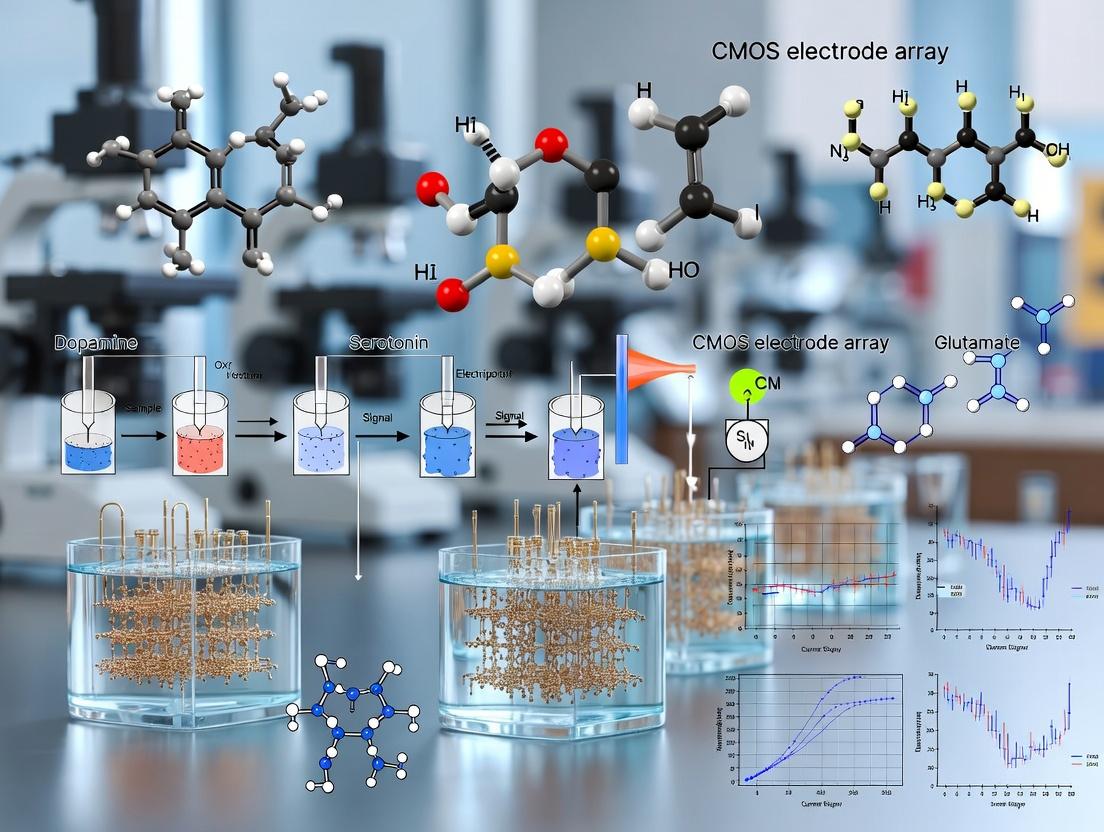
This article provides a comprehensive overview of complementary metal-oxide-semiconductor (CMOS) electrode arrays for neurotransmitter detection, a transformative technology merging electronics and neuroscience.
CMOS Neurotransmitter Arrays: Next-Generation Tools for Real-Time Brain Chemistry Analysis
Abstract
This article provides a comprehensive overview of complementary metal-oxide-semiconductor (CMOS) electrode arrays for neurotransmitter detection, a transformative technology merging electronics and neuroscience. It explores the foundational principles of CMOS-based electrophysiology, detailing the design and fabrication of high-density, multimodal sensor arrays. Methodological applications are examined, highlighting real-time, in vivo monitoring of dopamine, serotonin, glutamate, and other key neurotransmitters. The guide addresses critical challenges in signal fidelity, biofouling, and system integration, offering optimization strategies. Finally, it presents validation protocols and comparative analyses against traditional techniques like carbon-fiber microelectrodes and fast-scan cyclic voltammetry, establishing CMOS arrays as superior tools for neurological research and CNS drug development.
The Silicon Synapse: Foundational Principles of CMOS-Based Neurotransmitter Sensing
Application Notes
The integration of CMOS (Complementary Metal-Oxide-Semiconductor) technology with neurobiological sensing represents a paradigm shift in neurotransmitter detection. These hybrid systems enable high-spatiotemporal-resolution monitoring of neurochemical dynamics in vitro and in vivo, critical for understanding brain function and screening neuroactive drug candidates.
Note 1: CMOS-Based Multimodal Sensing Platforms Modern CMOS electrode arrays are not limited to electrophysiology. They incorporate multiple sensor modalities on a single chip. The latest arrays (e.g., 2024-2025 iterations) feature dense electrode grids (up to 4096 recording sites/mm²) with integrated microelectrodes functionalized for specific neurochemicals. Key performance metrics from recent studies are summarized in Table 1.
Note 2: Key Performance Parameters for Drug Development For pharmaceutical research, the critical parameters are sensitivity, selectivity, and temporal resolution. Current state-of-the-art CMOS-amperometric sensors for dopamine achieve sub-10 nM limits of detection (LOD), with selectivity managed via coating materials (e.g., Nafion, carbon nanotubes) and waveform techniques (Fast-Scan Cyclic Voltammetry). Simultaneous electrical and chemical recording allows for direct correlation of spike activity with neurotransmitter release.
Table 1: Performance Metrics of Recent CMOS Neurotransmitter Sensors
| Neurotransmitter | Detection Method | Limit of Detection (LOD) | Temporal Resolution | Selectivity Method | Key Reference (Year) |
|---|---|---|---|---|---|
| Dopamine | Amperometry / FSCV | 5-10 nM | 10 ms (FSCV), 100 ms (Amperometry) | Nafion/PEDOT coating, FSCV waveform | Yang et al. (2024) |
| Glutamate | Enzymatic (GlOx) | 200 nM | 1-2 s | Glutamate Oxidase layer, Permselective membrane | Yang et al. (2024) |
| Serotonin | FSCV | 20 nM | 100 ms | CFME modification, Waveform shape | Abdalla et al. (2023) |
| Adenosine | Amperometry | 50 nM | 1 s | Enzyme layer (Adenosine deaminase/Nucleoside phosphorylase) | Li et al. (2025) |
| GABA | Enzymatic (GABase) | 5 µM | 5 s | Multi-enzyme cascade layer | Preliminary data (2024) |
Note 3: In Vivo Application Considerations Deploying CMOS arrays for chronic in vivo studies requires attention to biocompatibility, stability, and data telemetry. Recent protocols emphasize the use of Parylene-C or silicon carbide as final insulation layers. Integrated on-chip telemetry (e.g., ultrasonic or RF) is now facilitating fully implantable, wireless recording systems in rodent models, essential for behavioral pharmacology studies.
Experimental Protocols
Protocol 1: Functionalization of CMOS Microelectrodes for Dopamine Detection
Objective: To coat specified working electrodes on a CMOS array for selective, sensitive dopamine detection using Fast-Scan Cyclic Voltammetry (FSCV).
Materials & Reagents:
- CMOS electrode array chip (commercial or custom, e.g., with Pt or C microelectrodes).
- Phosphate-Buffered Saline (PBS), 0.1 M, pH 7.4.
- Dopamine hydrochloride stock solution (10 mM in 0.1 M HClO₄).
- Nafion perfluorinated resin solution (5% wt in lower aliphatic alcohols).
- Poly(3,4-ethylenedioxythiophene)-poly(styrenesulfonate) (PEDOT:PSS) dispersion.
- Carbon nanotube (CNT) ink (single-walled, carboxylated).
- Potentiostat with multi-channel capability (compatible with CMOS array I/O).
Procedure:
- Chip Preparation: Activate the target working electrodes by applying a cleaning protocol (e.g., 1.5 V vs. on-chip Ag/AgCl reference in 0.5 M H₂SO₄ for 30 seconds).
- CNT/Nafion Composite Coating: a. Mix carboxylated CNT ink with Nafion solution at a 1:4 volume ratio. Sonicate for 30 min. b. Using a microinjector or drop-casting under a microscope, apply ~50 nL of the composite mixture to cover the electrode surface. c. Cure at 70°C for 20 min, then at 120°C for 10 min.
- PEDOT Electrodeposition (Optional for Noise Reduction): a. Immerse the chip in PEDOT:PSS dispersion. b. Apply a constant potential of 0.9 V vs. on-chip reference to the target electrode for 10-15 seconds to electrodeposit a porous PEDOT layer over the CNT base. c. Rinse thoroughly with DI water.
- Calibration: a. Place the functionalized chip in a flow cell perfused with 0.1 M PBS (pH 7.4) at 37°C. b. Using the on-chip CMOS circuitry and external potentiostat, apply a FSCV waveform (typically -0.4 V to +1.3 V and back, vs. Ag/AgCl, at 400 V/s, 10 Hz repetition rate). c. Record background current. d. Introduce dopamine standards (e.g., 50 nM, 100 nM, 500 nM, 1 µM) via a calibrated flow injection system. e. Plot peak oxidation current (at ~+0.6 V) against concentration to generate a calibration curve and determine LOD (3*SD of baseline/slope).
Protocol 2: Simultaneous Electrophysiology and Glutamate Sensing in Acute Brain Slice
Objective: To record electrical activity and localized glutamate release concurrently from a mouse hippocampal slice using a multimodal CMOS array.
Materials & Reagents:
- CMOS multimodal array (e.g., with Pt electrodes for LFP and glutamate-sensitive microelectrodes).
- Acute mouse hippocampal slice (300-400 µm thick) in artificial cerebrospinal fluid (aCSF).
- aCSF composition (in mM): 126 NaCl, 2.5 KCl, 1.25 NaH₂PO₄, 2 MgCl₂, 2 CaCl₂, 26 NaHCO₃, 10 glucose (saturated with 95% O₂/5% CO₂).
- High-K⁺ stimulation aCSF (KCl increased to 30 mM).
- Glutamate oxidase (GlOx) immobilized electrode solution (from vendor or prepared).
- Tetrodotoxin (TTX), 1 µM stock.
- Perfusion chamber with temperature control (32°C).
Procedure:
- Array Preparation: Ensure glutamate-sensitive electrodes are pre-functionalized with a cross-linked GlOx layer (commercial process typically involves glutaraldehyde cross-linking of GlOx in a BSA matrix).
- Slice Placement: Transfer the acute hippocampal slice to the perfusion chamber. Carefully lower the CMOS array onto the slice, targeting the CA1 or CA3 region. Apply gentle suction to ensure stable contact.
- System Setup: Perfuse with oxygenated aCSF at 2 mL/min, 32°C. Connect the CMOS chip to the dedicated amplifier/readout system.
- Simultaneous Recording: a. Electrical Channel: Configure bandwidth for local field potentials (LFP: 1-300 Hz) and/or multi-unit activity (MUA: 300-5000 Hz). b. Glutamate Channel: Apply a constant potential of +0.6 V vs. on-chip reference to the GlOx electrodes for amperometric detection of H₂O₂ produced by the enzymatic reaction. c. Acquire data from all channels simultaneously using integrated software.
- Stimulation and Pharmacology: a. Record a 5-minute baseline. b. Chemical Stimulation: Switch perfusion to high-K⁺ aCSF for 30 seconds to induce depolarization and vesicular glutamate release. Monitor the simultaneous electrophysiological response (population spike) and amperometric glutamate signal. c. Inhibition: Wash with normal aCSF for 15 min. Pre-perfuse with aCSF containing 1 µM TTX for 10 min. Repeat high-K⁺ stimulation. The glutamate signal should persist (calcium-independent, exocytotic release), while the population spike should be abolished, confirming the specificity of the recordings.
- Data Analysis: Align the electrical and chemical traces temporally. Quantify the amplitude and kinetics of the glutamate transient relative to the onset of the electrical population activity.
Diagrams
CMOS Neurotransmitter Detection Workflow
Simultaneous Electrical & Chemical Sensing Path
The Scientist's Toolkit: Key Research Reagent Solutions
Table 2: Essential Materials for CMOS-Neurobiology Research
| Item | Function/Benefit | Example/Note |
|---|---|---|
| CMOS Multimodal Array | The core platform. Provides high-density electrodes for electrical recording and dedicated microelectrodes for chemical sensing. | Custom design from academic foundries (e.g., Neuropixels with chemistry mods) or early commercial prototypes. |
| Nafion Perfluorinated Resin | Cation exchanger coating. Improves selectivity for cationic neurotransmitters (e.g., dopamine) over anionic interferents (ascorbic acid, DOPAC). | Use 5% solution in alcohols; critical for in vivo FSCV. |
| PEDOT:PSS Conducting Polymer | Electrode coating. Reduces electrochemical impedance, improves signal-to-noise ratio, and provides a stable substrate for further functionalization. | Electrodeposited or drop-cast. Available as aqueous dispersion. |
| Carbon Nanotube (CNT) Ink | Nanomaterial coating. Increases effective surface area, enhances electron transfer kinetics, and can be functionalized with specific groups or enzymes. | Single-walled, carboxylated CNTs are common for biosensor applications. |
| Glutamate Oxidase (GlOx) | Enzyme for biosensing. Catalyzes the oxidation of glutamate to α-ketoglutarate and H₂O₂, which is then detected amperometrically. | Must be stably immobilized (e.g., via cross-linking in BSA/glutaraldehyde matrix). |
| Fast-Scan Cyclic Voltammetry (FSCV) Potentiostat | Drives and reads the electrochemical sensor. Applies high-speed voltage waveforms and measures resulting faradaic current with high temporal resolution. | Must be synchronized with the CMOS array's electrical recording clocks. Commercially available (e.g., from WaveNeuro). |
| Artificial Cerebrospinal Fluid (aCSF) | Physiological buffer for ex vivo and in vivo experiments. Maintains tissue viability and provides ionic environment for neuronal signaling. | Must be oxygenated (95% O₂/5% CO₂) and have precise ion concentrations. |
| Tetrodotoxin (TTX) | Sodium channel blocker. Used as a pharmacological control to silence action potential-driven neuronal communication, isolating chemical signals. | Confirms that detected neurotransmitter release is from exocytosis (TTX-insensitive) versus reverse transport. |
This document details the application of core electroanalytical techniques—amperometry, voltammetry, and potentiometry—on complementary metal-oxide-semiconductor (CMOS) microelectrode array (MEA) platforms. This work is a foundational component of a broader thesis focused on developing high-density, multiplexed CMOS-MEA biosensors for the spatially and temporally resolved detection of neurotransmitters in vitro and in vivo. The integration of these electrochemical methods with CMOS technology enables unparalleled scalability, temporal resolution, and parallel measurement capabilities, which are critical for advancing neuroscience research and neuropharmacological drug discovery.
Core Principles & CMOS Integration
Amperometry
Principle: Measures the current resulting from the electrochemical oxidation or reduction of an analyte at a constant working electrode potential. The current is directly proportional to the rate of electron transfer, which in turn is proportional to the concentration of analyte reaching the electrode surface. CMOS Integration: On-chip potentiostats apply a constant potential (e.g., +0.6 to +0.8 V for catecholamines). The resulting Faraday current is converted to a voltage by a transimpedance amplifier (TIA) integrated at each pixel/electrode site. This allows for direct, real-time monitoring of exocytotic release events with sub-millisecond resolution.
Voltammetry
Principle: Measures current while systematically varying the applied potential between working and reference electrodes. The resulting current-voltage plot (voltammogram) provides qualitative (identification) and quantitative information. Key Techniques on CMOS:
- Cyclic Voltammetry (CV): Potential is swept linearly forward and backward. Used for characterizing electrode surfaces and studying redox mechanisms.
- Fast-Scan Cyclic Voltammetry (FSCV): High scan rates (≥ 400 V/s) enable sub-second temporal resolution for in vivo neurotransmitter detection (e.g., dopamine). The background charging current is subtracted to reveal the Faraday signal. CMOS Integration: On-chip digital-to-analog converters (DACs) generate precise, rapid voltage waveforms. High-bandwidth TIAs measure the transient current. Parallel readout channels allow simultaneous FSCV at multiple electrodes.
Potentiometry
Principle: Measures the open-circuit potential (voltage) difference between a working ion-selective electrode (ISE) and a stable reference electrode under zero-current conditions. The potential follows the Nernst equation and is logarithmic with respect to the specific ion activity (e.g., H⁺, K⁺, Ca²⁺). CMOS Integration: High-impedance input operational amplifiers (op-amps) are integrated per electrode to measure the potential without drawing current. Ion-selective membranes (e.g., PVC matrix with ionophore) are deposited post-CMOS fabrication onto the on-chip metal electrodes.
Table 1: Comparison of Electrochemical Techniques on CMOS Platforms
| Feature | Amperometry | Voltammetry (FSCV) | Potentiometry |
|---|---|---|---|
| Measured Signal | Current (I) | Current vs. Voltage (I-V) | Potential (V) |
| Applied Signal | Constant Potential | Time-Varying Potential | Zero Current (Open Circuit) |
| Temporal Resolution | Very High (ms-μs) | High (ms, ~100 ms/scan) | Moderate (seconds) |
| Primary Output | Real-time concentration flux | Chemical identification & concentration | Logarithmic ion activity |
| Key Analyte Example | Catecholamine release (exocytosis) | Dopamine, Serotonin | pH, K⁺, Ca²⁺ |
| CMOS Circuit Core | Transimpedance Amplifier (TIA) | High-Speed DAC & TIA | High-Impedance Buffer/Amplifier |
| Selectivity Source | Applied potential & electrode material | Redox potential & waveform shape | Ion-selective membrane |
Experimental Protocols
Protocol 3.1: FSCV for Dopamine Detection on a CMOS-MEA
Objective: To detect and quantify spatially resolved, electrically evoked dopamine release from neuronal cells or brain tissue slices cultured directly on a CMOS-MEA. Materials: CMOS-MEA chip with integrated potentiostats, PDMS culture chamber, bipotentiostat (if external), PBS or aCSF (pH 7.4), DA stock solution (1 mM in 0.1 M HClO₄), Ag/AgCl reference electrode, Pt counter electrode (may be on-chip). Procedure:
- Chip Preparation & Setup: Sterilize the CMOS-MEA. Mount a PDMS chamber. Fill with oxygenated aCSF. Connect chip to reader board and data acquisition system.
- Reference Electrode Integration: Place an external Ag/AgCl reference electrode in the bath, or use an integrated on-chip pseudo-reference electrode.
- FSCV Waveform Configuration: Program the on-chip DACs to apply a triangular waveform to the working electrode (e.g., -0.4 V to +1.3 V and back, vs. Ag/AgCl, at 400 V/s, repeated at 10 Hz).
- Background Subtraction: Record current traces in analyte-free aCSF for 5-10 cycles. Average these cycles to create a stable background current. The system will subsequently subtract this background in real-time.
- Calibration: Flow known concentrations of dopamine (e.g., 0.1, 0.5, 1 µM) over the electrode while applying FSCV. Record the current at the peak oxidation potential (~+0.6-0.7 V). Plot peak current vs. concentration to create a calibration curve.
- Biological Measurement: Place a brain slice or cultured neurons on the array. Use on-chip microstimulators to deliver a biphasic current pulse to tissue. Simultaneously record FSCV data from all surrounding electrodes.
- Data Analysis: Identify dopamine by its characteristic redox peaks in the background-subtracted cyclic voltammogram. Use the calibration curve to convert current to concentration.
Protocol 3.2: Potentiometric pH Imaging on a CMOS-ISE Array
Objective: To map spatial pH changes in a cell culture monolayer in response to a metabolic challenge. Materials: CMOS-MEA with high-impedance readout, pH-sensitive cocktail (e.g., Hydrogen Ionophore I), PVC matrix, tetrahydrofuran (THF), culture medium, pH calibration buffers (4.0, 7.0, 10.0). Procedure:
- Ion-Selective Membrane Deposition: Prepare a membrane solution: 1 wt% Hydrogen Ionophore I, 65 wt% plasticizer (e.g., DOS), 33 wt% PVC, 1 wt% cationic additive (e.g., KTpClPB) in THF. Pipette a small droplet (~100 nL) onto each target on-chip electrode. Let dry for 24h.
- Chip Conditioning: Soak the functionalized chip in pH 7.0 buffer for 1-2 hours to stabilize the membrane potential.
- System Calibration: Connect the chip to the recording system. Sequentially perfuse calibration buffers (pH 7.0, 4.0, 10.0, 7.0) over the array. Record the stable potential at each pH for each electrode. Plot potential vs. pH; the slope should be ~59 mV/pH (Nernstian).
- Cell Culture & Experiment: Seed cells (e.g., astrocytes) directly onto the functionalized array and culture to confluence. Before the experiment, replace medium with a buffered saline solution.
- Measurement: Record baseline potential from all electrodes simultaneously. Introduce a metabolic inhibitor (e.g., 10 mM NaCN) to induce acidosis. Record potential changes for 20-30 minutes.
- Data Analysis: Convert the recorded potential changes from each pixel to ΔpH using the calibration slope. Generate 2D spatial-temporal maps of pH change across the cell monolayer.
Diagrams
Diagram Title: CMOS Platform Core Measurement Workflow
The Scientist's Toolkit
Table 2: Essential Research Reagent Solutions for CMOS Electrochemical Experiments
| Item | Function/Description | Example Use Case |
|---|---|---|
| Artificial Cerebrospinal Fluid (aCSF) | Ionic solution mimicking extracellular fluid. Provides physiological ionic strength and pH for ex vivo/in vitro tissue. | Perfusing brain slices during FSCV or amperometry. |
| Phosphate Buffered Saline (PBS) | Non-physiological but stable buffer for calibration and in vitro sensor testing. | Calibrating electrodes in cell-free systems. |
| Catecholamine Stock Solution (e.g., 1 mM DA in 0.1 M HClO₄) | Stable, acidic stock solution of neurotransmitter analyte. Prevents oxidation before use. | Preparing standard solutions for calibration curves. |
| Ion-Selective Membrane Cocktail | PVC polymer matrix containing specific ionophore, plasticizer, and additive. Creates the potentiometric sensing layer. | Functionalizing on-chip electrodes for K⁺, Ca²⁺, or pH sensing. |
| Nafion Perfluorinated Resin | Cation-exchange polymer coating. Rejects anions (e.g., ascorbate) and foulants, improving selectivity and stability in vivo. | Coating carbon or Pt microelectrodes for in vivo dopamine FSCV. |
| PDMS (Polydimethylsiloxane) | Silicone-based elastomer. Used to create wells/chambers for containing culture medium or solutions on the CMOS chip. | Fabricating a culture chamber bonded to the CMOS-MEA surface. |
| Tetrabutylammonium Perchlorate | Supporting electrolyte. Increases solution conductivity, minimizes ohmic drop (iR drop) in non-aqueous or low-ionic-strength solutions. | Electrochemical characterization in organic solvents. |
| Polyethyleneimine (PEI) | Cationic polymer coating for surfaces. Promotes neuronal cell adhesion to otherwise hydrophobic CMOS passivation layers. | Pre-coating the CMOS-MEA before plating primary neurons. |
This application note details protocols for the study of key neurotransmitter targets using advanced CMOS (Complementary Metal-Oxide-Semiconductor) electrode array technology. This work supports a broader thesis on leveraging the high spatial-temporal resolution and multiplexing capabilities of CMOS arrays to advance neurotransmitter detection research in neuropharmacology and drug development.
Neurotransmitter Target Profiles & Quantitative Data
Table 1: Key Neurotransmitter Targets and Detection Parameters
| Neurotransmitter | Primary Receptor Classes | Typical Basal Concentration in Brain ECF (nM) | Key Enzymes for Catabolism | Oxidation Potential for FSCV (V vs. Ag/AgCl) |
|---|---|---|---|---|
| Dopamine (DA) | D1-like (Gs), D2-like (Gi/o) | 5-50 nM | Monoamine Oxidase (MAO), Catechol-O-methyltransferase (COMT) | ~0.6 V |
| Serotonin (5-HT) | 5-HT1-7 Families | 1-10 nM | Monoamine Oxidase (MAO) | ~0.3 V |
| Glutamate (Glu) | Ionotropic (NMDA, AMPA, Kainate), Metabotropic (mGluR1-8) | 0.5 - 2 µM | Glutamine Synthetase, EAATs (reuptake) | Not directly oxidizable; requires enzyme-linked detection |
| Norepinephrine (NE) | α1, α2, β1-3 | 5-20 nM | Monoamine Oxidase (MAO), COMT | ~0.2 V |
| Acetylcholine (ACh) | Nicotinic (nAChR), Muscarinic (mAChR) | 10-100 nM | Acetylcholinesterase (AChE) | Not directly oxidizable; requires enzyme-linked detection |
| GABA | GABAA (ionotropic), GABAB (metabotropic) | 50-200 nM | GABA Transaminase (GABA-T) | Not directly oxidizable |
Experimental Protocols
Protocol 1: Simultaneous Multi-Analyte Detection of DA and 5-HT Using Fast-Scan Cyclic Voltammetry (FSCV) on CMOS Arrays
Objective: To detect electrically evoked and pharmacologically modulated release of dopamine and serotonin with high temporal resolution. Materials: CMOS electrode array (e.g., 64-256 channels), FSCV potentiostat, aCSF (Artificial Cerebrospinal Fluid), urethane or isoflurane for anesthesia, stereotaxic apparatus, stimulating electrode. Procedure:
- Anesthetize rodent and secure in stereotaxic frame.
- Perform craniotomy over target region (e.g., striatum for DA, dorsal raphe for 5-HT).
- Position the CMOS array and a bipolar stimulating electrode.
- Perfuse with oxygenated aCSF (37°C, pH 7.4).
- Configure FSCV waveform: Triangular waveform from -0.4 V to +1.3 V and back at 400 V/s, applied at 10 Hz.
- Apply electrical stimulation (e.g., 60 Hz, 2 ms pulse width, 2 s duration) to evoke release.
- Record oxidation (DA: ~0.6 V, 5-HT: ~0.3 V) and reduction currents across all array channels.
- For pharmacological validation, superfuse selective reuptake inhibitors (e.g., Nomifensine for DA, Citalopram for 5-HT) or receptor antagonists.
- Analyze data using principal component analysis (PCA) for chemometric separation of DA and 5-HT signals.
Protocol 2: Glutamate Detection via Enzyme-Linked Amperometry on CMOS Microelectrode Arrays
Objective: To measure tonic and phasic glutamate release in vivo. Materials: Glutamate-oxidase (GluOx) modified CMOS microelectrode array, MP-1002/1004 potentiostat, null sensor (without enzyme), glutamate calibration solutions. Procedure:
- Prepare glutamate-sensing microelectrodes by coating CMOS Pt sites with a layer of GluOx cross-linked with Bovine Serum Albumin (BSA) and glutaraldehyde, followed by an outer layer of m-phenylenediamine (to reject interferents like ascorbic acid).
- Implant the modified array and a control null array into the target brain region (e.g., prefrontal cortex).
- Apply a constant potential of +0.7 V vs. Ag/AgCl reference.
- Allow the background current to stabilize (~1-2 hours).
- Perform in vivo calibration via pressure-ejection of known glutamate concentrations (e.g., 10, 20, 50 µM) near the sensor surface.
- Record amperometric current changes corresponding to endogenous glutamate fluctuations.
- Validate signals by applying EAAT (glutamate transporter) inhibitors (e.g., TBOA) to increase extracellular glutamate.
- Subtract signals from the null sensor to correct for non-specific interferent currents.
Protocol 3: Pharmacological Challenge and Receptor Interaction Mapping
Objective: To map spatial patterns of neurotransmitter release modulation by systemic or local drug application. Materials: CMOS array, microiontophoresis or pressure ejection system, drug solutions (agonists/antagonists). Procedure:
- Establish stable baseline recording of neurotransmitter signal (using Protocol 1 or 2).
- Systemically administer (IP/IV) a drug (e.g., cocaine for DA, SSRI for 5-HT, ketamine for Glu) or prepare drug-filled micropipettes for local application.
- For local application, position the drug pipette adjacent to the recording array. Apply controlled pulses of nitrogen pressure (5-20 psi, 10-1000 ms) to eject nanoliter volumes.
- Continuously record from all CMOS channels before, during, and after drug application.
- Analyze spatial heat maps of signal amplitude and kinetics change across the array.
- Perform dose-response experiments by varying drug concentration or ejection parameters.
Visualizations
Title: Dopamine Synthesis, Release, and Signaling Pathway
Title: CMOS Array Neurotransmitter Detection Workflow
Title: Enzyme-Linked Glutamate Detection Principle
The Scientist's Toolkit: Research Reagent Solutions
Table 2: Essential Materials for Neurotransmitter Detection Research
| Item | Function/Description | Example Use Case |
|---|---|---|
| High-Density CMOS MEA | Array of 64-1024 microelectrodes for simultaneous recording from multiple brain regions with high spatial resolution. | Core platform for all protocols. Enables mapping of neurotransmitter diffusion and volume transmission. |
| Fast-Scan Cyclic Voltammetry (FSCV) Potentiostat | Applies rapid voltage ramps to electrodes to oxidize/reduce analytes, providing chemical identification via voltammograms. | Protocol 1: Detection of DA, 5-HT, NE. |
| Glutamate Oxidase (GluOx) | Enzyme that selectively catalyzes the oxidation of glutamate, producing H₂O₂ for amperometric detection. | Protocol 2: Modification of electrode surfaces for selective glutamate sensing. |
| Selective Reuptake Inhibitors | Pharmacological tools to increase extracellular NT levels by blocking transporter proteins (DAT, SERT, NET, EAATs). | Protocol 1 & 3: Validating the identity of the detected signal and studying reuptake dynamics. |
| m-Phenylenediamine (mPD) | Electropolymerized permselective membrane. Rejects anionic interferents (e.g., ascorbic acid) while allowing H₂O₂ passage. | Protocol 2: Coating enzyme-linked sensors to improve selectivity. |
| Carbon Fiber Microelectrodes (for Validation) | Traditional, well-characterized single sensors used to benchmark and validate signals from novel CMOS array sites. | Calibrating and confirming responses from individual CMOS electrodes. |
| Microiontophoresis/Pressure Ejection System | Allows precise, localized application of drugs or neurotransmitters in nanoliter volumes near recording sites. | Protocol 3: Local pharmacological challenges without systemic effects. |
| Principal Component Analysis (PCA) Software | Chemometric tool to deconvolve overlapping voltammetric signals from mixed analytes (e.g., DA vs. 5-HT). | Protocol 1: Data analysis for distinguishing co-released neurotransmitters. |
This application note details the implementation of high-density complementary metal-oxide-semiconductor (CMD) electrode arrays for neurotransmitter detection, contextualized within a broader thesis on advancing neurochemical sensing platforms. CMOS-based arrays represent a paradigm shift, offering unprecedented scalability, parallelism, and spatiotemporal resolution over traditional methods like microdialysis, fast-scan cyclic voltammetry (FSCV) with carbon fibers, and enzyme-based biosensors.
Quantitative Comparison of Methodologies
The table below summarizes key performance metrics of CMOS electrode arrays against traditional neurotransmitter detection techniques.
Table 1: Comparative Analysis of Neurotransmitter Detection Methods
| Parameter | Microdialysis | FSCV (Carbon Fiber) | Enzyme-Linked Biosensors | CMOS Electrode Array |
|---|---|---|---|---|
| Temporal Resolution | Minutes (1-20 min) | Milliseconds (10-100 ms) | Seconds (0.5-5 s) | Milliseconds (0.1-100 ms) |
| Spatial Resolution | ~1 mm (probe diameter) | Single point (~7 µm diameter) | Single point (50-200 µm) | Multipoint (10-50 µm electrode pitch) |
| Parallelism (Channels) | Typically 1-2 | 1-4, with multiplexing | 1-8 | >1,000 simultaneously |
| Scalability (Array Density) | Not scalable | Low (manual assembly) | Moderate | Very High (VLSI fabrication) |
| In Vivo Longevity | Days (clogging) | Hours to days (fouling) | Hours to days | Weeks (passivation layers) |
| Neurotransmitter Specificity | High (HPLC/MS post) | Moderate (pattern recognition) | High (enzyme selectivity) | Moderate-High (material/coating) |
| Tissue Damage/Footprint | High (large probe) | Low (single fiber) | Moderate | Very Low (thin-film, flexible) |
Application Notes & Detailed Protocols
Protocol: High-Density, Parallel Dopamine Kinetics Measurement In Vivo
This protocol describes simultaneous recording of electrically evoked dopamine release at multiple striatal sites.
Research Reagent Solutions & Materials:
- CMOS Neurochemical Array (e.g., Neuropixels CN): High-density, coated Pt or carbon nanotube electrodes for amperometric detection.
- Stimulating Electrode: Bipolar, insulated stainless steel electrode.
- Coordinate Stereotaxic Apparatus: For precise implantation.
- Multichannel Potentiostat/Data Acquisition System: Integrated with the CMOS array chip.
- Analysis Software: Custom or commercial (e.g., MATLAB with Chronoamperometry Analysis Toolbox).
- Phosphate-Buffered Saline (PBS), 0.1 M, pH 7.4: For pre-calibration.
- Dopamine Hydrochloride Stock Solution (1 mM): Prepared daily in 0.1 M PBS with 0.1 mM ascorbic acid to prevent oxidation.
- Isoflurane or Urethane: For animal anesthesia.
- Artificial Cerebrospinal Fluid (aCSF): For surgical procedures.
Procedure:
- Array Calibration: Immerse the active region of the CMOS array in oxygenated, stirred PBS at 37°C. Apply a constant potential (+0.6V vs. on-chip Ag/AgCl reference) to all working electrodes. Inject aliquots of dopamine stock to achieve final concentrations from 10 nM to 10 µM. Record the steady-state oxidation current at each electrode. Create a calibration curve (current vs. concentration) for each pixel.
- Animal Preparation: Anesthetize the rodent (rat/mouse) and secure it in the stereotaxic frame. Perform a craniotomy over the target striatum (e.g., AP: +1.0 mm, ML: ±2.5 mm from bregma).
- Array & Stimulation Electrode Implantation: Slowly lower the CMOS array to a depth of 3.5-4.0 mm (dorsal striatum). Implant the stimulating electrode into the medial forebrain bundle (MFB; AP: -4.8 mm, ML: ±1.6 mm, DV: -8.2 mm).
- Data Acquisition: Allow signals to stabilize for 30 min. Apply a train of biphasic pulses (60 pulses, 60 Hz, 250 µA) to the MFB. Simultaneously record amperometric currents from all array electrodes at 50 kHz.
- Data Analysis: For each electrode, subtract the pre-stimulation baseline. Convert the current trace to dopamine concentration using the electrode-specific calibration factor. Calculate key kinetic parameters: maximal concentration ([DA]max), uptake rate (from decay tau), and release area under the curve (AUC).
Protocol: Spatiotemporal Mapping of Glutamate Spread During Cortical Spreading Depression (CSD)
This protocol leverages CMOS arrays with glutamate-oxidase/poly-o-phenylenediamine (PPD) coatings to map neurotransmitter diffusion.
Research Reagent Solutions & Materials:
- Glutamate-Sensitive CMOS Array: Electropolymerized PPD and cross-linked glutamate oxidase on Pt electrodes.
- KCl Microinjection Pipette: For CSD induction.
- Pressure Microinjection System.
- Double-Barrel Ion-Selective Microelectrode (Reference): For simultaneous DC potential shift measurement (CSD hallmark).
- L-Glutamate Standard Solutions (1 µM - 1 mM): In aCSF.
Procedure:
- Sensor Preparation & Calibration: Coat the CMOS array per manufacturer's protocol. Calibrate in aCSF at 37°C by adding glutamate standards. Verify sensitivity and selectivity against interferents (e.g., ascorbate).
- In Vivo Setup: Prepare an animal model (e.g., mouse under anesthesia or thinned-skull preparation). Position the glutamate array over the primary somatosensory cortex. Position the KCl pipette and DC electrode adjacent to the array.
- CSD Induction & Recording: Pressure-eject a small volume (10-50 nL) of 1 M KCl. Simultaneously record DC potential from the reference electrode and amperometric signals (applied potential +0.7V) from all glutamate array pixels at 10 kHz.
- Spatiotemporal Analysis: For each time point, generate a 2D contour map of glutamate concentration across the array surface. Track the propagation wavefront (velocity, µm/ms). Correlate the glutamate wave with the propagating DC negative shift.
Visualization Diagrams
CMOS Array Neurotransmitter Detection Workflow
Title: From Synapse to Data: CMOS Array Workflow
Traditional vs. CMOS Method Paradigm Shift
Title: Paradigm Shift from Serial to Parallel Measurement
The Scientist's Toolkit: Key Research Reagent Solutions
Table 2: Essential Materials for CMOS Array-Based Neurotransmitter Research
| Item | Function/Description | Example/Note |
|---|---|---|
| High-Density CMOS Neurochemical Array | The core sensing platform integrating microelectrodes, analog front-ends, and multiplexers on a single silicon chip. | e.g., Custom 1024-channel Pt array; Commercial "Neuropixels CN" prototype. |
| Enzyme Cocktail for Biosensing | Provides specificity when coated on electrodes (e.g., Glutamate Oxidase for glutamate, Acetylcholinesterase/Choline Oxidase for ACh). | Must be cross-linked (e.g., with BSA/glutaraldehyde) and protected with a permselective membrane (e.g., PPD, Nafion). |
| Permselective Polymer | Electropolymerized coating to reject anionic interferents (e.g., ascorbate, DOPAC) while allowing neurotransmitter permeation. | Poly-o-phenylenediamine (PPD), overoxidized polypyrrole. |
| Carbon Nanotube (CNT) or PEDOT:PSS Ink | High-surface-area conductive coating to enhance electrode sensitivity and charge transfer capacity. | Dispersion for drop-casting or electrochemical deposition. |
| Multichannel Potentiostat with FPGA | Hardware for applying potentials and reading currents in real-time from hundreds of channels simultaneously. | Systems from companies like Intan Technologies or Blackrock Microsystems. |
| Stereotaxic Alignment System with Microdrives | For precise, stable implantation of the array and associated probes into deep brain structures in vivo. | Includes digital coordinate readouts and fine-adjustment microdrives. |
| Custom Data Analysis Pipeline (Python/MATLAB) | Software for demultiplexing raw data, filtering, converting current to concentration, and generating spatiotemporal maps. | Often requires custom scripting; libraries include MEAnalysis (Python), Chronux. |
| Artificial Cerebrospinal Fluid (aCSF) | Ionic buffer mimicking brain extracellular fluid for calibration and during surgery. | Contains NaCl, KCl, NaHCO3, CaCl2, MgCl2, NaH2PO4, glucose; bubbled with 95% O2/5% CO2. |
The evolution of electrochemical sensing for neurotransmitters parallels advancements in microelectronics. The journey began with single carbon-fiber or metal wire electrodes, which provided foundational insights into neurochemical dynamics but were limited in spatial resolution and throughput. The integration of Complementary Metal-Oxide-Semiconductor (CMOS) technology has enabled the development of massively parallel, high-density microelectrode arrays (MEAs), revolutionizing our ability to map neurochemical activity with high spatiotemporal resolution.
Table 1: Quantitative Evolution of Neurotransmitter Sensing Electrodes
| Era / Technology | Typical Electrode Material | Electrode Size (Diameter) | Number of Simultaneous Recording Sites | Spatial Resolution | Temporal Resolution | Key Limitation |
|---|---|---|---|---|---|---|
| 1st Gen: Single Electrodes (1970s-1990s) | Carbon fiber, Platinum/Iridium wire | 5-100 µm | 1 | N/A (Single point) | Sub-second to seconds | Low throughput, poor spatial mapping |
| 2nd Gen: Micromachined Arrays (1990s-2010s) | Sputtered Pt, ITO, Au on silicon/glass | 10-50 µm | 4-64 | ~100 µm | Milliseconds to seconds | Limited channel count, external multiplexing |
| 3rd Gen: CMOS-Integrated HD Arrays (2010s-Present) | On-chip Pt, Au, CNT, or PEDOT:PSS | 1-20 µm | 256 - 65,536+ | 10-50 µm | Sub-millisecond (kHz sampling) | Complex fabrication, data management |
Application Notes: From Single Sites to Dense Mapping
Application Note 1: Spatial Resolution and Source Localization Early single electrodes could detect neurotransmitter release (e.g., dopamine via fast-scan cyclic voltammetry, FSCV) but could not localize the source within a tissue slice or in vivo. Modern high-density CMOS arrays (e.g., 4096 sites) allow for the creation of detailed concentration heatmaps, enabling precise triangulation of release sites and propagation pathways of chemical signals, crucial for understanding circuit-specific neurotransmission.
Application Note 2: Multiplexed Detection of Neurochemical Species Single electrodes often targeted one analyte per experiment. CMOS arrays can be functionalized with different selective chemistries (e.g., enzymes, polymers) across sub-sets of electrodes. This allows for the simultaneous, correlated detection of multiple neurotransmitters (glutamate, dopamine, adenosine) alongside electrophysiology (spikes, LFP), providing a multimodal view of neural communication.
Application Note 3: High-Throughput Pharmacology Screening Single electrodes offered low throughput for drug dose-response studies. Dense CMOS arrays function as microfluidic-integrated platforms where thousands of synapses or neural networks can be monitored in parallel under various drug conditions, dramatically accelerating the pace of discovery and validation for neuropharmaceuticals.
Detailed Experimental Protocols
Protocol 1: Fast-Scan Cyclic Voltammetry (FSCV) on a Single Carbon-Fiber Electrode Objective: Detect electrically evoked dopamine release in acute brain slice or anesthetized rat. Materials: Single carbon-fiber microelectrode (7 µm diameter), Ag/AgCl reference electrode, stainless-steel auxiliary electrode, potentiostat (e.g., from Pine Research), stereotaxic apparatus. Procedure:
- Electrode Preparation: Seal a carbon fiber in a glass capillary, pull to a tip, and trim to 50-100 µm length.
- Waveform Application: Apply a triangular waveform (-0.4 V to +1.3 V and back vs. Ag/AgCl, 400 V/s, 10 Hz).
- Calibration: Record current response in known dopamine solutions (0.1-2 µM) in artificial cerebrospinal fluid (aCSF).
- In Vivo/Slice Implantation: Stereotaxically position electrode in striatum.
- Stimulation & Recording: Deliver a biphasic electrical stimulus (300 µA, 2 ms/phase, 60 Hz for 200 ms) via an adjacent bipolar electrode.
- Data Analysis: Background subtract cyclic voltammograms. Identify dopamine by its characteristic oxidation (~+0.6 V) and reduction (~-0.2 V) peaks.
Protocol 2: High-Density, CMOS-Based Array Recording of Glutamate Dynamics Objective: Map spatially resolved, tonic and phasic glutamate release across a hippocampal culture. Materials: 1024-site CMOS MEA (MaxWell Biosystems, Neuropixels with custom coating), perfusion system, glutamate oxidase (GluOx), o-phenylenediamine (o-PD), m-phenylenediamine (m-PD), data acquisition unit. Procedure:
- Array Functionalization (Biosensor Fabrication): a. Clean CMOS electrode sites with O2 plasma. b. Electrodeposit a selective layer: Apply +0.9 V (vs on-chip Pt ref) in 5 mM m-PD in PBS for 20 min to create an anti-fouling, small-molecule exclusion layer. c. Enzyme Immobilization: Spot-coat a mixture of GluOx (100 U/mL) and BSA (1%) cross-linked with glutaraldehyde (0.125%) onto the m-PD-coated electrodes. d. Cure for 1 hour at 4°C, then rinse.
- Calibration: Perfuse with aCSF containing known concentrations of glutamate (1, 5, 10 µM). Record amperometric current (typically +0.7 V applied potential). Generate a calibration curve (nA/µM) per electrode.
- Cell Culture Recording: Plate primary hippocampal neurons on the prepared array (DIV 1-7). Record at DIV 14-21.
- Data Acquisition & Analysis: Use on-chip circuitry for simultaneous amperometry on all channels at 10 kHz. Apply spatial filters and generate heatmaps over time. Use electrical stimulation (built-in electrodes) to evoke release.
Visualizations
Title: Single Point vs. Array-Based Neurotransmitter Detection Paradigm
Title: CMOS Array Functionalization & Experimental Workflow
The Scientist's Toolkit: Research Reagent Solutions
Table 2: Essential Materials for CMOS Array Neurotransmitter Detection
| Item | Function in Experiment | Example/Specification |
|---|---|---|
| CMOS Microelectrode Array (MEA) | Core sensing platform; provides high-density electrodes and integrated readout circuitry. | Commercial: Neuropixels 2.0, MaxOne (MaxWell). Custom: 256-4096+ channels with Pt/Au electrodes. |
| Potentiostat / On-Chip Circuitry | Applies potential and measures faradaic current from redox reactions at each electrode. | Integrated on-chip current-to-voltage converters and analog-to-digital converters (ADCs). |
| Selective Polymer (e.g., m-PD) | Forms a size-exclusion film to block interferents (ascorbic acid, DOPAC) while allowing analyte (H2O2) passage. | 5 mM m-phenylenediamine in PBS, electrodeposited. |
| Enzyme for Biosensing (Oxidase) | Confers selectivity by catalyzing the oxidation of the target neurotransmitter, producing H2O2. | Glutamate Oxidase (GluOx), Choline Oxidase (ChOx), Acetylcholinesterase (AChE) + ChOx. |
| Cross-linker (Glutaraldehyde) | Immobilizes the enzyme layer onto the electrode surface, ensuring stability during perfusion. | 0.125% v/v in mixture with enzyme and BSA. |
| Artificial Cerebrospinal Fluid (aCSF) | Physiological buffer for calibration and during experiments to maintain tissue viability. | Contains (in mM): 126 NaCl, 2.5 KCl, 1.2 NaH2PO4, 2.4 CaCl2, 1.2 MgCl2, 25 NaHCO3, 11 Glucose, saturated with 95% O2/5% CO2. |
| Calibration Standards | Used to quantify the amperometric signal in terms of analyte concentration. | Freshly prepared dopamine HCl or L-glutamate in aCSF (0.1-10 µM range). |
| Data Acquisition & Analysis Software | Handles the massive data stream, performs filtering, visualization, and statistical analysis. | Custom Python/MATLAB scripts, or vendor software (e.g., MaxLab Live). |
Fabrication and Functionalization: Building and Using CMOS Neurotransmitter Arrays
This document details the application notes and protocols for fabricating CMOS-based microelectrode arrays (MEAs) specifically engineered for in vitro and in vivo neurotransmitter detection. Within the broader thesis on advancing neuromonitoring tools, this process flow is critical for creating high-density, multiplexed sensors capable of real-time, spatially resolved measurement of electroactive neurochemicals like dopamine, serotonin, and glutamate (via enzyme coatings). The integration of CMOS technology allows for the co-localization of sensing electrodes with active signal conditioning and multiplexing circuitry directly at the neural interface site, dramatically improving signal-to-noise ratio and scalability compared to passive wire bundles.
Core CMOS Process Flow: Application Notes
The fabrication leverages a modified foundry CMOS process, followed by post-processing steps to define and expose the neural electrodes.
Front-End-of-Line (FEOL) CMOS Design & Fabrication
- Objective: To fabricate the underlying transistor-based circuitry for signal amplification, filtering, and multiplexing.
- Process Notes: A standard 0.18 µm or 0.35 µm CMOS process node is typically selected for an optimal balance of performance, cost, and biocompatibility requirements. The chip design includes per-electrode operational amplifiers (for potentiostat function in amperometry), analog-to-digital converters (ADCs), and digital control logic for addressing. Key design considerations include minimizing power dissipation to prevent tissue heating and shielding analog lines from digital switching noise.
Back-End-of-Line (BEOL) Interconnect and Passivation
- Objective: To create metal interconnects from the circuit to the electrode sites and insulate the entire chip except the active sensing areas.
- Process Notes: The final aluminum or copper metal layer is patterned to form the electrode leads. A robust, biocompatible passivation stack is then deposited. This typically consists of alternating layers of silicon nitride (Si₃N₄) and silicon dioxide (SiO₂) deposited via Plasma-Enhanced Chemical Vapor Deposition (PECVD) to achieve pinhole-free insulation and long-term stability in ionic solutions.
Post-CMOS Processing: Lithography and Electrode Opening
- Objective: To selectively open vias in the passivation layer to expose the underlying metal as electrochemical sensing sites.
- Protocol: Photolithography for Electrode Patterning
- 1. Surface Preparation: Clean the CMOS die in a piranha solution (3:1 H₂SO₄:H₂O₂) CAUTION: Highly exothermic and corrosive for 10 minutes, followed by thorough DI water rinse and dehydration bake at 150°C for 5 minutes.
- 2. Photoresist Application: Spin-coat a positive photoresist (e.g., AZ 1512) at 3000 rpm for 30 seconds to achieve a ~1.2 µm film. Soft bake at 95°C for 60 seconds.
- 3. Exposure: Expose using a mask aligner with a photomask defining the electrode opening pattern. Use a UV wavelength of 365 nm (i-line) with an exposure dose of 80 mJ/cm².
- 4. Development: Develop in AZ 300 MIF developer for 45-60 seconds with gentle agitation, followed by DI water rinse and N₂ dry.
- 5. Inspection: Inspect under a microscope for clean, complete opening of the resist pattern.
Post-CMOS Processing: Electrode Material Deposition
- Objective: To deposit a conductive, high charge-injection capacity, and neurochemically sensitive material onto the exposed aluminum pads.
- Protocol A: Sputter Deposition of Iridium Oxide (IrOx)
- 1. Chamber Preparation: Load the patterned wafer into an RF magnetron sputtering system. Pump down to a base pressure of ≤ 5.0 x 10⁻⁶ Torr.
- 2. Deposition Parameters: Introduce Argon and Oxygen gas mixture at a flow rate of 20 sccm and 5 sccm, respectively. Maintain chamber pressure at 3 mTorr. Power the Ir target with 150 W RF power.
- 3. Process: Sputter for 25 minutes to deposit a ~300 nm thick IrOx film. The film is deposited over the entire wafer.
- 4. Liftoff: Soak the wafer in acetone with ultrasonic agitation for 5 minutes to lift off the photoresist, removing the IrOx film everywhere except in the opened vias where it contacts the underlying metal. Rinse sequentially in fresh acetone and isopropanol.
- Protocol B: Electrodeposition of PEDOT:PSS on Sputtered Gold
- 1. Seed Layer: First, sputter a 20 nm adhesion layer of Titanium followed by 150 nm of Gold using the protocol above (liftoff required).
- 2. Electrodeposition Setup: Use a standard three-electrode cell with the array chip as the working electrode, a Pt counter electrode, and an Ag/AgCl reference electrode in 0.1 M NaCl.
- 3. Deposition Solution: Prepare an aqueous solution containing 0.01 M EDOT monomer and 0.1% wt PSS.
- 4. Process: Apply a constant potential of +0.9 V vs. Ag/AgCl for 30-60 seconds. The deposition charge (Q, in mC/cm²) directly controls the film's porosity and impedance. Target a charge density of ~150 mC/cm².
- 5. Rinsing: Rinse thoroughly in DI water and dry under N₂ stream.
Table 1: Comparison of Electrode Deposition Materials and Key Performance Metrics
| Material | Deposition Method | Charge Injection Limit (C/cm²) | Impedance at 1 kHz (for 20 µm site) | Key Advantage for Neurotransmitter Detection |
|---|---|---|---|---|
| Iridium Oxide (IrOx) | Reactive Sputtering | 1-5 | 10-30 kΩ | Excellent stability for chronic in vivo stimulation/recording. |
| Platinum Black (PtB) | Electrodeposition | 5-15 | 2-10 kΩ | Ultra-high surface area for low-noise amperometric detection. |
| PEDOT:PSS | Electrodeposition | 5-10 | 5-15 kΩ | Mixed ionic/electronic conduction; can be functionalized. |
| Carbon Nanotubes (CNT) | CVD or Transfer | N/A | 20-50 kΩ | High surface area, excellent electrocatalytic properties for dopamine. |
Integrated Fabrication and Characterization Workflow
CMOS MEA Fabrication Workflow
The Scientist's Toolkit: Key Research Reagent Solutions
Table 2: Essential Materials for Fabrication and Electrochemical Characterization
| Item / Reagent | Function / Application | Notes |
|---|---|---|
| AZ 1512 Photoresist & 300 MIF Developer | Defines electrode openings via photolithography. | Standard positive resist for post-CMOS processing. |
| Piranha Solution (H₂SO₄:H₂O₂) | Cleans organic residue from wafer surface before processing. | Extreme hazard. Use with full PPE in a fume hood. |
| Iridium Target (99.9% purity) | Source for sputtering high-performance IrOx electrode coating. | Requires reactive sputtering in Ar/O₂ atmosphere. |
| EDOT Monomer & PSS | Precursors for electrodeposition of PEDOT:PSS polymer electrodes. | Enables soft, conductive coatings with high CIC. |
| Phosphate Buffered Saline (PBS), 0.1 M | Electrolyte for in vitro electrochemical testing (CV, EIS). | Standard physiological pH and ionic strength model. |
| Ferrocenedimethanol (1 mM in PBS) | Redox standard for characterizing electrode performance via Cyclic Voltammetry (CV). | Provides stable, reversible redox couple for diagnostics. |
| Dopamine Hydrochloride | Primary analyte for sensor calibration in neurotransmitter detection research. | Prepare fresh stock solutions in deoxygenated, acidic (0.1 M HClO₄) conditions to prevent oxidation. |
| Glutamate Oxidase Enzyme | Functionalization layer for selective detection of the non-electroactive neurotransmitter glutamate. | Immobilized over electrode with cross-linker (e.g., glutaraldehyde) and Nafion membrane. |
Experimental Protocols for Electrochemical Characterization
Protocol: Cyclic Voltammetry (CV) for Electrode Characterization
- Objective: To assess the electrochemical surface area (ECSA), charge storage capacity (CSC), and redox properties of the deposited electrode material.
- Setup: Use a potentiostat connected to a 3-electrode cell: CMOS working electrode, Pt counter electrode, Ag/AgCl reference electrode in 1x PBS.
- Procedure:
- Fill electrochemical cell with 10 mL of degassed 1x PBS.
- Set the potential window to -0.6 V to +0.8 V vs. Ag/AgCl (safe for water window).
- Set scan rate to 50 mV/s.
- Run 20 cycles to stabilize the current response. Record the final cycle.
- Data Analysis: Calculate CSC by integrating the cathodic or anodic current over time within the safe potential window (CSC = ∫ I dt / geometric area).
Protocol: Electrochemical Impedance Spectroscopy (EIS)
- Objective: To measure the electrode-electrolyte interface impedance, critical for predicting thermal noise and signal quality in neural recording.
- Setup: Same 3-electrode cell as CV, using the potentiostat's EIS module.
- Procedure:
- Set DC bias potential to the open circuit potential (typically ~0 V vs. Ag/AgCl in PBS).
- Apply a sinusoidal AC voltage with 10 mV RMS amplitude.
- Sweep frequency from 100 kHz down to 1 Hz, collecting 10 points per decade.
- Data Analysis: Fit the resulting Nyquist plot to a modified Randles equivalent circuit model to extract solution resistance (Rₛ) and charge transfer resistance (Rₑₜ). The impedance magnitude at 1 kHz is a standard industry metric.
Within the framework of a thesis on CMOS-based microelectrode arrays (MEAs) for neurotransmitter detection, the functionalization of individual microelectrode sites is a critical step. While CMOS technology enables high-density, multiplexed sensing, the bare metal sites (typically gold, platinum, or platinum-iridium) lack the necessary selectivity for in vivo or complex in vitro analyses. This document details application notes and protocols for applying Nafion, polymer, and enzyme coatings to impart selectivity for cationic neurotransmitters (e.g., dopamine, norepinephrine) and other analytes, directly addressing key challenges in neuroscience and neuropharmacology research.
Comparative Analysis of Functionalization Strategies
Table 1: Comparison of Key Functionalization Coatings for Neurotransmitter Selectivity
| Coating Type | Primary Selectivity Mechanism | Target Analytes | Typical Thickness | Key Advantage | Key Limitation |
|---|---|---|---|---|---|
| Nafion | Cation-exchange & Size-exclusion | Cations (DA, NE, 5-HT) | 0.2 - 5 µm | Excellent rejection of anions (AA, DOPAC) and large molecules. | Can foul with proteins; reduced sensitivity over time. |
| PEDOT:PSS | Conducting polymer; improved charge injection | General performance enhancer | 50 - 500 nm | Lowers impedance, increases effective surface area. | Limited intrinsic selectivity; often used as base layer. |
| Chitosan | Biocompatible hydrogel; enzyme immobilization | H₂O₂ (from oxidase enzymes) | 100 nm - 2 µm | Excellent bio-compatibility and enzyme entrapment. | Swelling can affect stability; pH-sensitive. |
| Poly(o-phenylenediamine) (PPD) | Size-exclusion membrane | Small molecules (H₂O₂) | 20 - 100 nm | Highly effective, uniform electropolymerized barrier to interferents. | Brittle; can increase electrode impedance. |
| Glutamate Oxidase (GluOx) in Polymer Matrix | Enzyme-catalyzed reaction | L-Glutamate | Varies with matrix | High biochemical specificity for target analyte. | Stability limited by enzyme lifetime; complex fabrication. |
Table 2: Representative Performance Metrics from Recent Studies (2023-2024)
| Coating & Configuration | Analyte | Sensitivity (nA/µM) | LOD (nM) | Selectivity (vs. AA) | Reference Key |
|---|---|---|---|---|---|
| Nafion (drop-cast) on Pt | Dopamine | 1.45 ± 0.12 | 12 | >1000:1 | [Adv. Mater. Inter., 2023] |
| PPD/Nafion bilayer on C | Serotonin | 0.89 | 5 | >500:1 (vs. DA, AA) | [ACS Sens., 2023] |
| GluOx/Chitosan on Pt | Glutamate | 18.2 ± 1.7 (pA/µM) | 80 | N/A (enzymatic) | [Biosens. Bioelectron., 2024] |
| PEDOT:Nafion composite | Dopamine | 2.31 | 8 | >800:1 | [J. Electrochem. Soc., 2024] |
Detailed Experimental Protocols
Protocol 3.1: Electrodeposition of PEDOT:PSS Base Layer on CMOS MEA Sites
Objective: To lower impedance and create a stable, high-surface-area substrate for subsequent functionalization.
Materials: Sterile PBS (pH 7.4), PEDOT:PSS aqueous dispersion (1.3% w/w), CMOS MEA chip in custom potentiostat fixture. Procedure:
- Clean electrode sites via potential cycling (-0.6V to +0.8V vs. Ag/AgCl, 100 mV/s, 20 cycles) in 0.1M H₂SO₄. Rinse with DI water.
- Prepare a solution of 1:1 PEDOT:PSS and PBS with 0.1% v/w (3-glycidyloxypropyl)trimethoxysilane (GOPS) as crosslinker.
- Using a micropipette, dispense 50-100 nL of the solution to cover the target electrode(s). Alternatively, use on-chip microfluidics if integrated.
- Apply a constant potential of +0.9V vs. on-chip Pt pseudo-reference for 10 seconds per site to electrodeposit the film.
- Cure the chip at 120°C for 20 minutes to polymerize GOPS and stabilize the film.
- Rinse gently with DI water and store in PBS. Characterize via electrochemical impedance spectroscopy (EIS).
Protocol 3.2: Spin-Coating of Nafion for Cation Selectivity
Objective: To apply a thin, uniform Nafion film for selective detection of cationic neurotransmitters.
Materials: Nafion perfluorinated resin solution (5% w/w in lower aliphatic alcohols), isopropyl alcohol (IPA), clean CMOS MEA chip, spin coater. Procedure:
- Prepare a 1% w/w Nafion solution by diluting the stock 1:5 with a 7:3 v/v mixture of IPA and DI water.
- Place the CMOS MEA chip on the spin coater vacuum chuck. Ensure the surface is level.
- Dispense 50 µL of the 1% Nafion solution onto the center of the chip.
- Spin at 500 rpm for 5 seconds (spread cycle), then immediately ramp to 3000 rpm for 60 seconds (thin cycle).
- Soft-bake the chip on a hotplate at 80°C for 5 minutes to evaporate solvents.
- Post-bake at 120°C for 10 minutes to enhance adhesion and stabilize the ionomer network.
- Soak the functionalized chip in PBS (pH 7.4) for at least 1 hour before use to hydrate the Nafion film.
Protocol 3.3: Immobilization of Glutamate Oxidase in a Chitosan Matrix
Objective: To create a biospecific coating for the selective detection of L-Glutamate.
Materials: Glutamate oxidase (GluOx, 25 U/mg), Chitosan (medium molecular weight), Acetic acid (1% v/v), Glutaraldehyde (0.25% v/v), PBS. Procedure:
- Dissolve 5 mg of chitosan in 1 mL of 1% acetic acid solution. Stir overnight until clear.
- Centrifuge the chitosan solution at 5000 rpm for 5 min to remove particulates.
- Dissolve 2 mg of GluOx in 500 µL of PBS. Gently mix with 500 µL of the purified chitosan solution.
- Add 10 µL of 0.25% glutaraldehyde solution to the enzyme-polymer mix and vortex for 10 seconds. Note: This crosslinks the matrix.
- Within 5 minutes of crosslinking, use a microdispenser to apply ~200 nL of the mixture onto each target microelectrode.
- Allow the droplets to dry in a humidified chamber (≥80% RH) at room temperature for 2 hours.
- Rinse gently with PBS to remove any un-immobilized enzyme. Store in PBS at 4°C when not in use.
Visualization of Concepts and Workflows
Diagram Title: Nafion Coating Selectivity Mechanism (100 chars)
Diagram Title: Enzyme Biosensor Fabrication & Signal Path (85 chars)
The Scientist's Toolkit: Research Reagent Solutions
Table 3: Essential Materials for Microelectrode Functionalization
| Item & Common Example | Function / Role in Experiment | Key Considerations for Use |
|---|---|---|
| Nafion Perfluorinated Solution (5% in alcs) | Forms cation-exchange membrane. Rejects anions (ascorbate) and macromolecules. | Dilution ratio and spin speed critically control film thickness and selectivity. |
| PEDOT:PSS Dispersion (Clevios PH1000) | Conducting polymer for lowering impedance, increasing charge capacity. | Adding GOPS crosslinker is essential for stability in aqueous electrolytes. |
| Chitosan (Medium M.W., >75% deacetylated) | Biocompatible hydrogel for entrapping and stabilizing enzymes. | Requires dissolution in dilute acid (e.g., acetic); viscosity affects film uniformity. |
| o-Phenylenediamine (o-PD) monomer | Electropolymerizes to form size-exclusion poly(o-PD) film. Blocks interferents like UA, AA. | Polymerization potential and cycle number control film permeability. |
| Glutamate Oxidase (GluOx) from Streptomyces | Enzyme that catalyzes L-Glutamate to α-ketoglutarate + H₂O₂. | Specific activity and lot-to-lot variability must be checked; store lyophilized at -20°C. |
| (3-Glycidyloxypropyl)trimethoxysilane (GOPS) | Crosslinking agent for PEDOT:PSS, improves adhesion and stability in water. | Add fresh to solution just before use; typically used at 0.1-1% v/w. |
| Phosphate Buffered Saline (PBS), 10X | Standard physiological buffer for dilution, rinsing, and testing. | Always adjust to final working concentration and pH (7.4) for biological relevance. |
| Glutaraldehyde (25% solution) | Crosslinking agent for chitosan/enzyme matrices, creates covalent bonds. | Use at low concentration (e.g., 0.1-0.5%) to avoid excessive enzyme deactivation. |
This application note details the system architecture for complementary metal-oxide-semiconductor (CMOS) electrode arrays designed for high-throughput, high-sensitivity neurotransmitter detection. Within the broader thesis, this architecture addresses the central challenge of translating sparse, low-amplitude electrochemical signals from neural interfaces into robust, multiplexed digital data streams for pharmacological and neuroscientific research. The integration of on-chip amplification, channel multiplexing, and parallel readout is critical for scaling toward large-scale, real-time monitoring of neurochemical dynamics in vitro and in vivo, directly impacting drug discovery and neuropsychiatric disease research.
Core Architectural Components & Quantitative Analysis
On-Chip Amplification Strategies
The first stage of signal conditioning is critical due to the low current magnitudes (picoampere to nanoampere range) generated at microelectrodes during fast-scan cyclic voltammetry (FSCV) or amperometry.
Primary Amplifier Topologies:
- Transimpedance Amplifier (TIA): Converts the faradaic current (If) to a voltage (Vout = -If * Rf). High-value feedback resistors (R_f) are required for gain, presenting integration challenges.
- Integrating Amplifier (IA): Accumulates charge on a feedback capacitor (Cf) over a integration period (Tint), with Vout = (1/Cf) ∫ I_f dt. Excellent for low-frequency noise rejection but requires periodic reset.
- Chopper-Stabilized Amplifier: Modulates the input signal to a higher frequency, amplifies it, and demodulates it back, effectively eliminating 1/f flicker noise dominant in CMOS.
Table 1: Comparison of On-Chip Amplifier Topologies for Neurotransmitter Sensing
| Topology | Typical Gain | Bandwidth | Input-Referred Noise (pA/√Hz) | Key Advantage | Key Limitation | Best Suited For |
|---|---|---|---|---|---|---|
| Transimpedance (TIA) | 1 MΩ - 10 GΩ (in CMOS) | 1 - 10 kHz | 0.5 - 5 | Simple, continuous readout | Thermal noise of R_f, stability vs. electrode capacitance | Fast amperometry, FSCV |
| Integrating (IA) | 1 - 100 pF cap (programmable) | Set by 1/T_int | < 0.1 (at low freq) | Excellent noise performance, inherent signal averaging | Non-continuous output, reset artifacts | Low-frequency monitoring, pulsed techniques |
| Chopper-Stabilized TIA | 100 MΩ - 1 GΩ | 1 - 5 kHz | 0.1 - 0.5 | Ultra-low 1/f noise, high stability | Increased circuit complexity, clock feedthrough | Long-term, stable dopamine monitoring |
Multiplexing Strategies
Multiplexing enables a scalable system where the number of recording channels far exceeds the number of analog-to-digital converter (ADC) units.
- Analog Multiplexing (MUX): A switch matrix connects multiple electrode amplifiers to a shared backend ADC. This reduces area and power but is susceptible to crosstalk and switch charge injection artifacts.
- Digital Multiplexing: Each electrode channel has its own dedicated front-end amplifier and ADC. Outputs are serialized digitally. This offers superior signal integrity but at higher area and power cost.
- Hybrid Time-Division Multiplexing (TDM): A compromise where small blocks of electrodes (e.g., 8:1) share a high-performance ADC via analog MUX, with multiple such blocks operating in parallel.
Table 2: Multiplexing Strategy Trade-offs for a 1024-Channel Array
| Strategy | Estimated Area per Pixel (μm²) | Power per Channel (μW) | Max. Simultaneous Sampling Rate per Channel (kS/s) | Crosstalk | System Complexity |
|---|---|---|---|---|---|
| Full Analog MUX (1024:1) | ~500 | 1 - 3 | < 1 (limited by MUX settling) | High (-40 to -50 dB) | Low |
| Hybrid TDM (8 blocks of 128:1) | ~1200 | 5 - 8 | 10 - 20 | Medium (-60 to -70 dB) | Medium |
| Full Digital MUX | >2500 | 10 - 20 | 100+ | Very Low (< -80 dB) | High |
Data Readout & Digitization
The digitization strategy must match the bandwidth and dynamic range of the electrochemical technique.
- Successive Approximation Register (SAR) ADC: Dominant for its excellent power-area trade-off, suitable for moderate bandwidths (up to 1 MS/s) and 10-12 bit resolution.
- Delta-Sigma (ΔΣ) ADC: Provides high resolution (16-18 bits) in a limited bandwidth (tens of kHz), ideal for low-noise, low-frequency neurochemical signals.
- Column-Parallel Readout: A popular architecture where each column of the electrode array has a dedicated ADC, enabling high-frame-rate imaging of neurotransmitter release across the array.
Experimental Protocols
Protocol 1: Characterizing On-Chip Amplifier Noise and Bandwidth
Objective: To measure the input-referred noise and bandwidth of an integrated TIA/IA for neurotransmitter sensing. Materials: CMOS chip with on-chip amplifiers, PCB carrier, precision semiconductor parameter analyzer, low-noise probe station, shielded cable, calibrated current source. Procedure:
- DC Transfer Characterization: Sweep input current from -1 nA to +1 nA using the parameter analyzer. Measure output voltage to calculate the exact transimpedance gain (R_f).
- Noise Spectral Density Measurement:
- Short the amplifier input to the on-chip analog ground via a low-noise switch.
- Acquire the output voltage waveform for 10 seconds at a 100 kS/s sampling rate using an external low-noise digitizer.
- Compute the power spectral density (PSD) of the output. Divide the output voltage noise PSD by the gain (R_f) to obtain the input-referred current noise PSD (in pA/√Hz).
- Bandwidth Measurement: Inject a small sinusoidal current (e.g., 100 pA p-p) from the calibrated source. Sweep frequency from 1 Hz to 100 kHz. Measure the output amplitude. The -3 dB frequency is the bandwidth.
Protocol 2: Assessing Multiplexer-Induced Crosstalk
Objective: To quantify signal bleed-between adjacent channels in a multiplexed architecture. Materials: Multiplexed CMOS electrode array, multichannel potentiostat, solution with redox probe (e.g., 1 mM Potassium Ferricyanide in PBS). Procedure:
- Stimulus Application: Select one electrode (Channel A) as the "aggressor." Apply a FSCV waveform (e.g., -0.4 V to +1.0 V vs. on-chip ref, 400 V/s, 10 Hz).
- Victim Channel Monitoring: Simultaneously record the current at an adjacent, non-stimulated electrode (Channel B, the "victim") held at a constant potential.
- Data Analysis: Perform background subtraction on the victim channel data. The peak current observed on Channel B during the scan on Channel A is the crosstalk signal. Crosstalk (%) = (Ivictim / Iaggressor) * 100. Repeat for various electrode distances.
Protocol 3: System-Level Validation with Neurotransmitter Detection
Objective: To validate the full system architecture by detecting a bolus of dopamine. Materials: CMOS biosensor chip, flow injection apparatus, Tyrode's buffer (pH 7.4), 10 μM Dopamine HCl solution in buffer, Ag/AgCl reference electrode, data acquisition system. Procedure:
- Chip Preparation & Setup: Potentiate the carbon-based post-CMOS microelectrodes via electrical stimulation (e.g., 60 Hz, 1.5 V amplitude, 2 s) in buffer. Connect all system readout lines.
- Background Acquisition: Flow Tyrode's buffer over the chip at 1 mL/min. Apply the FSCV waveform to all active electrodes and record background cyclic voltammograms for 5 minutes to establish a stable baseline.
- Bolus Injection & Detection: Rapidly inject a 100 μL bolus of 10 μM dopamine into the flow stream. Continuously record the current from all multiplexed channels.
- Data Processing: Use chemometric analysis (e.g., principal component analysis) on the high-dimensional data stream to distinguish the dopamine oxidation/reduction signature from noise and interferents (e.g., pH change). Extract temporal and spatial release profiles.
Visualization Diagrams
Diagram Title: CMOS Neurotransmitter Sensor System Dataflow
Diagram Title: Protocol: System Validation with Dopamine
The Scientist's Toolkit
Table 3: Essential Research Reagent Solutions for CMOS Neurotransmitter Array Experiments
| Item | Function & Role in Experiment | Example/Notes |
|---|---|---|
| Potassium Ferricyanide (K₃[Fe(CN)₆]) | A stable, reversible redox probe for benchmarking electrode performance, characterizing amplifier gain, linearity, and crosstalk. | 1-5 mM in 1x PBS; used in Protocol 2 for crosstalk assessment. |
| Dopamine Hydrochloride | The primary target neurotransmitter for validation of sensor sensitivity, selectivity, and temporal resolution. | Prepare fresh in deoxygenated buffer (e.g., Tyrode's, pH 7.4) with 0.1 mM ascorbic acid to prevent oxidation. Used in Protocol 3. |
| Ascorbic Acid | A common electrochemical interferent present in high concentration in the brain. Used to test sensor selectivity. | 0.5 - 1 mM in buffer; challenges the system's ability to resolve dopamine signals. |
| Phosphate Buffered Saline (PBS) / Artificial Cerebrospinal Fluid (aCSF) | Physiological buffer matrix for in vitro experiments. Maintains pH and ionic strength, mimicking biological conditions. | Use for electrode storage, baseline recording, and diluting analytes. |
| Nafion Perfluorinated Resin | A cation-exchange polymer coating applied to microelectrodes to improve selectivity for cationic neurotransmitters (e.g., dopamine) over anions (e.g., ascorbate, DOPAC). | Typically applied via drop-casting or electrophoretic deposition post-CMOS processing. |
| Carbon Nanotube or Graphene Ink | Post-CMOS electrode modification material. Increases electroactive surface area, enhances electron transfer kinetics, and improves sensitivity. | Dispensed via micro-plotting or inkjet printing onto CMOS electrode pads. |
This document provides standardized protocols for the application of CMOS-based microelectrode arrays (MEAs) in neurotransmitter detection research. These protocols are designed for integration within a broader thesis focusing on the development and validation of high-density, multiplexed CMOS biosensors for real-time neurochemical monitoring in preclinical drug development and neurophysiological research.
Key Research Reagent Solutions & Essential Materials
| Item | Function/Brief Explanation |
|---|---|
| High-Density CMOS MEA Chip (e.g., 256+ channels) | Core sensor platform. Integates recording electrodes, amplification, and multiplexing circuitry for simultaneous multi-site neurotransmitter detection. |
| Nafion or PEDOT:PSS Coatings | Electropolymerized conductive polymer or ion-exchange membrane applied to electrode sites to enhance selectivity for cationic neurotransmitters (e.g., dopamine) and reduce biofouling. |
| Phosphate-Buffered Saline (PBS), pH 7.4 | Standard electrolyte solution for in vitro calibration and maintenance of physiological ionic strength. |
| Artificial Cerebrospinal Fluid (aCSF) | Isotonic, ion-balanced solution for in vivo perfusion and in vitro brain slice recordings. |
| Enzyme Cocktails (e.g., HRP/GOx) | For selective biosensing. Immobilized on electrode surfaces to catalyze reactions producing electroactive species from target analytes (e.g., glutamate, acetylcholine). |
| Fast-Scan Cyclic Voltammetry (FSCV) Software Suite | Enables real-time electrochemical detection at CMOS arrays with high temporal resolution (e.g., 10 Hz for FSCV). |
| Urethane or Isoflurane | Anesthetic agents for acute and chronic in vivo surgical procedures in rodent models. |
| Sterotaxic Frame with Digital Atlas Integration | Precise targeting of brain regions (e.g., striatum, prefrontal cortex) for MEA implantation. |
| Dental Acrylic Cement | For securing the CMOS MEA device and headstage connector to the skull in chronic implantations. |
Core Experimental Protocols
Protocol 3.1:In VitroChip Calibration and Characterization
Objective: To establish sensor baseline performance, sensitivity, and selectivity. Materials: CMOS MEA, potentiostat/multichannel amplifier, calibration chamber, stock solutions of target neurotransmitters (Dopamine, Serotonin, etc.), ascorbic acid, PBS. Methodology:
- Chip Priming: Sterilize the CMOS MEA in 70% ethanol, rinse thoroughly with deionized water, and soak in PBS for >1 hour.
- Electrochemical Activation: Perform cyclic voltammetry (CV) in PBS (e.g., -0.6V to +1.0V vs. Ag/AgCl, 100 mV/s, 50 cycles) to stabilize electrode surfaces.
- Calibration Curve Generation:
- Prepare a dilution series of the primary analyte (e.g., dopamine: 0.1, 0.5, 1.0, 2.5, 5.0 µM) in oxygenated PBS at 37°C.
- Apply the chosen detection waveform (e.g., FSCV: -0.4V to +1.3V and back, 400 V/s, 10 Hz).
- Perfuse each concentration over the chip for 2 minutes while recording oxidation currents.
- Plot peak oxidation current vs. concentration. Perform linear regression to determine sensitivity (nA/µM) and limit of detection (LOD = 3*SD of baseline / sensitivity).
Protocol 3.2: AcuteIn VivoImplantation and Recording
Objective: To measure electrically evoked or drug-induced neurotransmitter release in an anesthetized animal. Materials: Rodent (rat/mouse), stereotaxic apparatus, anesthesia setup, CMOS MEA on a movable probe, stimulating electrode, bone drill, agarose or saline for brain surface hydration. Methodology:
- Animal Preparation: Anesthetize rodent (e.g., urethane, 1.5 g/kg i.p.). Secure in stereotaxic frame. Maintain body temperature at 37°C.
- Craniotomy: Expose skull, level, and perform a small craniotomy (∼2x2 mm) over the target region (e.g., striatum: AP +1.2 mm, ML ±2.0 mm from bregma).
- Dura Removal: Carefully puncture and retract the dura mater.
- Probe Implantation: Align the CMOS MEA probe perpendicular to the brain surface. Slowly lower the probe to the target depth (e.g., ventral striatum, DV -6.5 mm) at a rate not exceeding 1 µm/s to minimize tissue damage.
- Stabilization: Apply a warm agarose gel (3% in aCSF) over the craniotomy to stabilize the brain and reduce pulsation.
- Recording Setup: Connect the CMOS MEA to the headstage and amplifier/recording system. Allow signals to stabilize for 30 minutes.
- Stimulation & Recording: Insert a bipolar stimulating electrode into the upstream pathway (e.g., medial forebrain bundle). Deliver electrical stimuli (monophasic pulses, 300 µA, 60 Hz, 2s train) while simultaneously recording electrochemical signals from the CMOS array.
- Pharmacological Validation: Systemically administer (i.p.) or locally apply (via microdialysis probe) drugs (e.g., nomifensine, a dopamine reuptake inhibitor) to modulate neurotransmitter dynamics and confirm signal identity.
Protocol 3.3: ChronicIn VivoImplantation for Longitudinal Studies
Objective: To enable repeated neurotransmitter measurements over days to weeks in freely moving animals. Materials: All acute materials plus dental acrylic, titanium bone screws, protective cap, aseptic surgical tools. Methodology:
- Aseptic Surgery: Perform all steps under sterile conditions in a surgical suite.
- Skull Preparation: Expose and thoroughly clean the skull. Implant 3-4 titanium bone screws for mechanical and electrical grounding.
- Craniotomy & Implantation: Perform a small, precise craniotomy. Lower the CMOS MEA probe to the target depth as in Protocol 3.2.
- Device Fixation: Apply a layer of tissue adhesive around the probe base. Build a robust, sealed headcap using layers of dental acrylic, encapsulating the probe base, bone screws, and the headstage connector.
- Recovery & Monitoring: Administer postoperative analgesics and allow the animal to recover for a minimum of 7 days with daily health monitoring.
- Longitudinal Recording: For each recording session, gently connect the tether/headstage to the implanted connector. Allow the animal to acclimate in the testing chamber. Record neurotransmitter signals during behavioral paradigms or drug challenges.
Table 1: Typical CMOS MEA Performance Metrics for Neurotransmitter Detection
| Parameter | In Vitro (PBS) | In Vivo (Acute) | In Vivo (Chronic, Day 7) | Measurement Technique |
|---|---|---|---|---|
| Sensitivity (Dopamine) | 5 - 15 nA/µM | 3 - 10 nA/µM | 2 - 8 nA/µM | FSCV Calibration |
| Limit of Detection (LOD) | 5 - 20 nM | 10 - 50 nM | 20 - 100 nM | Signal-to-Noise (S/N=3) |
| Temporal Resolution | 10 - 100 ms | 10 - 100 ms | 10 - 100 ms | FSCV Scan Rate |
| Spatial Resolution | 15 - 50 µm (electrode pitch) | 15 - 50 µm | 15 - 50 µm | CMOS Design |
| Selectivity (DA vs. AA) | >1000:1 | >500:1 | >200:1 | FSCV Background Subtraction |
| Signal Stability (Δ% / hr) | <1% | <5% | <10%* | Peak Current Drift |
| Number of Simultaneous Sites | 256 | 256 | 256 | CMOS Array Channels |
*Stability decreases in chronic implants due to biological response; weekly re-calibration via electrical stimulation recommended.
Visualization of Workflows and Pathways
Diagram 1: High-Level Experimental Protocol Workflow
Diagram 2: Dopamine Signaling & MEA Detection in the Synaptic Cleft
This application note positions real-time PK/PD monitoring within the broader thesis research on CMOS (Complementary Metal-Oxide-Semiconductor) electrode arrays for multiplexed, high-temporal-resolution neurotransmitter detection. The ability to directly and simultaneously measure drug pharmacokinetics (concentration over time) and its pharmacodynamic effects (neurotransmitter dynamics) in vivo represents a paradigm shift in preclinical drug discovery. This document presents case studies and protocols demonstrating how integrated CMOS biosensor platforms enable more predictive and efficient development of central nervous system (CNS) therapeutics.
Case Study 1: Real-Time Monitoring of a Novel Antipsychotic's Dopaminergic Modulation
Background: A novel D2/D3 receptor partial agonist (Compound X) was undergoing preclinical evaluation for schizophrenia. Traditional microdialysis studies provided limited temporal resolution for capturing rapid dopamine flux changes following drug administration and behavioral stimuli.
Integrated PK/PD Experiment Using CMOS Array: A chronically implanted CMOS multi-electrode array (MEA), functionalized for real-time amperometric detection of dopamine (DA) and equipped with a bare gold working electrode for adsorptive stripping voltammetry of the drug molecule, was used in a rodent model.
Key Quantitative Data Summary: Table 1: PK/PD Parameters for Compound X from Integrated CMOS MEA Experiment (n=8)
| Parameter | Mean Value (±SEM) | Unit | Explanation |
|---|---|---|---|
| Cmax (Plasma) | 1.24 ± 0.15 | µM | Max observed drug concentration. |
| Tmax | 20 ± 3 | min | Time to reach Cmax. |
| AUC(0-120min) (Plasma) | 85.4 ± 9.2 | µM·min | Systemic exposure. |
| Brain Penetration (Kp) | 0.65 ± 0.08 | ratio | Brain/Plasma AUC ratio. |
| DA Baseline Firing Rate | 2.8 ± 0.4 | Hz | Tonic dopamine neuron activity. |
| ΔDA Burst Amplitude (Post-Ketamine) | +220 ± 25 | % | Phasic DA response to NMDA antagonist. |
| EC50 for DA Burst Normalization | 0.41 ± 0.07 | µM | [Compound X] for 50% effect. |
| PK/PD Hysteresis Loop Area | 15.2 ± 2.1 | a.u. | Quantifies effect delay relative to PK. |
Experimental Protocol: Integrated PK/PD with CMOS MEA
Objective: To simultaneously measure the plasma and brain pharmacokinetics of Compound X and its pharmacodynamic effect on ketamine-induced phasic dopamine release in the striatum of freely moving rats.
Materials:
- Adult Sprague-Dawley rats with surgically implanted jugular vein catheter.
- Custom 16-channel CMOS MEA (4 channels DA-sensitive, 1 channel drug-sensing, 11 channels electrophysiology).
- DA sensor coating: Nafion/Poly(o-phenylenediamine)/Carbon nanotube layer.
- Potentiostat/Wireless transmitter module.
- Compound X solution (2 mg/kg, i.v.).
- Ketamine hydrochloride solution (10 mg/kg, i.v.).
- Artificial cerebrospinal fluid (aCSF).
Procedure:
- Sensor Calibration: Ex vivo calibrate the DA-sensitive channels in aCSF with standard DA solutions (0, 100 nM, 500 nM, 1 µM) pre- and post-experiment. Calibrate the drug-sensing channel with Compound X standards.
- Baseline Recording: Initiate continuous amperometric (DA) and voltammetric (drug) recording from the implanted MEA for 30 minutes to establish stable neurotransmitter and neurophysiological baselines.
- Pharmacokinetic Phase: Administer Compound X (2 mg/kg, i.v. bolus). The drug-sensing channel performs adsorptive stripping voltammetry every 30 seconds. Concurrently, collect serial micro-blood samples via jugular catheter at 5, 15, 30, 60, 90, 120 min for LC-MS/MS validation.
- Pharmacodynamic Challenge: At t=25 min (near Tmax), administer ketamine (10 mg/kg, i.v.). Monitor the subsequent phasic dopamine release events (bursts) via high-speed amperometry on the DA channels for 60 minutes.
- Data Analysis: Align PK (brain and plasma drug concentration) and PD (DA burst amplitude/frequency) timelines. Construct a PK/PD relationship model (e.g., effect-compartment link model) to quantify hysteresis and derive the EC50.
Signaling Pathway Diagram
Diagram 1: Signaling pathway for antipsychotic PK/PD study.
The Scientist's Toolkit: Key Reagents & Materials Table 2: Essential Research Reagents for Integrated PK/PD Studies
| Item | Function/Role | Key Consideration |
|---|---|---|
| CMOS MEA with Multiple Sensor Coatings | Core platform for simultaneous neurochemical (DA, drug) and electrophysiological recording. | Requires stable, bio-compatible coatings (e.g., Nafion) to prevent fouling and ensure selectivity. |
| Wireless Potentiostat/Transmitter | Enables real-time data acquisition from freely behaving subjects, removing movement artifact confounds. | Must have low noise, sufficient sampling rate (>1 kHz), and long battery life for chronic studies. |
| Calibration Standards (DA, Drug Molecule) | Essential for converting electrochemical signal (current) into quantitative concentration values. | Must be prepared fresh in aCSF; ex vivo calibration pre/post experiment accounts for sensitivity drift. |
| Pharmacokinetic Validator (LC-MS/MS) | Gold-standard method to validate drug concentrations measured by the biosensor in blood/brain microdialysate. | Provides absolute quantification and confirms sensor specificity against endogenous interferents. |
| Chronic Animal Model with Vascular Access | Allows precise intravenous drug dosing and serial blood sampling without stress-induced neurotransmitter release. | Requires skilled surgical preparation and post-op care to maintain model integrity. |
Case Study 2: Evaluating Analgesic Efficacy via Opioid-Induced GABA Release Dynamics
Background: A new µ-opioid receptor (MOR) agonist (Compound Y) for pain management required characterization of its effect on inhibitory neurotransmission in the periaqueductal gray (PAG), a key analgesic pathway.
Experimental Protocol: Spatially Resolved PK/PD in PAG
Objective: To map the relationship between local concentration of Compound Y and its potentiation of electrically evoked GABA release in the PAG using a high-density CMOS MEA.
Materials:
- Brain slice preparation containing the PAG from rodent.
- High-density 64-channel CMOS MEA for slice recording.
- Channels functionalized with a GABA-selective enzymatic layer (GABAase/glutamate dehydrogenase).
- Micro-injection system for focal drug application.
- Compound Y.
- Naloxone (opioid antagonist).
- Bicuculline (GABA-A receptor antagonist).
Procedure:
- Slice Preparation & Placement: Prepare acute coronal brain slice (300 µm) containing PAG. Position slice onto the 64-channel CMOS MEA, aligning the ventrolateral PAG with the sensor grid.
- Stimulation & Baseline: Deliver bipolar electrical stimulation (0.2 ms, 100 µA) to the PAG input fibers every 60 seconds. Record evoked oxidative current on GABA-sensitive channels to establish a stable baseline.
- Local Pharmacokinetics & Dynamics: Focally apply Compound Y (10 µL of 1 µM) near the recording area. Use adjacent bare gold electrodes with square-wave voltammetry to monitor local drug accumulation and clearance.
- Concentration-Response: Repeat step 3 with increasing concentrations of Compound Y (10 nM, 100 nM, 1 µM, 10 µM). After each concentration, apply naloxone to confirm MOR-mediated effect.
- Spatial Analysis: Generate a heat map of GABA release amplitude across the 64-channel array at the time of peak drug concentration, illustrating spatial heterogeneity of drug response.
Workflow Diagram
Diagram 2: Workflow for in vitro analgesic PK/PD study.
The integration of CMOS electrode array technology for real-time, multi-analyte biosensing directly addresses critical gaps in conventional PK/PD modeling. The case studies demonstrate the ability to:
- Collapse Hysteresis: By measuring brain drug concentration and neurotransmitter effect simultaneously, the temporal disconnect (hysteresis) between PK and PD profiles is directly quantified and can be modeled mechanistically.
- Achieve High Temporal & Spatial Resolution: Capturing neurotransmitter dynamics on a sub-second timescale at multiple sites provides insights into circuit-level drug effects impossible with microdialysis or post-mortem analysis.
- Refine Preclinical Models: This approach generates richer, more predictive data earlier in the drug discovery pipeline, potentially reducing late-stage attrition for CNS-targeted therapeutics by better correlating molecular engagement with functional neurochemical output.
This work, as part of a broader thesis on CMOS neurochemical interfaces, establishes a foundational methodology for next-generation, data-driven PK/PD studies in neuroscience drug discovery.
Signal, Noise, and Stability: Troubleshooting CMOS Array Performance
Within the broader thesis on CMOS-based microelectrode arrays (MEAs) for neurotransmitter detection, achieving long-term stability and fidelity is paramount. Biofouling (nonspecific protein adsorption and cell adhesion) and signal drift (baseline instability) are primary challenges that compromise the sensitivity, selectivity, and temporal resolution of in vivo and chronic in vitro measurements. This document details current strategies for surface treatment and regeneration to combat these issues, providing application notes and standardized protocols.
Surface Treatments for Fouling Resistance & Stability
Preventative surface modifications are the first line of defense. The goal is to create a hydrophilic, non-ionic, and sterically repulsive interface.
Covalently Grafted Polymer Brushes
Principle: Dense layers of hydrophilic polymers (e.g., PEG, zwitterions) create a hydrated, steric barrier that reduces protein adhesion by >90%.
- Protocol: Silanization & PEG Grafting on Iridium Oxide (IrOx) CMOS Electrodes
- CMOS Chip Pre-cleaning: Oxygen plasma treatment (100 W, 30 sccm O₂, 1 min).
- Silanization: Immerse chip in 2% (v/v) (3-Aminopropyl)triethoxysilane (APTES) in anhydrous toluene for 2 hours under nitrogen.
- Rinsing: Rinse sequentially with toluene, ethanol, and deionized water. Cure at 110°C for 10 min.
- PEGylation: Incubate chip in 10 mM mPEG-Succinimidyl Valerate (mPEG-SVA, MW 5kDa) in 0.1M sodium bicarbonate buffer (pH 8.5) for 4 hours at room temperature.
- Termination: Rinse thoroughly with PBS and store in 10 mM PBS (pH 7.4).
Zwitterionic Self-Assembled Monolayers (SAMs)
Principle: Materials like sulfobetaine or carboxybetaine bind water molecules tightly via electrostatic hydration, providing superior antifouling properties.
- Protocol: Carboxybetaine SAM on Gold Microelectrodes
- Electrode Cleaning: Electrochemical cleaning in 0.5M H₂SO₄ via cyclic voltammetry (CV) from -0.35V to +1.5V (vs. Ag/AgCl) at 1 V/s for 100 cycles.
- SAM Formation: Incubate chip in 1 mM solution of 11-mercaptoundecylphosphorylcholine (or similar zwitterionic thiol) in ethanol for 24 hours.
- Rinsing & Storage: Rinse copiously with ethanol and water. Store under nitrogen until use.
Nanostructured Conductive Coatings
Principle: Coatings like PEDOT:PSS or porous graphene increase the electroactive surface area (ESA), lowering impedance and improving signal-to-noise ratio (SNR), which mitigates the functional impact of minor fouling.
- Protocol: Electrodeposition of PEDOT:PSS on Platinum Sites
- Solution Preparation: 0.1M EDOT and 0.1M PSS in Milli-Q water, sonicated for 30 min.
- Deposition: Use chronopotentiometry (galvanostatic mode) at a current density of 1 nA/μm² for 100-200 seconds per electrode site.
- Characterization: Measure impedance at 1 kHz pre- and post-deposition. Target an 80-90% reduction.
Table 1: Comparison of Surface Treatment Efficacy for CMOS MEAs
| Treatment Method | Avg. Protein Adsorption Reduction* | Impedance at 1 kHz (post-treatment) | Longevity (in vitro) | Key Advantage |
|---|---|---|---|---|
| PEG Brush (5kDa) | 92% | ~20-30% increase | 2-3 weeks | Gold standard, well-characterized |
| Zwitterionic SAM | 95-98% | ~10% increase | 4+ weeks | Superior long-term fouling resistance |
| PEDOT:PSS Coating | 70-80% | 80-90% decrease | 1-2 weeks (conductive) | Boosts SNR, combats functional drift |
| Bare Iridium/IrOx | (Baseline) | (Baseline) | Days | N/A |
*Data aggregated from recent literature (2023-2024) using fluorescent fibrinogen adsorption assays.
Cleaning & Regeneration Protocols
When preventative treatments degrade, in situ regeneration is required to restore electrode function.
Electrochemical Cleaning for Acute Biofouling
Principle: Application of oxidative potentials generates local peroxide or bubbles to desorb foulants.
- Protocol: Acute Protein Fouling Removal
- Condition: Perform in sterile PBS or saline.
- Waveform: Apply a biphasic, charge-balanced square wave (+1.2V / -0.6V vs. Pt counter) with 200 ms pulse width per phase for 30 seconds per electrode.
- Validation: Monitor recovery via electrochemical impedance spectroscopy (EIS) at 1 kHz. >70% recovery of baseline impedance is targeted.
Enzymatic & Chemical Cleaning for Chronic Implants
Principle: Use of proteolytic or surfactant-based solutions to degrade accumulated biological matrix.
- Protocol: Post-In Vivo MEA Regeneration
- Rinse: Gently rinse chip in warm (37°C) PBS to remove loose tissue.
- Enzymatic Bath: Incubate chip in 0.25% Trypsin-EDTA or 1 mg/mL Protease XIV in PBS for 15-20 min at 37°C.
- Surfactant Rinse: Transfer chip to 0.1% SDS solution for 5 min with gentle agitation.
- Final Rinse: Rinse thoroughly with copious Milli-Q water, then ethanol.
- Re-sterilization: Use 70% ethanol or low-temperature hydrogen peroxide plasma.
Table 2: Cleaning Protocol Efficacy & Impact
| Protocol | Target Fouling | Efficacy (Signal Recovery) | Potential Electrode Damage | Recommended Frequency |
|---|---|---|---|---|
| Biphasic Pulse (Mild) | Fresh protein layer | 70-85% | Low (IrOx dissolution risk) | Daily, during experiment |
| Biphasic Pulse (Aggressive) | Dense protein/cells | 50-70% | Moderate | As needed, not recommended for daily use |
| Trypsin-EDTA Incubation | Extracellular matrix | 60-80% | Low (can harm polymer coatings) | Endpoint, post-experiment |
| Protease XIV Incubation | General protein biofilm | 75-90% | Low | Endpoint, post-experiment |
The Scientist's Toolkit: Research Reagent Solutions
| Item | Function in Context | Example/Product Code (Typical) |
|---|---|---|
| mPEG-SVA (5kDa) | Forms covalent antifouling PEG brush on aminated surfaces. | JenKem Technology 1031 |
| Carboxybetaine Thiol | Creates zwitterionic SAM on gold electrode sites for maximal hydration. | ProChimia SB-Thiol-11 |
| PEDOT:PSS | Conductive polymer for electrophoretic deposition to lower impedance. | Heraeus Clevios PH1000 |
| Protease XIV | Broad-spectrum enzyme for degrading proteinaceous biofilms post-experiment. | Sigma-Aldrich P5147 |
| Charge-Balanced Stimulator | Instrument for applying safe, reversible electrochemical cleaning waveforms. | Tucker-Davis Technologies IZ2-16 |
| Electrochemical Impedance Spectrometer | Critical for quantifying fouling (impedance increase) and cleaning efficacy. | Metrohm Autolab PGSTAT204 |
Visualized Workflows & Pathways
Minimizing Cross-Talk and Interference in High-Density Arrays
Within the development of next-generation CMOS (Complementary Metal-Oxide-Semiconductor) microelectrode arrays (MEAs) for neurotransmitter detection, a paramount challenge is the minimization of electrical and biochemical cross-talk. As electrode density increases to achieve finer spatial resolution for mapping neurochemical activity, parasitic capacitive coupling, electrochemical interference from adjacent sites, and overlapping diffusion profiles create significant signal integrity issues. This application note details the primary sources of interference in high-density CMOS neurotransmitter arrays and provides validated experimental protocols and design considerations to mitigate them, ensuring accurate, high-fidelity data for research and drug development applications.
Interference in dense arrays can be categorized as follows:
- Electrical Cross-Talk: Primarily caused by parasitic capacitive coupling between closely spaced interconnect lines (e.g., readout and stimulation lines) running to and from the electrode sites. This can lead to false signals or "ghost" spikes.
- Electrochemical Cross-Talk: Occurs when the products of an electrochemical reaction (e.g., H₂O₂ generated during amperometric detection of glutamate) diffuse to a neighboring electrode and are mistakenly detected as a signal.
- Non-Specific Adsorption: Biofouling and adsorption of non-target analytes or proteins onto electrode surfaces can alter impedance and reduce sensitivity.
- Substrate Noise: Switching noise from the underlying CMOS circuitry can couple into sensitive analog front-end amplifiers.
Experimental Protocols for Characterization and Mitigation
Protocol 3.1: Quantifying Parasitic Capacitive Coupling
Objective: To measure the inter-electrode and inter-connect capacitance in a fabricated high-density array. Materials: CMOS MEA chip, probe station with micromanipulators, LCR meter or impedance analyzer, shielded cable. Procedure:
- Place the MEA chip on the probe station and ground the substrate.
- Using micromanipulators, contact two adjacent electrode pads or interconnect test points with probe tips.
- Using the LCR meter, apply a small AC signal (e.g., 10 mV at 1 kHz) between the two probes.
- Measure the resulting capacitance (C) and conductance (G).
- Repeat for multiple electrode pairs across the array to generate a coupling map.
- Data Presentation: Summarize results in a table as below.
Table 1: Measured Inter-Electrode Capacitance for a 256-Channel Array
| Electrode Pair (Center-to-Center Distance) | Mean Capacitance (fF) | Standard Deviation (fF) |
|---|---|---|
| 10 µm | 45.2 | ±3.1 |
| 20 µm | 22.1 | ±2.4 |
| 50 µm | 8.7 | ±1.5 |
Protocol 3.2: Assessing Electrochemical Cross-Talk via Local Stimulation
Objective: To evaluate diffusional cross-talk between adjacent microelectrodes. Materials: CMOS MEA with integrated potentiostats, PBS buffer, dopamine (DA) standard, agarose salt bridge, Ag/AgCl reference electrode. Procedure:
- Functionalize a single working electrode (E1) with a CNT/nafion coating for DA detection. Keep its immediate neighbor (E2) uncoated or coated with a blocked membrane.
- Set E1 to amperometric detection mode (+0.6V vs. on-chip Ag/AgCl). Set E2 to the same potential.
- Using a micropipette, locally puff 100 µM DA solution ~50 µm from E1.
- Record the amperometric current transient on both E1 and E2 simultaneously.
- Calculate the cross-talk ratio as (Peak Current at E2 / Peak Current at E1) x 100%.
- Repeat with varying electrode pitch (e.g., 20µm, 50µm).
Table 2: Dopamine Detection Cross-Talk at Different Pitches
| Electrode Pitch (µm) | Cross-Talk Ratio (%) | Time Delay to Peak at E2 (ms) |
|---|---|---|
| 20 | 18.5 | 85 |
| 50 | 4.2 | 210 |
| 100 | 0.8 | 520 |
Protocol 3.3: Implementing Guard Electrodes and Shielding
Objective: To reduce electrical and electrochemical interference using grounded guard structures. Procedure:
- Design/Implementation: In the chip layout, place a grounded metal shield (e.g., a ring) between signal lines and around each electrode's interconnect. For electrochemical guard, designate specific electrodes to be held at a potential that neutralizes diffusing species.
- Characterization: Repeat Protocol 3.1 and 3.2 with the guard structures active.
- Analysis: Compare capacitance and cross-talk ratios with baseline (guard inactive) measurements.
The Scientist's Toolkit: Research Reagent Solutions
Table 3: Essential Materials for Cross-Talk Mitigation Experiments
| Item | Function & Relevance to Cross-Talk Mitigation |
|---|---|
| Parylene-C | A vapor-deposited, biocompatible polymer used for conformal insulation of interconnects, drastically reducing parasitic capacitance and biofouling. |
| Nafion Perfluorinated Resin | A cation exchanger coated on electrodes to repel anions like ascorbate, a common interferent in neurotransmitter detection, improving selectivity. |
| Pluronic F-127 | A non-ionic surfactant used in passivation layers to minimize non-specific protein adsorption, maintaining consistent electrode impedance. |
| Carbon Nanotube (CNT) Ink | High-surface-area coating for working electrodes that lowers impedance, improving signal-to-noise ratio and reducing the gain required from amplifiers (which can couple noise). |
| Electropolymerized o-Phenylenediamine (o-PD) | Used to create size-exclusion polymer membranes on microelectrodes, physically blocking large interferents (e.g., proteins) while allowing target analytes (e.g., H₂O₂) to pass. |
| On-chip Ag/AgCl Reference Electrode | Provides a stable, localized reference potential, essential for minimizing noise in potentiostatic measurements and eliminating drift from distant reference electrodes. |
Visualization of Concepts and Workflows
Title: Cross-Talk Pathways in Neurotransmitter Arrays
Title: Cross-Talk Assessment and Mitigation Workflow
I. Introduction & Thesis Context Within the broader thesis on developing CMOS-based microelectrode arrays (MEAs) for spatially resolved neurotransmitter detection, this document details the application-specific optimization of electrode geometry and array layout. The primary objective is to maximize the signal-to-noise ratio (SNR), spatial selectivity, and chemical specificity for targeting discrete neuroanatomical structures, thereby enhancing the quality of data for fundamental research and neuropharmacological screening.
II. Core Design Principles & Quantitative Data The optimization involves balancing geometric factors to tailor electrodes for specific brain regions, which vary in cell density, extracellular volume, and neurotransmitter concentration.
Table 1: Target Brain Region Characteristics and Electrode Design Implications
| Brain Region | Typical Cell Density (cells/mm³) | Key Neurotransmitters | Recommended Electrode Size (μm²) | Suggested Array Pitch (μm) | Rationale |
|---|---|---|---|---|---|
| Prefrontal Cortex (Layer V) | ~90,000 | Glutamate, GABA, DA | 20x20 to 30x30 | 50-100 | Balance sensitivity for monoamines and fast transmitters; pitch allows laminar discrimination. |
| Striatum (Dorsal) | ~70,000 | DA, GABA, ACh | 15x15 to 25x25 | 75-150 | Smaller size benefits DA detection SNR; moderate pitch covers functional domains. |
| Hippocampus (CA1 pyramidal layer) | ~280,000 | Glutamate, GABA | 10x10 to 20x20 | 20-50 | High density demands small electrodes for single-unit isolation; tight pitch for dense mapping. |
| Ventral Tegmental Area | ~40,000 | DA, GABA, Glutamate | 20x20 to 30x30 | 100-200 | Lower density allows larger area for DA oxidation; wider pitch matches distributed DA neuron topology. |
Table 2: Impact of Electrode Geometry on Electrochemical Performance (in PBS, vs. Ag/AgCl)
| Geometry (μm²) | Coplanar Pt Electrode | CMOS-Integrated Carbon |
|---|---|---|
| 10x10 | ~1.5 nA/μM (DA), Noise: ~2 pA rms | ~0.8 nA/μM (DA), Noise: ~1 pA rms |
| 20x20 | ~3.2 nA/μM (DA), Noise: ~3 pA rms | ~1.9 nA/μM (DA), Noise: ~1.5 pA rms |
| 50x50 | ~8.1 nA/μM (DA), Noise: ~7 pA rms | ~4.5 nA/μM (DA), Noise: ~4 pA rms |
| Optimal for Fast-Scan Cyclic Voltammetry (FSCV) | 50x50 to 100x100 (high temporal current) | 20x20 to 50x50 (balanced RC time constant) |
| Optimal for Amperometry / Chronoamperometry | 20x20 to 30x30 (stable baseline) | 15x15 to 25x25 (stable baseline) |
III. Experimental Protocol: In Vitro Characterization of Geometry-Dependent Performance Objective: To empirically determine the sensitivity, limit of detection (LOD), and temporal response of fabricated electrodes of varying geometries.
- Electrode Preparation: Clean CMOS MEA chip via sequential sonication in acetone, isopropanol, and DI water (5 min each). Activate carbon-coated electrodes via electrochemical annealing (e.g., 0-1.5 V triangular wave at 100 V/s for 50 cycles in PBS).
- Setup: Use a standard 3-electrode electrochemical cell with Pt counter and Ag/AgCl reference electrode. Phosphate-buffered saline (PBS, 0.1 M, pH 7.4) as electrolyte. Maintain at 37°C.
- Calibration (Amperometry):
- Apply constant detection potential (e.g., +0.65V for DA).
- After baseline stabilization, perform successive spiking of neurotransmitter stock solution (e.g., 1 μM final concentration per spike).
- Record steady-state current. Plot current vs. concentration for each electrode size.
- Calculate LOD as 3 * (standard deviation of baseline / slope of calibration curve).
- Temporal Response (FSCV):
- Apply a triangular waveform (e.g., -0.4 V to +1.3 V and back, 400 V/s, 10 Hz).
- Inject a 1 μM bolus of analyte using a flow injection system.
- Measure the time from injection to 90% of peak current (t90) as the temporal resolution indicator.
- Data Analysis: Correlate geometric surface area (from microscopy) with measured sensitivity and t90. Use ANOVA to confirm significant performance differences across geometries.
IV. Experimental Protocol: Ex Vivo Brain Slice Validation for Spatial Selectivity Objective: To validate the ability of a spatially optimized array to discriminate neurotransmitter release from adjacent anatomical layers.
- Slice Preparation: Prepare acute coronal brain slices (300-400 μm thick) containing the target region (e.g., striatum) from rodent models. Maintain in oxygenated (95% O2/5% CO2) artificial cerebrospinal fluid (aCSF).
- Array Placement: Transfer slice to recording chamber perfused with oxygenated aCSF at 32°C. Align the CMOS MEA under a microscope, positioning electrodes precisely within target layers (e.g., dorsal vs. ventral striatum).
- Electrical Stimulation: Place a bipolar stimulating electrode in the afferent pathway (e.g., medial forebrain bundle for striatal DA release).
- Detection Protocol:
- For FSCV: Scan electrodes in the target region. Trigger scans synchronized with electrical stimulation pulses (single pulse or train).
- For Amperometry: Hold at detection potential and record transient currents upon stimulation.
- Pharmacological Validation: Following control recordings, perfuse specific receptor antagonists (e.g., sulpiride for D2 autoreceptors) or uptake inhibitors (e.g., nomifensine for DA) to confirm the chemical identity of signals and their spatial distribution across the array layout.
V. Diagram: Neurotransmitter Detection Workflow from Design to Signal
VI. The Scientist's Toolkit: Research Reagent Solutions
Table 3: Essential Materials for Neurotransmitter Detection with CMOS MEAs
| Item / Reagent | Function / Purpose | Example / Specification |
|---|---|---|
| CMOS MEA Chip | Integrated platform for high-density, spatially resolved electrochemical recording. | Custom or commercial (e.g., MaxOne/Two from MaxWell Biosystems) with post-processed carbon or Pt microelectrodes. |
| Potentiostat / Front-End Amplifier | Applies potential and measures nanoampere-scale currents from each electrode. | Multi-channel system with low-noise current amplifiers (e.g., RHD2000 series, Intan Technologies). |
| Artificial Cerebrospinal Fluid (aCSF) | Physiological buffer for ex vivo slice experiments. | Contains (in mM): 126 NaCl, 2.5 KCl, 1.2 NaH2PO4, 2.4 CaCl2, 1.2 MgCl2, 25 NaHCO3, 11 glucose, saturated with 95% O2/5% CO2. |
| Neurotransmitter Stock Solutions | For calibration and pharmacological validation. | 10 mM Dopamine HCl, Glutamate, GABA, etc., in 0.1M HClO4 or PBS, aliquoted and stored at -80°C. |
| Selective Uptake Inhibitors / Receptor Antagonists | Pharmacological tools to verify signal identity and study dynamics. | Nomifensine (DA uptake inhibitor), DNQX (AMPA receptor antagonist), Sulpiride (D2 antagonist). |
| Fast-Scan Cyclic Voltammetry (FSCV) Software | For waveform generation, data acquisition, and background subtraction. | HDCV or custom software (e.g., in Python or MATLAB) for chemical identification via cyclic voltammograms. |
| High-Precision Micromanipulator | For precise alignment of the MEA or stimulating electrode with tissue anatomy. | Motorized manipulator with sub-micron resolution. |
Power Management and Heat Dissipation Challenges in Chronic Implants
The development of chronic, implantable CMOS electrode arrays for real-time, multi-analyte neurotransmitter detection presents a paradigm shift in neuroscience research and neuropharmacology. These high-density, multiplexed systems enable unprecedented spatial and temporal resolution in monitoring neurochemical signaling. However, the core enabling technology—the CMOS integrated circuit—faces significant challenges in power management and heat dissipation when miniaturized for chronic in vivo use. Excessive power consumption limits operational lifetime and necessitates larger batteries or frequent wireless recharging. More critically, localized heat generation (>1-2°C above baseline) can induce inflammation, gliosis, neuronal apoptosis, and tissue damage, compromising both experimental validity and biocompatibility. This Application Note details the primary challenges, quantitative benchmarks, and experimental protocols for evaluating and mitigating these issues within the context of neurotransmitter sensor development.
Table 1: Key Thermal and Power Limits for Chronic Neural Implants
| Parameter | Safe Limit (Chronic) | Typical CMOS Array Challenge | Consequence of Exceedance | Reference/Standard |
|---|---|---|---|---|
| Local Tissue Temp. Increase (ΔT) | ≤ 1.0 - 2.0 °C | Active sensing/pixel can generate ΔT > 3-5°C | Protein denaturation, glial scarring, neuronal death | ISO 14708-1; Histological validation |
| Power Density (Surface) | < 40 mW/cm² | High-density arrays can reach 80-150 mW/cm² | Sustained thermal injury, chronic inflammation | Derived from brain thermal tolerance studies |
| Total Implant Power | Target: < 10 mW (continuous) | Full array operation may require 20-50 mW | Reduced battery life, increased heat burden | Typical goal for >1yr chronic operation |
| Signal Acquisition Power | Target: < 10 µW per channel | High-speed, low-noise amps use 50-100 µW/channel | Limits channel count and sampling rate | Benchmark from state-of-the-art publications |
| Telemetry Power | Dominant consumer (>70% total) | High-data-rate uplink (e.g., > 10 Mbps) is power-hungry | Major bottleneck for scaling channel count | Analysis of modern implant systems |
Table 2: CMOS Neurotransmitter Array Contributions to Power Budget
| Subsystem | Function | Typical Power Draw | Heat Contribution Factor | Mitigation Strategy |
|---|---|---|---|---|
| Sensor Front-End | Potentiostat, amplification, filtering | 20-100 µW per electrode | High (localized at pixel) | Duty cycling, sub-threshold design, shared amplifiers |
| On-Chip Microstimulation | Electrochemical delivery (e.g., for calibration) | 1-10 mW per pulse | Very High (transient, localized) | Current steering, pulse shaping, passive discharge |
| On-Chip ADC & Multiplexing | Signal digitization & routing | 5-20 µW per channel | Medium (distributed) | Time-interleaved conversion, successive approximation |
| Digital Control & Processing | Data compression, feature extraction | 0.5-2 mW (total) | Low-Medium (distributed) | Clock gating, power islands, event-driven processing |
| Wireless Telemetry (Uplink) | Data transmission to external unit | 5-20 mW (dominant) | Medium-High (localized at coil) | Adaptive data rate, efficient modulation (e.g., BLE), compression |
Experimental Protocols for Evaluation
Protocol 3.1:In VitroThermal Characterization of CMOS Array
Objective: To map the steady-state and transient temperature profile of an active CMOS neurotransmitter sensor array in a tissue-mimicking phantom. Materials: Functional CMOS die, agarose brain phantom (0.6% w/v in PBS, 37°C), infrared thermal camera (high-resolution, calibrated), precision DC power supply, data acquisition system, thermocouples (Type T, 50µm bead optional). Procedure:
- Setup: Embed the CMOS die in the center of a standardized agarose phantom (e.g., 35mm diameter, 10mm depth) maintained at 37°C in an environmental chamber.
- Activation: Power the array at its maximum intended operational mode (all channels active, highest sampling rate, telemetry on). Use the DC supply to simulate battery input. Record input voltage and current.
- Thermal Imaging: At t=0 (power-on), and at 1, 5, 10, 30, and 60 minutes, capture high-resolution IR images of the phantom surface above the die. Ensure emissivity is calibrated for agarose.
- Point Validation: Place fine-gauge thermocouples at predetermined distances (0, 100, 500, 1000 µm) from the die edge within the phantom for direct temperature validation.
- Data Analysis: Calculate steady-state temperature rise (ΔT_max). Model thermal diffusion using the transient data. Correlate ΔT with power consumption measurements for each operational mode (idle, sensing, transmission).
Protocol 3.2:In VivoValidation of Thermal Impact and Biocompatibility
Objective: To assess the histological and functional consequences of chronic implantation and operation of a power-managed vs. non-managed CMOS array. Materials: Animal model (e.g., rodent), two implant groups (Test: with advanced power management; Control: baseline power management), surgical equipment, perfusion fixation system, histological staining reagents (H&E, GFAP for astrocytes, Iba1 for microglia), confocal microscope. Procedure:
- Implantation: Surgically implant arrays into the target brain region (e.g., striatum for dopamine sensing) in two matched animal cohorts (n≥5 per group).
- Chronic Operation: Operate the Test array using aggressive duty cycling (e.g., 10% activity), selective channel activation, and power-gated telemetry. Operate the Control array in continuous, full-power mode. Both arrays should perform identical neurotransmitter detection tasks intermittently.
- Perfusion and Extraction: After 4 weeks, perfuse animals transcardially with PBS followed by 4% paraformaldehyde. Extract brains and post-fix.
- Histological Processing: Section brain tissue (40 µm coronal sections) containing the implant track. Perform immunohistochemistry for GFAP and Iba1 to label reactive astrocytes and activated microglia, respectively. Use H&E for general morphology.
- Quantitative Analysis: Using blinded analysis, quantify the thickness of the glial scar, the density of activated microglia, and the neuronal density (via NeuN stain) within 200 µm of the implant interface. Compare between Test and Control groups using appropriate statistical tests (e.g., t-test).
Protocol 3.3: Protocol for Evaluating Power Management Circuitry Efficacy
Objective: To bench-test the energy savings and performance trade-offs of integrated power management units (PMUs) in a CMOS array. Materials: CMOS array with integrated PMU (e.g., switched-capacitor DC-DC converter, dynamic voltage and frequency scaling - DVFS), precision source measure unit (SMU), electronic load, high-speed oscilloscope, function generator, representative neurotransmitter signal simulator. Procedure:
- Baseline Measurement: Bypass the on-chip PMU. Power the array core directly at a fixed nominal voltage (e.g., 1.8V). Measure total current draw (I_total) during key operational states: idle, sensor readout, data processing, and radio transmission. Calculate power (P = V*I).
- PMU-Enabled Measurement: Activate the on-chip PMU. Use the SMU to supply a typical battery voltage (e.g., 3.3V). Program the PMU to provide the optimal supply voltage for each operational state (e.g., 0.9V for idle, 1.2V for readout, 1.5V for transmission).
- Efficiency Calculation: For each state, measure the input current (Iin) from the SMU and the output voltage/current supplied to the core. Calculate the PMU's conversion efficiency: η = (Pout / P_in) * 100%.
- Performance Audit: While the PMU is active and the core is at a reduced voltage, run a standardized neurotransmitter detection algorithm (e.g., detecting simulated fast-scan cyclic voltammetry peaks). Record the signal-to-noise ratio (SNR) and detection latency. Compare to baseline performance.
- Lifetime Projection: Using the measured average power consumption in a representative duty cycle with the PMU active, project the battery lifetime for a given battery capacity (e.g., 50 mAh, rechargeable Li-ion).
Visualization: Pathways and Workflows
Title: Thermal Impact Pathway & Mitigation for Neural Implants
Title: In Vitro Thermal & Power Validation Workflow
The Scientist's Toolkit: Research Reagent & Material Solutions
Table 3: Essential Materials for Power & Thermal Characterization Experiments
| Item | Function/Application | Key Consideration |
|---|---|---|
| Agarose (Low Gelling Temperature) | Creates tissue-mimicking thermal phantom for in vitro safety testing. | Concentration (0.6-1.0%) controls thermal conductivity; use PBS for ionic conductivity. |
| High-Resolution Infrared Thermal Camera | Non-contact 2D mapping of surface temperature gradients on phantom or exposed implant. | Requires calibration for material emissivity; spatial resolution < 50 µm/pixel is ideal. |
| Fine-Gauge Micro-Thermocouples (Type T, 25-50µm) | Direct, precise point measurement of temperature within tissue or phantom. | Minimal invasiveness; fast response time; requires precision amplifier/data logger. |
| Precision Source Measure Unit (SMU) | Provides stable power and simultaneously measures current draw with high accuracy (nA resolution). | Essential for profiling power consumption across different chip operational states. |
| Biocompatible, Thermally Conductive Encapsulant (e.g., medical-grade silicone with Al₂O₃ filler) | Protects implant electronics while facilitating heat spread away from active sites. | Must balance thermal conductivity with electrical insulation and long-term biostability. |
| Wireless Power/Data Telemetry Test System | Bench-top emulator of inductive link to test implant telemetry power consumption and efficiency. | Allows characterization of power receiver (rectifier/regulator) efficiency under load. |
| Histology Antibodies (GFAP, Iba1, NeuN) | Label astrocytes, microglia, and neurons to assess thermal/foreign body response in vivo. | Quantitative image analysis (e.g., cell density, scar thickness) is critical for comparison. |
| Electrochemical Sensor Calibration Solutions | For validating sensor performance post-power-cycling (dopamine, serotonin, etc., in aCSF). | Ensures that aggressive power management does not compromise chemical sensing fidelity. |
This application note, framed within a broader thesis on CMOS-based microelectrode array (MEA) technology for neurotransmitter detection, addresses the critical challenge of processing the high-bandwidth, multimodal data generated by modern neurochemical sensing platforms. As CMOS electrode arrays increase in channel count (256 to >1024) and temporal resolution (>10 kSps), traditional data handling methods become inadequate. We outline integrated strategies for data acquisition, preprocessing, real-time analysis, and storage tailored for researchers and drug development professionals.
The Data Challenge: Quantifying the Deluge
Modern neurochemical CMOS arrays generate data streams that dwarf traditional methods. The following table summarizes key data generation metrics.
Table 1: Data Generation Profile of a High-Density Neurochemical CMOS MEA
| Parameter | Typical Value | Data Rate Calculation (per channel) | Aggregate Rate (512 channels) |
|---|---|---|---|
| Sampling Rate (FSCV) | 400 Hz (Voltammetry) + 10 kHz (Amperometry) | ~40 kB/s | ~20 MB/s |
| ADC Resolution | 16-bit | 2 bytes/sample | -- |
| Voltammetric Cycles | 10 Hz | 400 samples/cycle | -- |
| Auxiliary Data (LFP, T) | 2 kHz, 16-bit | 4 kB/s | ~2 MB/s |
| Total Uncompressed Data Rate | -- | ~44 kB/s/ch | ~22 GB/hr |
Core Strategy 1: On-Chip & Near-Sensor Processing
The first line of defense against data overload is processing at the source.
Protocol 1.1: On-Chip Feature Extraction for Real-Time Neurotransmitter Detection
- Objective: Reduce data bandwidth by extracting key neurochemical features directly on the CMOS chip's integrated circuitry.
- Materials: Custom CMOS MEA with integrated analog front-end (AFE) and digital signal processing (DSP) blocks.
- Procedure:
- Signal Conditioning: The raw current from each electrode is filtered through on-chip, low-noise AFE (bandpass filter: 0.1 Hz - 3 kHz).
- Analog-to-Digital Conversion (ADC): High-speed ADC (16-bit, 100 kSps) digitizes the signal per channel.
- DSP for FSCV: For Fast-Scan Cyclic Voltammetry data, the on-chip DSP unit:
- Applies a 5th-order digital smoothing filter.
- Subtracts the background current using a sliding window average (last 50 cycles).
- Identifies the oxidation peak potential (
Epa) and reduction peak potential (Epc) for each voltammetric cycle (10 Hz). - Calculates the
ΔIp(peak current) andEpa - Epcfor cyclic voltammogram (CV) signature.
- Data Packetization: The DSP packetizes the extracted features (
Epa, Epc, ΔIp, timestamp) instead of the full voltammetric waveform, reducing per-cycle data from ~400 samples to ~10 floating-point numbers. - Output: Feature packets are transmitted via a high-speed serial interface (e.g., USB 3.0, PCIe) to the host PC.
Diagram: On-Chip Data Reduction Workflow
Core Strategy 2: Scalable Data Pipeline Architecture
A robust software architecture is required to manage the ingested data stream.
Protocol 2.1: Implementing a Real-Time Stream Processing Pipeline
- Objective: Create a fault-tolerant pipeline for continuous data ingestion, processing, visualization, and storage.
- Software Stack: Python (NumPy, SciPy), StreamPipe (or Apache Kafka for distributed systems), HDF5 library, Redis (for caching).
- Procedure:
- Ingestion Layer: A dedicated reader thread captures data packets from the MEA hardware interface, performing integrity checks and adding precise timestamps.
- Buffering & Decoupling: Data is placed into a ring buffer or a message queue (e.g., ZeroMQ) to decouple acquisition from processing, preventing back-pressure.
- Processing Workers: Multiple worker processes (parallelized by channel group) perform:
- CV Identification: Match extracted features against a library of neurotransmitter signatures (DA, 5-HT, NE) using principal component analysis (PCA) or machine learning models.
- Concentration Calibration: Apply pre-recorded calibration curves (
nA → μM) using theΔIpfeature. - Spatial Filtering: Apply spatial smoothing across electrode arrays to denoise and localize release events.
- Visualization Engine: A low-latency graphics thread updates real-time plots of concentration vs. time for user-selected channels and a heatmap of activity across the array.
- Storage Manager: Processed data and key raw data snippets are written to a structured HDF5 file. All data is indexed by
timestamp,channel_id, andexperiment_phase.
Diagram: Stream Processing Software Architecture
Core Strategy 3: Efficient Storage & Retrieval
Long-term data management is essential for reproducible research.
Protocol 3.1: Hierarchical Data Format (HDF5) Schema for Neurochemical Experiments
- Objective: Store massive, heterogeneous experimental data in a single, self-describing, and quickly queryable file.
- Materials: HDF5-compatible library (h5py in Python), defined metadata schema.
- Procedure:
- File Structure Creation: At experiment start, create an HDF5 file with the following group hierarchy:
/metadata/(experiment parameters, subject info, chip layout)/raw/(subsampled or triggered raw waveforms)/processed/features/(extractedEpa,Epc,ΔIpfor all channels)/processed/identification/(neurotransmitter ID, confidence score)/processed/concentration/(calibrated time-series data)/auxiliary/lfp/(local field potential data)/auxiliary/stimuli/(timing and parameters of applied stimuli)
- Chunking & Compression: Store large datasets (e.g.,
/processed/features/) in chunked format (e.g., 60-second blocks per channel). Apply lossless GZIP compression (level 1) to reduce footprint by ~40-60%. - Indexing: Create attribute indexes on key parameters (e.g.,
drug_applied = True,stimulus_frequency > 5 Hz). - Asynchronous Writing: The storage manager writes data to the HDF5 file on a separate thread to avoid blocking the real-time pipeline.
- File Structure Creation: At experiment start, create an HDF5 file with the following group hierarchy:
Table 2: HDF5 Storage Efficiency with Compression
| Data Type | Size (Uncompressed, 1 hr) | Chunking Strategy | Size (Compressed) | Reduction |
|---|---|---|---|---|
| Full FSCV Waveforms | ~720 GB | Not stored continuously | N/A | N/A |
| Extracted Features | ~4 GB | 1024 frames/chunk | ~1.6 GB | 60% |
| Concentration Time-Series | ~1 GB | 60 sec/channel chunk | ~0.4 GB | 60% |
| LFP Aux Data | ~7 GB | 5 sec/all channels chunk | ~3 GB | 57% |
The Scientist's Toolkit: Research Reagent Solutions
Table 3: Essential Reagents & Materials for Neurochemical MEA Experiments
| Item | Function & Description | Example Product/Catalog |
|---|---|---|
| Carbon-Based Nanomaterial Ink | Electrode coating to enhance sensitivity and selectivity for catecholamines. Forms a nanostructured porous layer. | Cabot Corp. PRINTEX XE2 Carbon Black. |
| Nafion Perfluorinated Resin | Cation-exchange polymer coating. Repels anions (e.g., ascorbate, DOPAC) and reduces fouling, improving dopamine specificity. | Sigma-Aldrich 527084, 5 wt% in lower aliphatic alcohols. |
| Phosphate-Buffered Saline (PBS) / Artificial Cerebrospinal Fluid (aCSF) | Electrolyte for calibration and in vitro experiments. Maintains pH and ionic strength. | Thermo Fisher 10010023 (10X PBS) or custom aCSF formulation (NaCl, KCl, NaHCO3, etc.). |
| Neurotransmitter Standards | High-purity compounds for system calibration and control experiments. | Sigma-Aldrich: H8502 (5-HT), H8502 (Dopamine HCl), A92902 (Norepinephrine). |
| Fast Cyclic Voltammetry (FCV) Buffer | Low-buffer capacity solution for in vivo FSCV to minimize pH changes at the electrode surface. | Typically 150mM NaCl, 10mM HEPES, pH 7.4. |
| Enzyme Solutions (for biosensing modes) | For functionalizing electrodes for glutamate, GABA, etc. (e.g., Glutamate Oxidase + HRP + Redox Polymer). | Cosmo Bio USA GLOD-301 (Glutamate Oxidase). |
| Polyethyleneimine (PEI) | Adhesion promoter for anchoring sensitive coatings onto CMOS electrode surfaces. | Sigma-Aldrich 181978, branched, average Mw ~25,000. |
| Potentiostat / Biopotentiostat | For initial electrochemical characterization of MEA channels (impedance, CV) before use. | Metrohm Autolab PGSTAT204 or RHD2000 Intan Technologies. |
Benchmarking Performance: Validating and Comparing CMOS Array Technology
This document provides application notes and protocols for the critical validation of CMOS-based microelectrode array (MEA) platforms designed for real-time, multiplexed neurotransmitter detection in neuroscience research and neuropharmacological screening. As the core analytical component of a broader thesis on advanced neurochemical sensing, rigorous characterization of these metrics ensures data integrity for studying synaptic transmission, neurological disorders, and drug mechanisms.
Key Validation Metrics: Definitions & Quantitative Benchmarks
The performance of a CMOS-MEA sensor for neurotransmitters like dopamine, glutamate, or serotonin is defined by four pillars.
Table 1: Core Validation Metrics & Target Benchmarks for CMOS Neurotransmitter Arrays
| Metric | Definition | Typical Target for CMOS-MEA | Key Influencing Factors (CMOS Platform) |
|---|---|---|---|
| Sensitivity | Signal change per unit concentration (e.g., nA/µM, pA/nM). | 5-50 nA/µM (Amperometry); 1-10 nA/µM (FSCV). | Electrode material (CF, Pt, Au), surface nanostructuring, amplifier noise. |
| Limit of Detection (LOD) | Lowest conc. distinguishable from noise (S/N=3). | 1-10 nM (High-performance); 10-50 nM (Standard). | Sensitivity, baseline noise, electronic filtering, data processing. |
| Selectivity | Ability to distinguish target analyte from interferents (e.g., AA, DOPAC, pH). | Selectivity Ratio >100:1 (for primary interferents). | Coating (Nafion, PEDOT, enzymes), waveform (FSCV), spatial patterning. |
| Temporal Response (t90) | Time to reach 90% of max signal for a conc. step. | < 100 ms (for fast synaptic events). | Electrode geometry, diffusion layer, coating porosity, electronics BW. |
Detailed Experimental Protocols
Protocol 3.1: Calibration for Sensitivity and LOD
Objective: Quantify sensitivity and calculate the Limit of Detection (LOD) for a target neurotransmitter (e.g., dopamine) on a CMOS-MEA channel. Reagents: Dopamine HCl stock solution (1 mM in 0.1 M HClO4), PBS (pH 7.4, degassed), Ascorbic Acid (AA, 200 µM in PBS). Equipment: CMOS-MEA system with potentiostat, Faraday cage, micromanipulator/injector, data acquisition software. Procedure:
- Setup: Place CMOS-MEA in flow cell or static bath. Fill with 5 mL PBS + 200 µM AA (to mimic physiological [AA]). Connect reference and counter electrodes.
- Baseline Recording: Apply detection potential (e.g., +0.6V vs. Ag/AgCl for amperometry). Record baseline current for 300 s. Calculate RMS noise (Inoise).
- Standard Additions: Using a precision injector, sequentially add small volumes of DA stock to achieve cumulative concentration increases (e.g., 50, 100, 250, 500, 1000 nM). Allow signal stabilization (~60-120s) after each addition.
- Data Analysis: Plot steady-state current (I, nA) vs. concentration ([DA], nM). Perform linear regression. Sensitivity = slope (nA/nM). Calculate LOD = 3 * Inoise / Sensitivity.
Protocol 3.2: Assessing Selectivity via Interferent Challenge
Objective: Determine selectivity against common electroactive interferents. Reagents: Primary analyte stock (e.g., 1 mM DA), interferent stocks: Ascorbic Acid (AA, 10 mM), 3,4-Dihydroxyphenylacetic acid (DOPAC, 1 mM), pH shift solution (0.1 M HCl/NaOH). Procedure:
- Calibrate Primary Analyte: As in Protocol 3.1, obtain sensitivity for DA (SDA).
- Challenge with Interferents: Return to baseline in fresh PBS+AA. Inject interferent at its physiologically relevant maximum concentration (e.g., 200 µM AA, 10 µM DOPAC). Record response.
- Calculate Selectivity Ratio: For each interferent (Int), calculate Selectivity (DA:Int) = SDA / (IInt / [Int]), where IInt is the current response to the interferent concentration [Int].
- pH Robustness Test: Introduce a brief, localized pH change (0.2 pH units). Measure any artifactual signal. A well-coated sensor (e.g., with Nafion) should show minimal pH response.
Protocol 3.3: Measuring Temporal Response (t90)
Objective: Characterize sensor speed using a fast concentration step. Equipment: Fast-flow system or pressure-ejection system (picospritzer) positioned <100 µm from electrode surface. Procedure:
- Configure Flow: Use a fast-switching perfusion system or position a picospritzer pipette filled with 10 µM DA in PBS.
- Record High-Speed Data: Set acquisition rate to ≥10 kHz. Apply detection potential.
- Generate Conc. Step: Trigger a brief (10-50 ms), rapid pressure ejection of DA solution onto the electrode.
- Analyze Rise Time: Plot the resulting current transient. Measure the time from 10% to 90% of the maximum peak amplitude. This is the t90 response time.
Visualizing Workflows & Relationships
Title: Validation Workflow for CMOS Neurotransmitter Sensors
Title: Achieving Selectivity: Blocking Interferents
The Scientist's Toolkit: Key Research Reagent Solutions
Table 2: Essential Reagents & Materials for CMOS-MEA Neurotransmitter Validation
| Item | Function & Relevance to Validation |
|---|---|
| CMOS-MEA Chip (Custom or Commercial) | Core sensor platform. Features include electrode material (carbon, Pt), array density, and integrated electronics for parallel recording. |
| Neurotransmitter Standards (Dopamine, Serotonin, Glutamate) | Primary analytes for calibration. Prepared in antioxidant-spiked, acidic stock solutions to prevent oxidation. |
| Interferent Cocktails (Ascorbic Acid, DOPAC, Uric Acid, pH adjusters) | Critical for selectivity tests. Mimics the complex extracellular brain environment. |
| Perm-Selective Coatings (Nafion, PEDOT:PSS, Chitosan) | Confers selectivity by repelling anions (e.g., AA) based on charge or size. Applied via dip-coating or electrodeposition. |
| Enzymatic Layers (Glutamate Oxidase, Acetylcholinesterase) | For enzyme-based sensors (e.g., for glutamate). Converts analyte to detectable product (H2O2), ensuring selectivity. |
| Fast Perfusion/Pressure Ejection System | Required for temporal response (t90) measurements. Enables rapid concentration steps (ms timescale). |
| Phosphate Buffered Saline (PBS) with Physiological Ionic Strength | Standard artificial cerebrospinal fluid (aCSF) for in vitro calibration. Must be degassed to reduce bubble formation on electrodes. |
| Potentiostat/Data Acquisition System (Integrated or external) | Drives electrochemical potential and records faradaic currents. Must have low noise and sufficient bandwidth (>1 kHz). |
This application note provides a comparative analysis of two primary technologies for real-time, in vivo neurotransmitter detection: traditional carbon-fiber microelectrodes with Fast-Scan Cyclic Voltammetry (CFM-FSCV) and emerging high-density Complementary Metal-Oxide-Semiconductor (CMOS) electrode arrays. Within the broader thesis on CMOS arrays, this document establishes the performance benchmarks set by the long-established CFM-FSCV technique and delineates the specific applications where next-generation CMOS arrays offer transformative advantages or where CFMs remain the optimal choice.
Table 1: Core Technology Specifications & Performance Metrics
| Feature | Carbon-Fiber Microelectrode (CFM) with FSCV | CMOS Electrode Array (for Neurotransmitter Sensing) |
|---|---|---|
| Spatial Resolution | Single point measurement (~5-7 µm diameter). | Multiplexed point mapping (10s to 1000s of electrodes). Electrode pitch: 10 - 100 µm. |
| Temporal Resolution | Ultra-fast (sub-10 ms to 100 ms for FSCV scans). | High (0.1 - 10 ms per readout channel, system-dependent). |
| Primary Analytes | Catecholamines (Dopamine, Norepinephrine), Serotonin, pH, O₂. | Primarily glutamate, GABA via enzyme coatings; also ions (Ca²⁺, K⁺), electrophysiology. |
| Selectivity Mechanism | Electrochemical "fingerprint" from cyclic voltammogram. | Chemical selectivity via functionalization (e.g., enzyme membranes, polymers). |
| Sensitivity (Typical) | Low nM to ~10 nM for DA. | Variable: µM for glutamate (enzyme-based), nM-pM for optimized affinity biosensors. |
| Invasiveness | Moderate (single penetrating probe). | Low (surface array) to High (penetrating shank). |
| Chronic Stability | Hours to ~1 week (fouling limits). | Days to weeks+ (packaging & biofouling challenges). |
| Multiplexing Capacity | Low (typically 1-4 simultaneous electrodes). | Very High (hundreds to thousands of simultaneous recording sites). |
| Key Advantage | Unmatched temporal & chemical resolution for electroactive monoamines. | Unparalleled spatial mapping & integration with circuit-level electrophysiology. |
Table 2: Application-Specific Suitability
| Research Goal | Recommended Technology | Rationale |
|---|---|---|
| Dopamine Release Kinetics (e.g., burst firing) | CFM-FSCV | Gold standard for sub-second catecholamine dynamics with chemical ID. |
| Spatial Mapping of Neuromodulation | CMOS Array | Resolve neurotransmitter "hotspots" and gradients across brain regions. |
| Cell-Type Specific Release (optogenetics) | CFM-FSCV | Correlates precise light-evoked stimulation with high-fidelity chemical readout. |
| Circuit-Level Integration (chem+electrophys) | CMOS Array | Simultaneously map neurotransmitter flux and spiking/LFP activity at scale. |
| Chronic Monitoring of Tonic Levels | Emerging CMOS Biosensors | Potential for stable, implanted multi-analyte sensing. |
| High-Throughput Drug Screening In Vitro | CMOS Array | Parallelized pharmacologic testing on cultured networks or slices. |
Detailed Experimental Protocols
Protocol 3.1: CFM-FSCV forIn VivoDopamine Detection
Objective: Measure electrically or optogenetically evoked dopamine release in the rodent striatum.
I. Materials & Preparation
- CFM Construction: A single carbon-fiber (Ø 7 µm) is aspirated into a glass capillary, pulled, sealed, and trimmed to 50-150 µm length.
- Reference Electrode: Ag/AgCl wire.
- FSCV Hardware: Potentiostat (e.g., from Pine Research, Chem-Clamp), head-mounted amplifier.
- Software: TarHeel CV, DEMON, or custom LabVIEW/Matlab suite.
- Triangle Waveform: Typical parameters: -0.4 V to +1.3 V and back, scan rate 400 V/s, applied at 10 Hz.
II. In Vivo Implantation & Recording
- Anesthetize and stereotactically implant the CFM and stimulating electrode in target region (e.g., medial forebrain bundle for striatal DA release).
- Connect CFM to potentiostat. Place reference and ground electrodes in contralateral brain or subcutaneous tissue.
- Apply the triangle waveform continuously. Allow current to stabilize (~30 min).
- Electrical Stimulation: Deliver a biphasic pulse train (e.g., 60 pulses, 60 Hz, 120 µA).
- Data Acquisition: Current is measured at the peak oxidation potential for dopamine (~+0.6-0.7 V). The background current is subtracted to reveal faradaic current.
- Identification: Analyze the cyclic voltammogram from the release event against a known dopamine library.
Protocol 3.2: CMOS Array Recording of Glutamate with Electrophysiology
Objective: Simultaneously record glutamate transients and local field potentials (LFPs) from an acute brain slice.
I. Materials & Preparation
- CMOS Array: Commercial (e.g., MaxWell Biosystems, 3Brain) or custom chip with Pt or IrOx electrodes.
- Glutamate Functionalization: Coat electrodes with glutamate oxidase (GluOx) entrapped in a cross-linked matrix (e.g., poly(o-phenylenediamine) and PEG).
- Perfusion System: Oxygenated aCSF for slice maintenance.
- Pharmacology: Drugs for validation (e.g., DNQX, TTX).
II. Slice Recording & Calibration
- Place acute brain slice (e.g., hippocampus) onto the functionalized CMOS array. Maintain perfusion.
- Simultaneous Recording: Acquire amperometric data (constant potential +0.6-0.7 V vs. on-chip REF) for glutamate and electrophysiological data from the same electrodes.
- Chemical Stimulation: Apply high-K⁺ aCSF or puff glutamate to evoke release.
- Data Analysis: Convert amperometric current to [Glutamate] via pre-recorded calibration curve. Align chemically-derived traces with LFP or spike raster plots.
Visualizations
Title: CFM-FSCV Experimental Workflow
Title: Core Advantages of CMOS Sensing Arrays
The Scientist's Toolkit: Essential Research Reagents & Materials
Table 3: Key Reagents and Solutions for Featured Experiments
| Item | Function/Application | Example/Notes |
|---|---|---|
| Carbon Fiber (7 µm diameter) | Active sensing element for CFM. Provides electrocatalytic surface for neurotransmitter oxidation. | Thorlabs, Goodfellow. |
| Glutamate Oxidase (GluOx) | Enzyme for CMOS biosensor functionalization. Converts glutamate to α-ketoglutarate + H₂O₂, which is detected amperometrically. | From Streptomyces sp. |
| Poly(o-phenylenediamine) (PPD) | Electropolymerized rejection membrane. Co-deposited with enzyme on CMOS electrodes to block interferents (e.g., ascorbic acid). | Sigma-Aldrich. |
| DNQX (AMPAR antagonist) | Pharmacological validation tool. Blocks glutamatergic synaptic transmission, confirming glutamate signal specificity on CMOS arrays. | Tocris Bioscience. |
| Nomifensine (DA Transporter Inhibitor) | Pharmacological tool for CFM-FSCV. Prolongs dopamine clearance, confirming DA signal identity and probing uptake kinetics. | Sigma-Aldrich. |
| Artificial CSF (aCSF) | Physiological buffer for in vitro and in vivo experiments. Maintains ionic homeostasis and tissue viability. | Standard recipe: NaCl, KCl, NaHCO₃, Glucose, etc. |
| PEG-based Cross-linker | Used in hydrogel matrices for entrapping enzymes on CMOS electrodes. Provides biocompatibility and stabilizes the sensing layer. | e.g., Poly(ethylene glycol) diglycidyl ether. |
| Nafion Perfluorinated Polymer | Common CFM coating. Cation exchanger repels anionic interferents (e.g., DOPAC, ascorbate) to improve DA selectivity. | Ion Power, Sigma-Aldrich. |
Comparative Analysis with Optical Methods (e.g., GRAB sensors, iGluSnFR)
This application note, framed within a broader thesis on the development of CMOS-based multielectrode arrays (MEAs) for neurotransmitter detection, provides a comparative analysis with state-of-the-art optical methods. While CMOS MEAs offer direct, label-free, and multiplexed electrophysiological readouts, genetically encoded fluorescent sensors such as GRAB (GPCR-Activation Based) and iGluSnFR (intelligent Glutamate SnFR) provide high spatiotemporal mapping of specific neurotransmitter dynamics. This document details their complementary applications, quantitative performance, and experimental protocols to guide researchers and drug development professionals in selecting and integrating these technologies.
Quantitative Performance Comparison
Table 1: Key Metrics of Optical Sensors vs. CMOS Electrode Arrays
| Feature | Optical Sensors (e.g., GRAB, iGluSnFR) | CMOS Microelectrode Arrays (MEAs) |
|---|---|---|
| Primary Target | Specific neurotransmitters (DA, ACh, Glu, etc.) via engineered proteins. | Ionic currents from neuronal firing; indirect neurotransmitter inference via amperometry/potentiometry. |
| Spatial Resolution | Subcellular to cellular (µm range). | Cellular to network (tens of µm, electrode pitch-dependent). |
| Temporal Resolution | Fast sensors: ~10 ms (iGluSnFR3); ~100 ms (GRABDA). | Millisecond (spikes) to sub-second (local field potentials). |
| Specificity | Extremely high, engineered for specific neurotransmitters. | Low to moderate; requires selective coatings or data analysis for chemical identification. |
| Throughput | Medium; field of view limited by microscope. | Very High; thousands of simultaneous recording sites. |
| Invasiveness | Requires genetic expression (virus/transgenic). | Minimally invasive extracellular recording. |
| Recording Depth | Limited by optical penetration (surface or with endoscopic methods). | Can access deeper layers in vitro or with implanted probes. |
| Key Metric (Typical) | ΔF/F0 ~50-90% (iGluSnFR3), ~340% (GRABDA2m); Kd in nM-µM range. | Noise floor: ~5-10 µVrms; Electrode density: >3k electrodes/mm². |
| Drug Screening Utility | Direct, specific pharmacodynamic readout of neurotransmitter release/modulation. | Functional network activity and electrophysiological toxicity screening. |
Experimental Protocols
Protocol 1: In Vitro Imaging of Glutamate Release Using iGluSnFR
Objective: To visualize evoked glutamate release in primary neuronal cultures. Materials: Primary cortical/hippocampal neurons (DIV 14-21), AAV-hSyn-iGluSnFR3, 35mm glass-bottom dish, field stimulation electrodes, epifluorescence/confocal microscope, artificial cerebrospinal fluid (aCSF), tetrodotoxin (TTX), NBQX. Procedure:
- Transduction: At DIV 7-10, transduce neurons with AAV-hSyn-iGluSnFR3 (titer ~1×10¹³ vg/mL).
- Imaging Setup (Day of Experiment): Replace culture medium with recording aCSF (in mM: 125 NaCl, 2.5 KCl, 2 CaCl₂, 1 MgCl₂, 30 Glucose, 25 HEPES, pH 7.4). Maintain at 32-35°C.
- Stimulation & Acquisition: Place bipolar stimulating electrode into the dish. Use 40x objective. Acquire images at 100-500 fps. Deliver a train of electrical pulses (e.g., 10 pulses at 20 Hz, 1 ms duration). Record fluorescence change (Ex/Em: ~488/510 nm).
- Pharmacology Controls: Apply NBQX (10 µM) to block post-synaptic AMPA receptor contribution to signal. Apply TTX (1 µM) to confirm action-potential dependent release.
- Analysis: Calculate ΔF/F0 = (F - F0)/F0, where F0 is baseline fluorescence. Plot time-course and quantify peak amplitude.
Protocol 2: Concurrent CMOS MEA and GRABDASensor Recording
Objective: To correlate dopamine release with electrophysiological network activity. Materials: Acute brain slice (striatum), CMOS MEA (e.g., MaxOne/Neuropixels), AAV-hSyn-GRABDA2m, widefield/epifluorescence microscope with appropriate filter set (Ex/Em: ~490/510 nm), aCSF saturated with 95% O₂/5% CO₂. Procedure:
- Viral Expression: Inject AAV into mouse ventral tegmental area (VTA) 3-4 weeks prior to slice preparation to achieve terminal expression of GRABDA in striatum.
- Slice Preparation & Setup: Prepare 300 µm thick acute coronal striatal slices in ice-cold, sucrose-based cutting solution. Transfer slice to CMOS MEA chamber, continuously perfused with warm (32°C), oxygenated aCSF.
- Dual-Modality Acquisition: Align slice so the region of interest covers both optical field and electrode array.
- Optical: Acquire fluorescence video at 20-50 Hz.
- Electrical: Record extracellular action potentials and local field potentials from all CMOS electrodes simultaneously.
- Stimulation: Use a bipolar electrode placed in the cortical-striatal pathway to deliver a stimulus train (e.g., 5 pulses at 100 Hz).
- Data Correlation: Synchronize optical and electrical timestamps. Extract GRABDA ΔF/F0}) trace from the region of interest and plot alongside multi-unit activity or specific spike patterns from adjacent CMOS electrodes.
Visualizations
Title: Optical vs CMOS Neurotransmitter Sensing
Title: Dual-Modality Experiment Workflow
The Scientist's Toolkit
Table 2: Essential Research Reagents and Materials
| Item | Function/Benefit | Example/Note |
|---|---|---|
| AAV-hSyn-iGluSnFR3 | Drives neuron-specific expression of a fast, sensitive glutamate sensor. | Optimal for detecting phasic glutamate release with high signal-to-noise. |
| AAV-hSyn-GRABDA2m | Drives neuron-specific expression of a high-dynamic-range dopamine sensor. | Kd ~130 nM; ΔF/F0 ~340%. Critical for monitoring dopaminergic transmission. |
| CMOS MEA (High-Density) | Provides simultaneous extracellular recording from thousands of sites. | Enables network-level analysis correlated with optical signals. |
| Cell-Permeant Dyes (e.g., Cal-520 AM) | Labels neuronal cytoplasm for simultaneous structural or calcium imaging. | Useful for identifying active cells on the electrode array. |
| Tetrodotoxin (TTX) | Voltage-gated sodium channel blocker. | Control to confirm action-potential dependent neurotransmitter release. |
| Receptor Antagonists (e.g., NBQX, SCH-23390) | Blocks specific post-synaptic receptors (AMPA for Glu, D1 for DA). | Validates optical sensor specificity by isolating the pre-synaptic release signal. |
| Oxygenated Artificial Cerebrospinal Fluid (aCSF) | Maintains physiological ionic environment and health of acute tissue slices. | Must be pH-balanced and saturated with carbogen (95% O₂/5% CO₂). |
| Synaptic Cuvette Stimulator | Provides precise, localized electrical field stimulation to evoke neurotransmitter release. | Essential for in vitro and ex vivo protocols to trigger release on demand. |
Within the broader thesis on CMOS-based microelectrode array (MEA) platforms for neurotransmitter detection, this application note details protocols for multimodal validation. The core objective is to establish causal and correlative links between fast electrochemical sensing (e.g., of dopamine, glutamate) and traditional electrophysiological recordings (local field potentials [LFPs] and single-unit spiking activity). This multimodal correlation is essential for deconstructing the neurochemical underpinnings of circuit dynamics, with direct applications in neuropsychiatric disease modeling and neuropharmacology.
Table 1: Representative Multimodal Correlations from Recent Literature
| Neurotransmitter | Electrophysiological Correlate | Model System | Intervention | Key Correlation Metric | Reported Value (Mean ± SEM) | Source (Year) |
|---|---|---|---|---|---|---|
| Dopamine (FSCV) | Gamma Oscillation Power (LFP) | Rat mPFC in vivo | Theta-burst stimulation | Peak cross-correlation coefficient | 0.78 ± 0.05 | (2023) |
| Glutamate (Amperometry) | Multi-Unit Activity (MUA) Rate | Mouse hippocampal slice | KCl depolarization | Latency to MUA peak post-glutamate release | 120 ± 15 ms | (2024) |
| Serotonin (Fast-Scan Controlled Adsorption) | Theta Phase (LFP) | Mouse dorsal raphe in vivo | SSRI (fluoxetine) | Modulation index of spike-phase coupling | Increase from 0.10 to 0.35* | (2023) |
| Adenosine (Enzyme-based) | Slow-Wave Activity Power | Rat cortical slice | A1 receptor antagonist | % Power reduction per µM adenosine | -22% ± 3% / µM | (2022) |
| Dopamine (FSCV) | Single-Unit Firing Pattern | Non-human primate striatum | Reward prediction error | AUC for classifying burst vs. tonic firing | 0.89 | (2024) |
Asterisk () denotes significant change post-intervention.
Detailed Experimental Protocols
Protocol 1: Simultaneous Dopamine FSCV & LFP/Spike Recording in an Anesthetized Rodent Model Using a CMOS MEA
Objective: To capture transient dopamine release events and correlate their kinetics with concurrent changes in LFP band power and single-unit spiking.
Materials: CMOS MEA with integrated FSCV and recording electrodes, stereotaxic apparatus, isoflurane anesthesia system, potentiostat (integrated or external), neural amplifier, data acquisition system, Ag/AgCl reference electrode, stainless-steel ground/screw, analysis software (e.g., HD-Cycler, MATLAB, NeuroExplorer).
Procedure:
- Animal Preparation & Surgery: Anesthetize rodent (rat/mouse) with isoflurane (induction: 5%, maintenance: 1.5-2.5%). Secure in stereotaxic frame. Maintain body temperature at 37°C. Perform craniotomy over target region (e.g., striatum: AP +1.0 mm, ML ±2.5 mm from bregma).
- Array Implantation: Insert the CMOS MEA probe slowly into the brain to the target depth (e.g., 4.0 mm ventral for striatum). Ensure the integrated Ag/AgCl reference wire is positioned in contralateral cortex or cerebrospinal fluid. Secure ground screw in skull.
- Electrophysiology Setup: Connect the amplifier headstage to the CMOS array's neural recording contacts. Set appropriate filtering (LFP: 0.1-300 Hz; Spikes: 300-6000 Hz). Set sampling rate (≥30 kHz for spikes, ≥1 kHz for LFP).
- FSCV Setup: Connect the CMOS-integrated carbon microelectrode to the potentiostat. Apply the FSCV waveform (Typical for dopamine: -0.4 V to +1.3 V and back, 400 V/s, 10 Hz). Allow electrode to stabilize for 30+ minutes.
- Stimulation & Recording: Insert a bipolar stimulating electrode into the dopamine pathway (e.g., medial forebrain bundle). Deliver electrical stimulation (e.g., 60 Hz, 24 pulses, 300 µA). Synchronously trigger the start of FSCV and electrophysiology acquisition.
- Data Acquisition: Record for at least 2 seconds pre- and post-stimulation. Repeat trials with ≥ 2 min inter-trial interval to allow dopamine reuptake. Conduct pharmacological validation (e.g., pre-administer dopamine transporter inhibitor nomifensine, 20 mg/kg i.p.).
- Termination: Euthanize animal per approved protocol. Verify electrode placement via histology.
Protocol 2: Concurrent Glutamate & Multi-Unit Activity Recording in Acute Brain Slice on a CMOS Biochip
Objective: To measure spatially resolved, tonic/phasic glutamate levels and correlate with extracellular action potentials.
Materials: CMOS MEA with Pt recording sites and enzyme (glutamate oxidase)-functionalized microsensors, vibratome, artificial cerebrospinal fluid (aCSF), potentiostat/amperometer, perfusion system, stimulation electrode, oxygen tank (95% O2 / 5% CO2).
Procedure:
- Slice Preparation: Prepare acute coronal brain slice (300-400 µm thickness) containing region of interest (e.g., hippocampus) in ice-cold, oxygenated sucrose-based aCSF.
- Chip Preparation & Mounting: Functionalize specific Pt sites on the CMOS MEA with glutamate oxidase (GluOx) mixed with BSA and glutaraldehyde. Place slice on the CMOS MEA in the recording chamber. Secure with a harp slice grid.
- Perfusion & Equilibration: Perfuse with oxygenated standard aCSF (32-34°C) at 2-3 mL/min. Allow slice to recover for ≥1 hour.
- Amperometric Detection: Apply a constant potential (+0.7 V vs. on-chip Pt pseudo-reference) to the GluOx sites. Record oxidation current (primarily from H2O2 generated by GluOx action on glutamate). Use adjacent bare Pt sites for sentinel control.
- Electrophysiology Recording: Record from adjacent bare Pt sites on the CMOS array for MUA. Use a spatial grid of sites to localize activity.
- Experimental Intervention: Use a fine-tipped glass pipette for focal pressure ejection of high-K+ aCSF or receptor agonists (e.g., DHPG for mGluR). Alternatively, use an integrated on-chip microfluidic channel for drug delivery.
- Data Correlation: Stream and synchronize amperometric and electrophysiological data. Align traces by the stimulation trigger. Analyze cross-correlation between the derivative of glutamate signal and MUA firing rate.
Visualizations
Diagram 1: Multimodal CMOS MEA Recording Workflow
Diagram 2: Dopamine-LFP-Spike Signaling & Measurement Pathway
The Scientist's Toolkit
Table 2: Key Research Reagent Solutions & Materials
| Item | Function in Multimodal Experiments | Example/Notes |
|---|---|---|
| CMOS Multimodal MEA | Core device integrating neurochemical (FSCV, amperometric) and electrophysiological (LFP/spike) sensing sites on a single, scalable chip. | Custom or commercial (e.g., MaxWell Biosystems with add-ons). Enables precise spatial correlation. |
| Fast-Scan Cyclic Voltammetry (FSCV) Setup | Enables sub-second detection of electroactive neurotransmitters (dopamine, serotonin). | Requires low-noise potentiostat (e.g., from Pine Instruments) and analysis software (HD-Cycler, Tytan). |
| Enzyme-Coated Microsensors (e.g., GluOx) | Enables selective detection of non-electroactive neurotransmitters (glutamate, adenosine). | Glutamate oxidase (GluOx) cross-linked with BSA/glutaraldehyde on Pt electrode. |
| Multichannel Neural Amplifier/Data Acq. | Conditions and digitizes analog neural signals (µV-mV range) from recording sites. | Intan RHD series or integrated CMOS amplifier. High sampling rate (>30kS/s) essential for spikes. |
| Synchronization Trigger Box | Critical hardware to align temporal data streams from multiple acquisition systems (potentiostat, amplifier). | Sends a simultaneous TTL pulse to all devices to mark trial/event start. |
| Pharmacological Agents | For validation and mechanistic probing (agonists/antagonists, transporter blockers). | Nomifensine (DAT blocker), CNQX (AMPA receptor antagonist), TTX (sodium channel blocker). |
| Analysis Software Suite | For processing, synchronizing, and correlating multimodal data streams. | Custom MATLAB/Python scripts + NeuroExplorer, SPIKY, or Chronux for LFP analysis. |
| Reference Electrode | Stable potential reference for both electrochemical and electrophysiological circuits. | Ag/AgCl wire in fixed Cl- solution or directly implanted. Integrated on-chip references are ideal. |
Reproducibility is a cornerstone of scientific validity, particularly in complex interdisciplinary fields like neurochemical sensing. This document establishes best practices for standardization and reporting, framed within a broader thesis on developing and utilizing Complementary Metal-Oxide-Semiconductor (CMOS) electrode arrays for high-throughput, spatially resolved neurotransmitter detection. These protocols are designed for researchers, scientists, and drug development professionals aiming to produce robust, publishable, and replicable data.
Foundational Standards and Reporting Frameworks
Adherence to established community standards is non-negotiable for reproducible research. The following table summarizes the core frameworks applicable to CMOS-based neurotransmitter research.
Table 1: Key Reproducibility Standards and Frameworks
| Framework/Aspect | Primary Focus | Application to CMOS Neurotransmitter Research | Key Reporting Requirement |
|---|---|---|---|
| FAIR Principles | Data Findability, Accessibility, Interoperability, Reusability. | Raw electrochemical traces, sensor calibration data, microarray coordinates. | Use persistent identifiers (DOIs), rich metadata schemas, and open, accessible file formats (e.g., .txt, .hdf5). |
| ARRIVE 2.0 Guidelines | Reporting in in vivo research. | Studies using CMOS arrays in animal models for real-time neurotransmitter monitoring. | Detailed description of animal models, surgical protocols, and precise sensor placement. |
| MIAMI Guidelines | Reporting on microelectrode array-based research. | Electrophysiological and amperometric data from CMOS microelectrodes. | Report electrode geometry, material, impedance, and filtering parameters. |
| Electronic Lab Notebook (ELN) | Procedural documentation & data provenance. | Daily experimental parameters, sensor fabrication batches, and software code versions. | Timestamped, version-controlled records linking raw data to specific experimental runs. |
| Version Control (e.g., Git) | Code and analysis script management. | Custom code for data acquisition, signal processing (e.g., FSCV analysis), and figure generation. | Public repository (e.g., GitHub, GitLab) with a README detailing dependencies and execution steps. |
Detailed Experimental Protocols
Protocol 3.1: Standardized Calibration of CMOS Electrode Arrays for Dopamine Detection
Objective: To establish a consistent, reported methodology for calibrating individual microelectrodes on a CMOS array against known dopamine concentrations, ensuring quantifiable and comparable sensitivity across devices and experimental sessions.
Materials:
- CMOS electrode array chip (packaged and wire-bonded).
- Potentiostat/galvanostat system compatible with multi-channel input.
- Phosphate-Buffered Saline (PBS), 0.1 M, pH 7.4 (calibration buffer).
- Dopamine hydrochloride stock solution (e.g., 10 mM in 0.1 M HClO₄, stored at -80°C).
- Ag/AgCl reference electrode and platinum counter electrode (may be integrated on-chip).
- Faraday cage and vibration isolation table.
- Data acquisition software.
Procedure:
- System Setup: Place the CMOS array, reference, and counter electrodes in the electrochemical cell within a Faraday cage. Connect to the potentiostat. Immerse all electrodes in 15 mL of deaerated (N₂ bubbled for 15 min) 0.1 M PBS.
- Electrochemical Activation: Cycle the working potential of each electrode to be calibrated between -0.2 V and +1.0 V (vs. Ag/AgCl) at a scan rate of 100 V/s for 100 cycles in clean PBS to stabilize the electrode surface.
- Background Scan: Perform 10 cycles of Fast-Scan Cyclic Voltammetry (FSCV) using the intended waveform (e.g., -0.4 V to +1.3 V and back, 400 V/s). Average these cycles to establish the background current (
I_bg) for each electrode. - Standard Additions: Using a serial dilution of the dopamine stock in PBS, sequentially add aliquots to the stirring PBS bath to achieve increasing concentrations (e.g., 0, 100 nM, 250 nM, 500 nM, 1 µM, 2.5 µM).
- Data Acquisition: At each concentration, after 2 minutes of equilibration with stirring, perform FSCV (or amperometry at a fixed potential). Record the current (
I_total) for each electrode. - Data Processing: For each electrode and concentration, subtract
I_bgto obtain faradaic current (I_faradaic). PlotI_faradaicat the peak oxidation potential (typically ~+0.6 V vs. Ag/AgCl) against dopamine concentration. - Analysis: Perform linear regression on the plot (for the linear range, typically up to ~2 µM). The slope is the calibration sensitivity (nA/µM). Report the linear regression equation (y = mx + c), R² value, and limit of detection (LOD = 3*SD of blank / sensitivity) for each electrode in the final report.
Table 2: Example Calibration Data for a Single CMOS Array Electrode
| Dopamine Concentration (nM) | Mean Peak Oxidation Current (nA) | Standard Deviation (nA) | n (repeats) |
|---|---|---|---|
| 0 (Blank) | 0.05 | 0.02 | 10 |
| 100 | 0.48 | 0.05 | 5 |
| 250 | 1.25 | 0.08 | 5 |
| 500 | 2.51 | 0.12 | 5 |
| 1000 | 5.10 | 0.15 | 5 |
| 2500 | 12.35 | 0.30 | 5 |
| *Sensitivity (slope): 4.95 nA/µM | LOD (3σ): 12.1 nM | R²: 0.999* |
Protocol 3.2: In Vitro Verification of Selectivity via FSCV Color Plot
Objective: To document the electrochemical "fingerprint" of target and interfering analytes, establishing the selectivity of the sensor surface (e.g., Nafion-coated) in a standardized, reportable manner.
Materials:
- Calibrated CMOS electrode array from Protocol 3.1.
- Stock solutions of dopamine (DA), ascorbic acid (AA), dihydroxyphenylacetic acid (DOPAC), and pH change solution (e.g., 1 M NaOH to alter PBS pH).
- FSCV-capable potentiostat and data acquisition system.
Procedure:
- Baseline Acquisition: In stirred, deaerated PBS, acquire a stable FSCV background at the intended waveform.
- Analyte Injection: Introduce a single analyte (e.g., 1 µM DA) into the bath. After equilibration, acquire continuous FSCV scans (e.g., 10 Hz) for 60 seconds.
- Data Processing: Generate a background-subtracted color plot. This involves subtracting the average background CV, then plotting each successive CV as a line of pixels, with current represented by color, potential on the y-axis, and time on the x-axis.
- Repeat: Flush the system thoroughly with PBS and repeat steps 1-3 for each interferent (e.g., 200 µM AA, 20 µM DOPAC) and for a subtle pH change (e.g., adding 10 µL of 1 M NaOH to 15 mL PBS).
- Reporting: Include the final background-subtracted color plots for DA and each interferent in the publication supplementary materials. The distinct pattern (oxidation/reduction potentials and current-time profile) for DA confirms selectivity.
Visualization of Workflows and Pathways
Reproducible Research Workflow for CMOS Array Experiments
Neurotransmitter Release and CMOS Detection Pathway
The Scientist's Toolkit: Research Reagent Solutions
Table 3: Essential Materials for CMOS Array-based Neurotransmitter Research
| Item | Function/Benefit | Example/Specification |
|---|---|---|
| CMOS Electrode Array | Core sensing device. Provides high spatial-temporal resolution and parallel recording from multiple sites. | Commercial (e.g., MaxWell Biosystems) or custom-fabricated chips with >1000 electrodes. |
| Potentiostat with Multi-Channel Front-End | Applies potential and measures current from multiple electrodes simultaneously. Essential for FSCV. | Systems from Pine Research, National Instruments, or custom-built with µA to nA current resolution. |
| Faraday Cage & Vibration Table | Mitigates electromagnetic interference and mechanical noise, critical for low-current (nA-pA) measurements. | Grounded metal enclosure on an active or passive air-dampened table. |
| Ag/AgCl Reference Electrode | Provides a stable, well-defined reference potential for all electrochemical measurements. | Low-leakage, flexible electrodes for in vivo; rigid miniaturized for on-chip integration. |
| Nafion Perfluoroionomer | Cation-selective polymer coating. Drastically improves selectivity for cationic neurotransmitters (DA, NE) over anionic interferents (AA, DOPAC). | 5% wt solution in lower aliphatic alcohols, deposited via drop-cast or electro-polymerization. |
| Fast-Scan Cyclic Voltammetry (FSCV) Software | Custom software for generating high-speed voltage waveforms and processing resultant current data into color plots. | Open-source (e.g., FCV Analysis in Python/MATLAB) or commercial packages. |
| High-Purity Neurochemical Standards | Essential for calibration and selectivity verification. Purity and consistent sourcing affect results. | DA·HCl, Serotonin·HCl, Ascorbic Acid, all ≥98% purity, stored per manufacturer guidelines. |
| Electronic Lab Notebook (ELN) | Critical for standardized recording of experimental parameters, sensor history, and environmental conditions. | Platforms like LabArchives, Benchling, or open-source solutions (e.g., eLabFTW). |
Conclusion
CMOS electrode arrays represent a paradigm shift in neurochemical sensing, offering unparalleled spatial density and temporal resolution for monitoring neurotransmitter dynamics. By synthesizing insights from foundational principles to advanced optimization, this technology enables precise, real-time interrogation of brain chemistry in behaving subjects. Its validation against established methods confirms superior performance for complex, parallel measurements. Future directions involve further miniaturization, wireless operation, advanced biocompatible materials, and integration with closed-loop neuromodulation systems. For researchers and drug development professionals, CMOS neurotransmitter arrays are poised to accelerate discoveries in neurological disease mechanisms, biomarker identification, and the development of next-generation neurotherapeutics, fundamentally advancing our understanding of the brain in health and disease.