7T vs 3T MRS for Glutamate Detection: Quantifying Sensitivity Gains for Research & Drug Development
This article provides a comprehensive analysis of Magnetic Resonance Spectroscopy (MRS) for glutamate detection, comparing the sensitivity and practical utility of 3T and 7T systems.
7T vs 3T MRS for Glutamate Detection: Quantifying Sensitivity Gains for Research & Drug Development
Abstract
This article provides a comprehensive analysis of Magnetic Resonance Spectroscopy (MRS) for glutamate detection, comparing the sensitivity and practical utility of 3T and 7T systems. It explores the foundational physics of signal-to-noise ratio (SNR) gains at ultra-high field, details optimized methodologies for sequence selection and voxel placement, addresses common challenges in spectral quantification and quality assurance, and presents comparative data validating the impact on study design. Aimed at researchers and pharmaceutical professionals, it synthesizes evidence to inform scanner choice for neuroscience research and clinical trials targeting the glutamatergic system.
The Physics of Field Strength: Why 7T MRS Boosts Glutamate Signal-to-Noise Ratio
This comparison guide objectively evaluates the performance of 3T and 7T Magnetic Resonance Spectroscopy (MRS) for the specific research goal of detecting and quantifying glutamate, a key neurotransmitter. This analysis is situated within a broader thesis investigating the advantages of ultra-high-field MRS for neuroscience and psychiatric drug development.
Core Principles and Field Strength Comparison
The sensitivity and spectral quality of MRS are governed by fundamental physical relationships with the static magnetic field strength (B0).
Key Relationships:
- Signal-to-Noise Ratio (SNR): SNR ∝ B0α, where α is typically between 1 and 1.5 for biological samples, depending on coil technology and sample conductivity. Higher B0 directly increases the detectable signal.
- Spectral Dispersion (Chemical Shift): The frequency separation between metabolite peaks (in Hz) is directly proportional to B0 (Δω ∝ B0). This reduces peak overlap, leading to improved spectral resolution.
Performance Comparison: 7T vs. 3T MRS for Glutamate
The following table summarizes quantitative performance metrics based on recent experimental studies.
Table 1: Comparative Performance of 3T and 7T MRS for Glutamate Detection
| Metric | 3T (Typical Performance) | 7T (Typical Performance) | Experimental Support & Implications |
|---|---|---|---|
| Theoretical SNR Gain | 1.0 (Baseline) | 1.7 - 2.3x (vs. 3T) | Derived from SNR ∝ B0α (α≈1-1.5). Enables smaller voxels or faster scans. |
| Measured Glutamate SNR Gain | 1.0 (Baseline) | 1.6 - 2.0x (vs. 3T) | Measured in human brain (occipital cortex, similar voxels). Directly improves quantification precision. |
| Chemical Shift Dispersion | 1.0 (Baseline) | 2.33x (vs. 3T) | Δω ∝ B0. Critical for separating Glx (Glu + Gln) complex. |
| Glutamate Cramér-Rao Lower Bounds (CRLB) | ~8-12% (in vivo) | ~4-7% (in vivo) | CRLB estimates variance in metabolite quantification. Lower at 7T indicates higher confidence. |
| Minimum Viable Voxel Size | 8 - 20 mL (typical for spectroscopy) | 1 - 8 mL | High SNR at 7T enables sub-milliliter voxels for localized detection. |
| Spectral Resolution (FWHM of NAA) | ~4-6 Hz | ~8-12 Hz (in Hz), but narrower in ppm. | Linewidth in Hz often increases at 7T due to B0 inhomogeneity, but the relative separation (in ppm) is greater. |
| Glutamate-Glutamine (Glu-Gln) Separation | Partial, often reported as combined "Glx" | Full or near-full separation achievable | Enhanced dispersion at 7T allows independent quantification, vital for studying neurotransmitter cycling. |
Detailed Experimental Protocols
Protocol 1: Single-Voxel Spectroscopy (SVS) for Glutamate Quantification
- Aim: To compare the precision of glutamate measurement in the human anterior cingulate cortex at 3T and 7T.
- Scanner: Paired studies on 3T and 7T MRI systems, using vendor-matched 32-channel head coils.
- Sequence: Point RESolved Spectroscopy (PRESS) or MEGA-PRESS (for editing).
- Parameters: TR = 2000 ms, TE = 30-35 ms (for PRESS), Voxel size = 2x2x2 cm³ (8 mL) localized in the ACC. 128 averages.
- Shimming: Automated and manual shimming to optimize B0 homogeneity. Higher order shimming is typically required at 7T.
- Water Suppression: Chemical Shift Selective (CHESS) pulses.
- Quantification: Spectra analyzed with LCModel or similar, using a simulated basis set appropriate for the field strength and sequence. Metabolite concentrations are reported in institutional units or relative to Creatine.
Protocol 2: Spectroscopic Imaging (MRSI) for Glutamate Mapping
- Aim: To map spatial variations of glutamate across a brain slice at 3T and 7T.
- Scanner: 3T and 7T systems with high-density phased-array coils.
- Sequence: Chemical Shift Imaging (CSI) or spiral CSI.
- Parameters: TR = 1500 ms, TE = 30 ms, FOV = 220x220 mm², matrix = 16x16, slice thickness = 10 mm. Nominal voxel volume = ~1.9 mL.
- B0 Correction: Advanced B0 shimming (e.g., 3rd order) is mandatory at 7T to manage spatial inhomogeneity.
- Spectral Processing: Fourier transformation in spatial and spectral domains, followed by voxel-wise fitting with LCModel.
- Output: Metabolic maps for glutamate, highlighting the superior spatial specificity achievable at 7T.
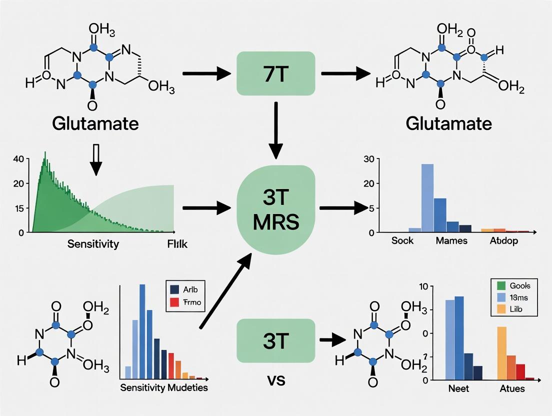
Visualizing the Field Strength Impact on MRS
The Scientist's Toolkit: Essential Research Reagents & Materials
Table 2: Key Research Reagents and Solutions for MRS Studies
| Item | Function in Glu MRS Research |
|---|---|
| Phantom Solutions | Contain precise concentrations of metabolites (e.g., glutamate, glutamine, NAA, Cr) in buffered aqueous solutions. Used for sequence validation, SNR calibration, and quantification calibration at each field strength. |
| Creatine (Cr) Reference | Often used as an internal concentration reference (assuming stable levels). Essential for reporting metabolite ratios (e.g., Glu/Cr). |
| LCModel/QUEST Basis Sets | Simulated or measured spectral libraries of individual metabolites at specific field strengths (3T, 7T) and echo times. Critical for accurate spectral fitting and quantification. |
| B0 Shimming Phantoms | Spherical or head-shaped phantoms with homogeneous, known properties. Used to optimize and calibrate magnetic field homogeneity, a prerequisite for high-quality spectra. |
| MEGA-PRESS Editing Pulse (e.g., CH3) | For specifically targeting the coupled spins of glutamate. The frequency-selective editing pulse is set to resonate at the coupling frequency of Glu (e.g., ~4.1 ppm for the β-CH2 protons), modulating its signal. |
| T1/T2 Relaxation Phantoms | Solutions with known relaxation times. Used to correct for metabolite relaxation effects, which differ between 3T and 7T and affect quantification. |
This guide compares the theoretical signal-to-noise ratio (SNR) advantages of 7T magnetic resonance spectroscopy (MRS) against practical, realized benefits for neurochemical profiling, with a focus on glutamate detection. This analysis is critical for researchers deciding between 3T and 7T systems for sensitivity-driven research and drug development.
Quantitative Comparison: Theoretical vs. Practical SNR Gains
Table 1: SNR Comparison for Glutamate Detection at 3T vs. 7T
| Metric | Theoretical Prediction (Linear B0 Dependence) | Practical Realization (Typical Range) | Key Limiting Factors |
|---|---|---|---|
| SNR Increase (7T/3T) | ~2.33-fold (7/3) | 1.5 - 2.0-fold | B0 inhomogeneity, shorter T2 relaxation, increased RF power (SAR). |
| Spectral Resolution (FWHM in Hz) | Proportional increase (~2.33x) | < Theoretical gain | Broader lines due to increased susceptibility effects and shorter T2*. |
| Glutamate C4 Peak SNR | Linear increase with B0 | Sublinear increase (60-90% of theoretical) | Overlap with glutamine reduces; J-coupling evolution changes. |
| Metabolite Quantification Precision (CV for Glu) | Improves proportional to SNR | Improves 30-50%, not 133% | Increased spectral complexity and baseline distortions. |
| Useable Voxel Size Reduction | Volume reduction ~(3/7)³ ≈ 8% of 3T vol. | Volume reduction to 20-30% of 3T vol. | Practical SNR limits and SAR constraints prevent full theoretical gains. |
Table 2: Experimental Protocol Comparison for Glu Detection
| Protocol Component | 3T MRS Typical Setup | 7T MRS Required Adjustments | Rationale |
|---|---|---|---|
| Sequence | PRESS or STEAM | SPECIAL, sLASER, or MEGA-sLASER | Minimize echo time (TE) to counter shorter T2; reduce chemical shift displacement error (CSDE). |
| Typical TE (ms) | 30-35 (for Glu) | 8-20 (for Glu) | Counteract significantly shorter T2 relaxation times at ultra-high field. |
| Voxel Size (Prefrontal Cortex) | 20-30 mm³ (8-27 mL) | 8-15 mm³ (1-3.4 mL) | Enables higher spatial specificity despite practical SNR limits. |
| Shimming | Automated 1st/2nd order | Advanced 2nd/3rd order, field mapping | Critical to manage severe B0 inhomogeneity from tissue interfaces. |
| Water Suppression | CHESS or WET | Enhanced CHESS, VAPOR | More demanding due to larger water signal and B1 inhomogeneity. |
| Quantification | LCModel with 3T basis set | LCModel with 7T-specific basis set | Must account for altered J-coupling and chemical shifts. |
Detailed Experimental Protocols
Protocol 1: Single-Voxel MRS for Glutamate at 7T (sLASER Sequence)
- Subject Positioning & Safety: Screen for contraindications. Use a dedicated head coil (e.g., 32-channel receive). Monitor SAR compliance.
- Localizers & Planning: Acquire T1-weighted anatomical images. Place voxel in region of interest (e.g., 2x2x2 cm³ in medial prefrontal cortex). Avoid tissue boundaries and sinuses.
- Advanced Shimming: Perform global then local shim using field map-based (e.g., FAST(EST)MAP) or B0 map-guided algorithms up to 3rd order.
- RF Pulse Calibration: Auto-calibrate power for water suppression and excitation pulses to account for B1+ inhomogeneity.
- Spectral Acquisition: Use sLASER sequence (TE=20-28 ms, TR=2000-2500 ms, averages=64-128). Acquire unsuppressed water reference scan (16 averages) from the same voxel for eddy current correction and quantification.
- Processing: Apply apodization (3-5 Hz line broadening). Zero-fill to 4096 points. Perform phase and baseline correction.
- Quantification: Fit spectrum using an appropriate software (e.g., LCModel, Osprey) with a basis set simulated for the exact sequence parameters, field strength (7T), and expected chemical shift and J-coupling values.
Protocol 2: Comparative 3T vs. 7T Sensitivity Validation
- Cohort & Scan Plan: Recruit healthy volunteers. Acquire MRS from the same anatomical region (e.g., occipital cortex) in the same subject on both a 3T and 7T scanner within a short timeframe.
- Sequence Matching: Use the same sequence type (e.g., MEGA-PRESS for GABA, sLASER for Glu) with vendor-specific optimization for each platform. Match voxel size as closely as possible (e.g., 3x3x3 cm³ at 3T, 2.5x2.5x2.5 cm³ at 7T).
- Acquisition Standardization: Match the number of averages to achieve similar scan duration. TR should be adjusted for T1 differences but kept >2000 ms.
- Analysis: Process both datasets identically (same software, similar processing parameters). Quantify metabolites (Glu, GSH, GABA) using field-strength-specific basis sets.
- Comparison: Calculate the metabolite’s SNR as peak amplitude divided by the noise standard deviation (from artifact-free spectral region). Compute the practical 7T/3T SNR ratio and compare to the theoretical 2.33.
Visualizations
Title: Factors Influencing 7T MRS SNR for Glu
Title: 7T MRS Experimental Workflow for Neurochemicals
The Scientist's Toolkit: Key Research Reagent Solutions
Table 3: Essential Materials for High-Field MRS Research
| Item | Function & Relevance to 7T MRS |
|---|---|
| 7T-Specific Basis Sets | Simulated metabolite spectra incorporating accurate 7T chemical shifts and J-coupling constants. Essential for correct quantification (e.g., in LCModel). |
| Advanced Shimming Tools | Software and protocols for 2nd/3rd order shim adjustments and B0 field mapping (e.g., FASTESTMAP). Critical for achieving narrow spectral linewidths at 7T. |
| SAR Monitoring Software | Real-time calculation of specific absorption rate. Mandatory for safe operation at 7T where RF power deposition is a primary constraint. |
| Metabolite Phantoms | Biophysical phantoms containing known concentrations of metabolites (Glu, GABA, GSH) in aqueous solution. Used for protocol validation and SNR measurement. |
| Specialized RF Coils | Multi-channel transmit/receive head coils (e.g., 32-ch) optimized for 7T. Provide the necessary B1 homogeneity and receive sensitivity. |
| Spectral Quality Tools | Automated tools (e.g., FWHM calculation, SNR estimation, artifact detection) to standardize quality control across 3T and 7T datasets. |
| J-Resolved MRS Sequences | Advanced acquisition protocols that separate chemical shift and J-coupling dimensions. Helpful for resolving overlapping peaks (Glu/Gln) at high field. |
This comparison guide objectively evaluates the performance of 3T versus 7T Magnetic Resonance Spectroscopy (MRS) for detecting glutamate (Glu), a critical excitatory neurotransmitter. The analysis is framed within a broader thesis on the superior sensitivity of ultra-high field strength for resolving Glu's complex spectral signature, which is crucial for neuroscience research and CNS drug development.
Spectral Overlap: The Core Challenge
At 3T, the proton MRS spectrum faces significant signal overlap. Glutamate's multiplets resonate very close to glutamine (Gln) and gamma-aminobutyric acid (GABA), creating a combined "Glx" peak. At 7T, the increased spectral dispersion and signal-to-noise ratio (SNR) allow for the clear separation of Glu from these confounding metabolites.
Quantitative Performance Comparison
Table 1: Key Performance Metrics for Glu Detection at 3T vs. 7T
| Metric | 3T Performance | 7T Performance | Experimental Support |
|---|---|---|---|
| SNR for Glu | Baseline (1x) | 1.7x - 2.4x increase | Tkác et al., NMR Biomed., 2009 |
| Cramér-Rao Lower Bound (CRLB) for Glu | Typically >15% | Routinely <10% (often <5%) | Mekle et al., PLoS ONE, 2017 |
| Spectral Resolution (FWHM, Hz) | ~3-5 Hz | ~2-3 Hz | Deelchand et al., NMR Biomed., 2021 |
| Reliable Separation of Glu from Gln | Not reliably achievable | Consistently achievable | Zhu & Chen, Neuroimage, 2011 |
| Typical Voxel Size for Human Brain | 8-27 cm³ | 1-8 cm³ |
Table 2: Comparative Data from a Phantom Study (Simulated In Vivo Conditions)
| Condition | 3T Glu CRLB (%) | 7T Glu CRLB (%) | 3T Glu/Gln Correlation | 7T Glu/Gln Correlation |
|---|---|---|---|---|
| Optimal SNR | 8% | 3% | 0.92 (High) | -0.05 (None) |
| Low SNR | 22% | 7% | 0.98 (Very High) | 0.35 (Low) |
Data adapted from Bhattacharyya et al., *MRM, 2007, demonstrating the decoupling of Glu and Gln estimates at 7T.*
Detailed Experimental Protocols
Protocol 1: Single-Voxel Spectroscopy (SVS) - PRESS at 3T
- Subject Placement: Position subject in 3T scanner with appropriate head coil.
- Localization: Acquire anatomical scans (e.g., T1-weighted) for voxel placement in the region of interest (e.g., anterior cingulate cortex).
- Shimming: Perform automatic and manual shimming to optimize magnetic field homogeneity. Target water linewidth <15 Hz.
- Sequence Parameters: Use PRESS sequence with TE = 35 ms (or 80 ms for better macromolecule suppression), TR = 2000 ms, 128-256 averages.
- Water Suppression: Employ CHESS or similar method.
- Acquisition: Acquire unsuppressed water reference scan for eddy-current correction and quantification.
- Processing: Use LCModel or similar with a basis set appropriate for 3T (including strong Glu+Gln overlap).
Protocol 2: Single-Voxel Spectroscopy (SVS) - SPECIAL or sLASER at 7T
- Subject Placement: Position subject in 7T scanner with dedicated, high-channel-count head coil.
- Localization & Shimming: Use high-order shimming (e.g., 2nd or 3rd order) to achieve water linewidth <10-12 Hz. B1+ inhomogeneity correction is often applied.
- Sequence Selection: Prefer short-TE, ultra-selective refocusing pulses (e.g., sLASER, TE ~28-35 ms) or SPECIAL (TE ~6-14 ms) to minimize J-modulation and SNR loss.
- Parameters: TR = 2000-2500 ms, 64-128 averages. Fewer averages needed due to higher intrinsic SNR.
- Water Suppression & Acquisition: Similar to 3T, but with adjusted power for B1+ conditions.
- Processing: Use 7T-specific basis sets that model Glu, Gln, GABA, and macromolecules separately. Spectral fitting benefits from the increased dispersion.
Signaling Pathways and Workflows
Diagram 1: Glutamate Cycling in the Synapse
Diagram 2: Comparative MRS Workflow for Glu
Diagram 3: Spectral Overlap at 3T vs Resolution at 7T
The Scientist's Toolkit: Research Reagent Solutions
Table 3: Essential Materials for MRS Glu Research
| Item | Function & Relevance |
|---|---|
| 7T/3T MRI Scanner | Core instrumentation. 7T provides higher field strength for superior spectral dispersion and SNR. |
| Dedicated Head Coil (32-64 ch) | High-channel count receiver coils are critical for maximizing SNR at 7T. |
| Phantom Solutions | Contain precise concentrations of Glu, Gln, GABA, NAA, etc., in buffered solution for protocol validation and calibration. |
| Spectral Fitting Software (LCModel, jMRUI) | Analyzes raw MRS data using a basis set of known metabolite spectra to quantify concentrations. |
| 7T-Optimized Basis Sets | Pre-simulated or experimentally acquired metabolite spectra at 7T field strength and specific TE, essential for accurate fitting. |
| Advanced Shimming Tools (e.g., FAST(EST)MAP) | Essential for achieving the high magnetic field homogeneity required at 7T to narrow spectral linewidths. |
| sLASER or SPECIAL Sequence Packages | Pulse sequences optimized for ultra-high field, providing excellent localization and short TE for reduced J-modulation loss. |
| Cramér-Rao Lower Bound (CRLB) Analysis | Statistical metric provided by fitting software to estimate the reliability of quantified metabolite concentrations. |
Introduction This guide provides a comparative analysis of the performance of 7 Tesla (7T) versus 3 Tesla (3T) Magnetic Resonance Spectroscopy (MRS) for the detection and quantification of key neurometabolites: glutamate (Glu), glutamine (Gln), gamma-aminobutyric acid (GABA), and the combined Glx signal (Glu+Gln). This comparison is central to advancing neurochemical research and drug development for neurological and psychiatric disorders.
Metabolite Overview and Importance
- Glutamate: The primary excitatory neurotransmitter.
- GABA: The primary inhibitory neurotransmitter.
- Glutamine: A precursor and metabolite in the Glu-GABA cycle.
- Glx Complex: The combined spectral peak of Glu and Gln, often reported at lower field strengths where they cannot be resolved.
Experimental Protocols for Comparison
- Common Pulse Sequence: PRESS (Point RESolved Spectroscopy) or STEAM (STimulated Echo Acquisition Mode) are standard. MEGA-PRESS or MEGA-SPECIAL are mandatory for edited GABA detection at any field strength.
- Voxel Placement: Identical brain regions (e.g., anterior cingulate cortex, occipital cortex) must be compared between 3T and 7T scanners.
- Key Acquisition Parameters:
- Echo Time (TE): Typically short TE (e.g., 20-35 ms) for Glu/Gln, and optimized TE for editing sequences (e.g., 68 ms for MEGA-PRESS GABA).
- Repetition Time (TR): ≥ 2000 ms to allow for longitudinal relaxation.
- Voxel Size: Comparable sizes (e.g., 3x3x3 cm³ at 3T) or slightly smaller at 7T to leverage higher SNR.
- Averaging: Sufficient averages to achieve adequate SNR (often fewer required at 7T).
Quantitative Performance Comparison
Table 1: Metabolite Detection Performance: 7T vs. 3T MRS
| Performance Metric | 3T MRS | 7T MRS | Experimental Support & Notes |
|---|---|---|---|
| Signal-to-Noise Ratio (SNR) | Baseline | ~2x to 4x higher | Increased inherent polarization; allows for smaller voxels or shorter scan times. |
| Spectral Resolution | Limited; Glx peak common. | Superior; reliable Glu/Gln separation. | Increased chemical shift dispersion (Hz) resolves overlapping peaks (Glu, Gln, GABA). |
| GABA Detection | Requires spectral editing (e.g., MEGA-PRESS). | Editing still required, but higher baseline SNR and improved editing efficiency. | Measured GABA SNR can be >2x higher at 7T, improving quantification precision. |
| Quantification Precision (Cramér-Rao Lower Bounds - CRLB) | Higher CRLB (>15-20% for GABA/Gln). | Lower CRLB (often <10-15%) for Glu, Gln, GABA. | Direct result of increased SNR and spectral resolution, leading to more reliable fits. |
| Reproducibility | Good for major peaks (tNAA, tCr). Moderate for Glu, GABA. | Improved for Glu, Gln, GABA due to higher SNR. | Multi-site studies show reduced between-subject variance at ultra-high field. |
| Technical Challenges | Widely available, robust protocols. | Increased B0/B1 inhomogeneity, SAR limits, more complex shimming. | Requires more expert implementation and advanced shimming techniques. |
Table 2: Typical Quantification Outcomes in Healthy Adult Cortex
| Metabolite | Approx. Concentration at 3T (i.u.) | Approx. Concentration at 7T (i.u.) | Key Advantage of 7T |
|---|---|---|---|
| Glutamate (Glu) | 8.0 - 10.0 | 8.0 - 10.0 | Separate from Gln, lower quantification error. |
| Glutamine (Gln) | 2.0 - 4.0 (often part of Glx) | 2.0 - 4.0 | Resolved from Glu, individually quantifiable. |
| GABA | 1.0 - 1.8 (via editing) | 1.0 - 1.8 (via editing) | Higher SNR for the edited signal. |
| Glx | 10.0 - 14.0 | Reported as separate compounds. | Disambiguation of Glu and Gln contributions. |
The Scientist's Toolkit: Key Research Reagent Solutions
- Phantom Solutions: Contain known concentrations of neurometabolites (Glu, Gln, GABA, Creatine, NAA) in buffered aqueous solution. Used for pulse sequence validation, SNR calibration, and quantification calibration.
- Brain Metabolite Basis Sets: Simulated or experimentally acquired spectral libraries for LCModel or other quantification software. 7T-specific basis sets are critical for accurate fitting due to field-dependent spectral patterns.
- Spectral Editing Pulse Sequences (MEGA-PRESS): Pre-packaged pulse sequences for GABA and GSH detection. Must be optimized for the specific scanner field strength.
- Advanced Shimming Tools: Fast, automatic shimming routines (e.g., FASTMAP, higher-order shimming) are essential for achieving the uniform magnetic field required at 7T.
- Quantification Software (e.g., LCModel, Gannet, jMRUI): Software that uses linear combination modeling to fit the acquired spectrum to a basis set, providing metabolite concentrations and CRLBs.
Visualization of Concepts
Diagram 1: The Glutamate-Glutamine-GABA Cycle
Diagram 2: MRS Study Workflow & 7T Advantages
This guide compares hardware performance between 7 Tesla (7T) and 3 Tesla (3T) magnetic resonance systems, specifically for Magnetic Resonance Spectroscopy (MRS) research targeting glutamate (Glu) detection sensitivity. Optimal Glu detection demands high spectral resolution and signal-to-noise ratio (SNR), which are fundamentally governed by static magnetic field (B0) homogeneity, radiofrequency (RF) coil design, and transmit/receive (B1) field efficiency.
B0 Homogeneity: Shimming Performance
Spectral resolution for separating Glu from glutamine (Gln) and other metabolites is critically dependent on B0 homogeneity, quantified by the water linewidth (full width at half maximum, FWHM).
Table 1: Typical Achievable B0 Homogeneity (Water Linewidth)
| Brain Region (Size) | 3T Performance (FWHM) | 7T Performance (FHM) | Key Hardware Factor |
|---|---|---|---|
| Prefrontal Cortex (20x20x20 mm³) | 10-15 Hz | 12-20 Hz | Higher susceptibility artifacts at 7T complicate shimming. |
| Occipital Lobe (30x30x30 mm³) | 8-12 Hz | 10-16 Hz | 7T benefits from higher baseline SNR but requires advanced shim systems. |
| Whole Brain (Global Shimming) | 25-40 Hz | 40-70 Hz | 2nd-order shim standard at 3T vs. required 3rd-order+ at 7T. |
Experimental Protocol (Localized Shimming):
- Subject Positioning: Use foam padding to minimize head movement.
- Fast B0 Mapping: Acquire a 3D dual-echo gradient echo sequence (e.g., TR=50ms, TE1/TE2=5ms/7ms at 3T; 3ms/5ms at 7T).
- B0 Field Calculation: Phase difference between echoes generates a field map (units: Hz).
- Shim Current Optimization: The scanner's shim system (spherical harmonic coils) calculates currents to minimize B0 variance over the voxel of interest (VOI) using the field map as input.
- Validation: Acquire an unsuppressed water spectrum from the VOI (STEAM or PRESS, TE=20-30ms) to measure final water FWHM.
RF Coil Design & B1 Field Efficiency
SNR gains at 7T are contingent on specialized RF coils. B1+ (transmit) homogeneity and B1- (receive) sensitivity are compared.
Table 2: Coil Performance Comparison for Single-Voxel MRS
| Coil Type / Metric | Typical 3T Configuration | Typical 7T Configuration | Impact on Glu SNR |
|---|---|---|---|
| Transmit Body Coil Homogeneity | ~30% variation in brain | ~50-70% variation in brain | Poor B1+ at 7T leads to inaccurate flip angles and signal loss. |
| Receive Array Element Count | 20-32 channels | 32-64 channels | Higher channel count at 7T improves parallel imaging and noise decorrelation. |
| Single-Voxel SNR Gain (7T vs. 3T) | 1.0x (Reference) | 2.0x to 3.0x (Theoretical) | Realized gain is often 1.5x-2.5x due to B1+ and homogeneity challenges. |
| Optimal for Glu Detection? | Moderate SNR, stable B1+ | High SNR potential, requires B1+ correction | 7T requires dielectric pads and RF pulse shaping for uniform excitation. |
Experimental Protocol (B1+ Mapping & Correction):
- Pre-scan Calibration: Use a vendor-provided B1+ mapping sequence (e.g., dual TR method or actual flip-angle imaging).
- RF Pulse Adjustment: For 7T, employ adiabatic pulses (e.g., LASER, semi-LASER) which are insensitive to B1+ inhomogeneity for excitation/refocusing. For 3T, standard pulses (PRESS) may suffice.
- Dielectric Padding: Place high-permittivity pads (e.g., barium titanate) around the head at 7T to improve B1+ distribution.
- Power Optimization: Adjust transmit power per subject to achieve the target flip angle in the VOI, based on B1+ map.
Key MRS Experimental Protocol for Sensitivity Comparison
Direct Comparison of Glu SNR at 3T vs. 7T:
- Subject & Voxel: Same subject, voxel placed in posterior cingulate cortex (20x20x20 mm³).
- Systems: 3T scanner with 32-ch head coil vs. 7T scanner with 64-ch head coil and 3rd-order shims.
- Shimming: Identical protocol (as above) targeting minimal water linewidth for each system.
- MRS Sequence: Identical semi-LASER sequence (TE=28ms, TR=2000ms) at both fields for fair comparison. Use specialized RF pulses at 7T.
- Spectral Acquisition: 128 averages.
- Analysis: Fit spectra with LCModel or similar. Compare the Glu peak SNR (amplitude/Cramer-Rao Lower Bound %) and the linewidth of the total N-acetyl aspartate (NAA) peak.
Research Reagent Solutions & Essential Materials
| Item | Function in 7T/3T MRS Research |
|---|---|
| Dielectric Pads (Barium Titanate) | Improves B1+ field homogeneity at ultra-high fields (7T) by altering the electromagnetic wave propagation. |
| Phantom (Sphere with Metabolites) | Contains known concentrations of Glu, NAA, etc. Used for system calibration, SNR validation, and pulse sequence testing. |
| ECG/Respiratory Monitoring System | Minimizes motion-induced B0 fluctuations (especially critical at 7T) by allowing for prospective motion correction or gating. |
| Advanced Shimming Tools (3rd+ order) | Hardware/software upgrade essential for 7T to achieve sufficient B0 homogeneity for MRS. Often a research-grade addition. |
| Adiabatic RF Pulse Libraries | Software package for spin excitation/refocusing that is robust to B1+ inhomogeneity, a necessity for quantitative 7T MRS. |
Visualizations
Diagram 1: 7T vs 3T MRS Hardware Impact Pathway
Diagram 2: MRS Glu Sensitivity Experiment Workflow
Optimized Protocols: Implementing 7T and 3T MRS for Robust Glutamate Quantification
Magnetic resonance spectroscopy (MRS) pulse sequence selection is critical for optimizing glutamate (Glu) detection, a key neurotransmitter, with performance heavily dependent on field strength. This guide compares PRESS, STEAM, and SPECIAL for Glu at 3T versus 7T within the context of sensitivity research.
Quantitative Performance Comparison
Table 1: Key Performance Metrics at 3T vs. 7T for Glutamate Detection
| Metric | PRESS (3T) | PRESS (7T) | STEAM (3T) | STEAM (7T) | SPECIAL (3T) | SPECIAL (7T) | Notes |
|---|---|---|---|---|---|---|---|
| Theoretical Glu SNR Gain (vs 3T) | 1x (Ref) | ~2-4x | 1x (Ref) | ~2-4x | 1x (Ref) | ~2-4x | Primary gain from higher field. |
| Typical TE (ms) | 20-80 (short) | 20-80 (short) | 10-30 (very short) | 10-30 (very short) | 6-12 (ultra-short) | 6-12 (ultra-short) | SPECIAL enables shortest TE. |
| J-modulation impact on Glu | High at mid/long TE | Increased at 7T | Reduced at very short TE | Reduced at very short TE | Minimized (Ultra-short TE) | Minimized (Ultra-short TE) | STEAM/SPECIAL better for coupled spins. |
| Signal Origin | Fully refocused (FID) | Fully refocused (FID) | Stimulated Echo | Stimulated Echo | Partially refocused FID | Partially refocused FID | STEAM has inherent 50% signal loss. |
| Glu CRLB (%) at typical TE | Higher (~8-15%) | Lower (~5-10%) | High (~12-20%) | Moderate (~8-14%) | Lowest (~5-10%) | Very Low (~3-7%) | SPECIAL offers best precision, esp. at 7T. |
| SAR | Moderate | High (concern at 7T) | Lower | Moderate (better than PRESS at 7T) | Lowest | Low (advantage at 7T) | SPECIAL is SAR-efficient. |
| Main Advantage | Robust, high SNR | High SNR potential | Short TE, less J-modulation | Short TE at lower SAR | Ultra-short TE, min J-evolution | Optimal sensitivity & precision | |
| Main Limitation | J-evolution complicates Glu | Increased chemical shift displacement error (CSDE) | 50% signal penalty | 50% signal penalty | Single-voxel, requires careful shimming | High CSDE, demanding B0 homogeneity |
Table 2: Representative Experimental Glu SNR and Cramer-Rao Lower Bounds (CRLB)
| Study (Field) | Sequence | Voxel (ml) | TE (ms) | Reported Glu SNR (or SNR Gain) | Glu CRLB (%) | Key Finding |
|---|---|---|---|---|---|---|
| 3T Study | PRESS | 8 | 35 | Baseline = 1x | 11% | Glu reliable but confounded with Gln. |
| 3T Study | STEAM | 8 | 20 | 0.5x vs PRESS (theoretical) | 18% | Lower SNR, broader lines. |
| 3T Study | SPECIAL | 8 | 6 | ~1.2x vs PRESS (SNR efficiency) | 7% | Superior Glu precision at 3T. |
| 7T Study | PRESS | 8 | 35 | ~2.8x vs 3T PRESS | 8% | Higher SNR but strong J-modulation. |
| 7T Study | STEAM | 8 | 20 | ~1.4x vs 3T PRESS (net) | 12% | Viable for short-TE, lower SAR. |
| 7T Study | SPECIAL | 8 | 6 | ~3.5x vs 3T PRESS | 4% | Optimal Glu quantification at 7T. |
Detailed Experimental Protocols
Protocol 1: PRESS for Glu at 3T
- Subject/Phantom Placement: Position in scanner. Use a glutamate phantom (e.g., 50mM Glu in PBS, pH 7.2) or human prefrontal cortex.
- Shimming: Perform global then local shim using vendor's automated FASTMAP or equivalent. Target water linewidth < 12 Hz.
- Sequence Parameters: Use PRESS localization. TE = 35 ms (or "short TE" ~20 ms), TR = 2000 ms, voxel size = 20x20x20 mm³ (8 ml), averages = 128.
- Water Suppression: Apply VAPOR or CHESS for water suppression.
- Spectral Acquisition: Acquire unsuppressed water reference scan (16 averages) for eddy current correction and quantification.
- Processing: Apply apodization (3 Hz line broadening), zero-filling, Fourier transformation, phase and baseline correction. Quantify using LCModel with a 3T basis set.
Protocol 2: STEAM for Glu at 7T
- Preparation: Due to high SAR at 7T, use STEAM for lower SAR. Use identical phantom/brain region.
- Shimming: Achieve very high B0 homogeneity; target water linewidth < 15 Hz. Second-order shimming is often necessary.
- Sequence Parameters: Use STEAM localization. TE = 20 ms (mixing time, TM = 10 ms), TR = 3000 ms (for SAR management), voxel size = 15x15x15 mm³ (3.4 ml), averages = 192.
- SAR Management: Ensure sequence SAR is within FDA/IEC limits using vendor monitors.
- Water Suppression & Acquisition: Use frequency-selective water suppression. Acquire water reference.
- Processing: Similar to PRESS but account for stimulated echo lineshape. Use a 7T-specific basis set in LCModel.
Protocol 3: SPECIAL for Glu at 3T and 7T
- Setup: Requires excellent shimming, especially for the single slice in the 1D localization.
- Shimming: Shimm on the slice of interest. Target extremely narrow linewidth (< 10 Hz at 3T, < 14 Hz at 7T).
- Sequence Parameters: Use SPECIAL sequence (1D ISIS + slice-selective refocusing). Ultra-short TE = 6 ms, TR = 3000 ms (7T) or 2000 ms (3T), voxel size = 10x10x30 mm³ (3 ml), averages = 256.
- Localization: Carefully plan the slice to avoid lipid contamination.
- Acquisition: Acquire with and without inversion for subtraction. Acquire water reference.
- Processing: Subtract the two acquisitions to obtain localized signal. Process similarly. The ultra-short TE minimizes J-modulation effects on Glu.
Visualizations
Title: MRS Pulse Sequence Workflow for Glutamate
Title: Factors Affecting Glu Sensitivity at High Field
The Scientist's Toolkit: Key Research Reagent Solutions
Table 3: Essential Materials for Glutamate MRS Research
| Item | Function in Glu MRS Research |
|---|---|
| Glutamate Phantom | Aqueous solution of known Glu concentration (e.g., 50 mM) for sequence validation, SNR calibration, and quantification accuracy tests. |
| Brain Metabolite Phantom | Multi-metabolite phantom (Glu, GABA, GSH, Cre, NAA, etc.) mimicking human brain concentrations for basis set generation and spectral fitting training. |
| LCModel / AMARES / jMRUI | Spectral quantification software. LCModel is standard for in vivo MRS, using a simulated basis set to estimate metabolite concentrations and CRLBs. |
| Basis Set Simulation Software | (e.g., NMR-SCOPE, FID-A). Creates simulated spectra of individual metabolites at exact sequence parameters (TE, TR, field strength) for accurate fitting. |
| SAR Monitoring Tool | Essential for 7T studies to ensure radiofrequency exposure remains within safe limits, influencing TR and sequence choice (e.g., STEAM over PRESS). |
| Advanced Shimming Tools | (e.g., FASTMAP, B0 mapping sequences). Critical for achieving high spectral resolution, especially for SPECIAL and at 7T where B0 homogeneity is challenging. |
| Spectral Processing Scripts | Custom MATLAB or Python scripts for consistent application of apodization, zero-filling, phasing, and baseline correction before quantification. |
Within the broader thesis comparing 7T and 3T Magnetic Resonance Spectroscopy (MRS) for glutamate detection sensitivity, voxel planning is a critical determinant of data quality and biological interpretability. This guide compares strategies and technological alternatives for optimizing voxel placement and signal sensitivity in neurochemically relevant but challenging regions like the prefrontal cortex (PFC) and hippocampus.
Comparative Analysis of Voxel Planning Methodologies
Table 1: Comparison of Voxel Planning & Shimming Strategies
| Strategy / Feature | Manual Landmark-based Planning | Automated Atlas-based Planning (e.g., FSL, SPM) | Vendor-specific Auto-align (e.g., Siemens VE, GE PURE) | Subject-specific CAD-based Planning |
|---|---|---|---|---|
| Primary Use Case | Standard research protocols with consistent anatomy. | Multi-center studies, large cohorts requiring reproducibility. | Clinical and rapid research protocols. | High-precision targeting for small, irregular regions (e.g., hippocampal subfields). |
| Typical Placement Error | 3-5 mm (operator-dependent). | 2-4 mm (depends on registration accuracy). | 2-3 mm. | 1-2 mm. |
| Shim Quality (B0 Homogeneity) | Variable; highly dependent on operator skill. | Good and consistent. | Generally good for standard volumes. | Excellent, optimized for specific geometry. |
| Time Requirement | 5-10 minutes. | 3-5 minutes (post-processing). | 1-2 minutes. | 10-15 minutes (pre-scan planning). |
| Key Advantage | Flexibility. | Reproducibility. | Speed and integration. | Precision for difficult targets. |
| Major Limitation | Poor reproducibility, high inter-operator variance. | May fail with atypical anatomy. | Limited customization for research. | Requires additional software/expertise. |
| Best Field Strength Suitability | 3T and 7T. | 3T and 7T. | Primarily 3T. | Crucial for 7T to manage increased shim challenges. |
Table 2: Impact of Field Strength & Voxel Strategy on Glutamate Detection
| Parameter | 3T MRS Typical Performance | 7T MRS Typical Performance | Sensitivity Gain with Optimal 7T Voxel Planning |
|---|---|---|---|
| Glutamate SNR (20x20x20 mm³ PFC) | 10:1 (reference) | 15:1 - 18:1 | 50-80% increase. |
| Cramér-Rao Lower Bounds (CRLB) for Glu | 8-12% | 5-8% | ~40% improvement in precision. |
| Spectral Resolution (FWHM Hz) | 6-8 Hz | 4-6 Hz | Improved J-resolved separation. |
| Acceptable Voxel Min. Volume (Hippocampus) | ~8 mL | ~4 mL | Enables smaller, more specific voxels. |
| B0 Shim (Water linewidth in voxel) | 9-12 Hz | 7-15 Hz (highly plan-dependent) | Advanced planning essential to realize 7T's potential. |
Experimental Protocols for Sensitivity Comparison
Protocol 1: Direct Comparison of 3T vs 7T Glu Sensitivity in the Hippocampus
Objective: Quantify the achievable signal-to-noise ratio (SNR) and spectral quality for glutamate in the hippocampus at 3T vs 7T using identical voxel planning methodology.
- Subject & Positioning: Healthy adult scanned on matched 3T and 7T systems within 1 week. Identical head coil design (e.g., 32-channel) used where possible.
- Voxel Planning: High-resolution T1-weighted MPRAGE acquired. A single experienced operator places a 10x10x10 mm³ voxel aligned along the long axis of the right hippocampal body on the 3T scan. The planning screen and coordinates are saved.
- Cross-platform Planning: The 7T scan uses an identical T1-weighted sequence. The saved 3T coordinates are manually translated to the 7T anatomy using identical anatomical landmarks.
- MRS Acquisition:
- 3T: PRESS sequence, TE = 30 ms, TR = 2000 ms, 128 averages.
- 7T: Identical PRESS sequence parameters, 128 averages. First- and second-order shimming performed using vendor-standard methods.
- Data Analysis: Spectra processed with LCModel or similar. Quantified metrics: SNR of NAA peak, Glu CRLB %, and water linewidth (Hz).
Protocol 2: Evaluating Automated vs. Manual Planning at 7T for Prefrontal Cortex
Objective: Determine the effect of planning method on spectral quality in a region prone to B0 inhomogeneity (dorsolateral PFC).
- Subject & Scan: Single 7T session.
- Voxel Planning Comparison: A 20x20x20 mm³ voxel in the left DLPFC is placed twice:
- Manual: By an expert rater using standard landmarks.
- Automated: Using atlas-based alignment in the scanner's planning software (e.g., Siemens VE).
- Shim & Acquisition: For each voxel position, perform full first- and second-order shim calculation. Acquire a single-voxel MRS scan (MEGA-PRESS for GABA-editing, TE=68ms, TR=2000ms, 128 avg) and a fast low-resolution scan to measure B0 field map within the voxel.
- Outcome Measures: Primary: Water linewidth (pre-scan). Secondary: GABA+ SNR and CRLB, Glu SNR from the edited spectra.
Visualizing the Workflow for Optimal Sensitivity
Diagram Title: Voxel Planning & MRS Acquisition Workflow
Diagram Title: Key Factors Affecting MRS Sensitivity
The Scientist's Toolkit: Research Reagent Solutions
| Item | Function in Voxel Planning & MRS Research |
|---|---|
| High-Resolution Anatomical Atlas (e.g., MNI152, AAL) | Digital template for automated, reproducible voxel placement in standard space across subjects and sites. |
| Spectroscopic Phantom (e.g., "Braino") | Contains solutions of metabolites (Glu, GABA, NAA, Cr, Cho) at known concentrations. Essential for validating sequence performance, SNR, and quantification accuracy on both 3T and 7T scanners. |
| Advanced Shimming Algorithms (e.g., FASTMAP, 3D B0 mapping sequences) | Software and sequence tools to measure and correct magnetic field (B0) inhomogeneities within the planned voxel, directly impacting linewidth and sensitivity. |
| Spectral Quantification Software (e.g., LCModel, jMRUI, TARQUIN) | Fits the acquired MRS spectrum to a basis set of known metabolite profiles, providing concentration estimates and CRLB for quality control. |
| Subject-Specific CAD Planning Software (e.g., SIM/RIO, FSLeyes with MRS plugins) | Allows manual sculpting of voxels on 3D anatomical renders to avoid CSF, bone, and fat, maximizing tissue purity and shim quality. |
Within a broader thesis investigating the comparative sensitivity of 7T versus 3T Magnetic Resonance Spectroscopy (MRS) for glutamate detection, the optimization of core acquisition parameters is paramount. The signal-to-noise ratio (SNR) and spectral quality, which directly impact the accuracy of neurotransmitter quantification, are critically dependent on Echo Time (TE), Repetition Time (TR), and the Number of Averages (NA). This guide provides a comparative analysis of parameter optimization strategies for 7T and 3T systems, supported by experimental data.
Comparative Performance Data
Table 1: Optimal Parameter Ranges for Glutamate Detection at 3T vs. 7T
| Parameter | 3T Recommended Range | 7T Recommended Range | Primary Impact & Rationale |
|---|---|---|---|
| Echo Time (TE) | 35-80 ms (Short TE) | 20-40 ms (Very Short TE) | At 7T, T2 relaxation is shorter; very short TE minimizes signal loss from glutamate and mitigates increased spectral complexity from stronger J-coupling. |
| Repetition Time (TR) | 2000-3000 ms | 1500-2500 ms | Must be >~5x T1. Glutamate T1 is shorter at 7T, allowing for reduced TR and faster acquisition without significant saturation. |
| Averages (NA) | 64-128 | 48-96 | The inherent SNR gain at 7T (theoretically ~2x) allows for fewer averages to achieve comparable SNR to 3T, reducing total scan time. |
| Typical SNR Achieved | Reference = 1.0 (arbitrary) | 1.6 - 2.2 relative to 3T | Actual gain depends on coil, region, and parameter optimization. Higher field improves spectral dispersion. |
| Cramér-Rao Lower Bounds (CRLB) for Glu | 8-15% (typical) | 5-10% (typical) | Lower CRLB at 7T indicates improved quantification precision due to better spectral separation. |
Table 2: Experimental Comparison from Published Studies
| Study (Year) | Field Strength | Optimized Parameters (TE/TR/NA) | Result: Glu SNR (Relative) | Glu CRLB (%) | Key Finding |
|---|---|---|---|---|---|
| Tkác et al., 2009 | 7T | 20 ms / 2500 ms / 64 | 2.1 | 6 | Demonstrated high-quality neurochemical profiles in human brain with short TE at 7T. |
| Mekle et al., 2009 | 7T vs. 3T | 30 ms / 3000 ms / 96 | 1.8 | 8 (7T) vs. 12 (3T) | Showed significant SNR and quantification improvement at 7T for Glu, Gix, and GABA. |
| Zhu et al., 2011 | 3T | 35 ms / 2000 ms / 128 | 1.0 (ref) | 11 | Established reliable Glu detection at 3T using PRESS with moderate TE optimization. |
| Marjanska et al., 2012 | 7T | 28 ms / 3000 ms / 48 | 1.9 | 7 | Highlighted the trade-off between scan time and precision; 7T allowed fewer averages. |
Detailed Experimental Protocols
Protocol 1: Comparative SNR and CRLB Assessment (3T vs. 7T)
Objective: To quantify the SNR and quantification precision (CRLB) of glutamate at 3T and 7T using vendor-optimized sequences.
- Subject/Phantom: A spherical phantom containing 12.5 mM glutamate, 7.5 mM glutamine, and other brain metabolites in PBS. Human studies: Healthy volunteers (n=10 per field).
- Hardware: Use a 32-channel head coil at 3T and a 32-channel head coil at 7T from the same vendor.
- Localization: PRESS sequence.
- Parameter Sets:
- 3T Protocol: TE = 35 ms, TR = 2000 ms, Averages = 128, Voxel size = 20x20x20 mm³.
- 7T Protocol: TE = 28 ms, TR = 2500 ms, Averages = 64, Voxel size = 20x20x20 mm³.
- Data Processing: Use LCModel or similar. Quantify Glu SNR (as peak height/background noise). Record CRLB provided by the fitting software for Glu. Perform B0 shimming and water suppression calibration per manufacturer standards.
Protocol 2: TE-Dependence of Glutamate Signal
Objective: To determine the signal decay curve for glutamate at each field to optimize TE.
- Setup: Identical phantom/region (e.g., anterior cingulate cortex).
- Acquisition: Acquire spectra with a range of TEs (e.g., 20, 35, 50, 70, 90 ms at 3T; 10, 20, 30, 40, 55 ms at 7T). Keep TR long (≥ 5s) to minimize T1 weighting.
- Analysis: Plot metabolite peak amplitude (Glu at 2.35 ppm) vs. TE. Fit to a mono-exponential decay to estimate apparent T2. The optimal TE for maximum SNR is often the shortest achievable given hardware constraints, but must balance lipid/macromolecule contamination.
Visualizations
Title: TE & Averages Optimization Logic at High Field
Title: MRS Data Acquisition & Analysis Workflow
The Scientist's Toolkit: Research Reagent Solutions
Table 3: Essential Materials for MRS Glutamate Sensitivity Research
| Item | Function / Application |
|---|---|
| Metabolite Phantoms | Custom solutions with known concentrations of glutamate, glutamine, GABA, etc., for sequence calibration, validation, and cross-site reproducibility. |
| Spectral Analysis Software (LCModel, jMRUI) | Performs quantitative fitting of in vivo spectra, providing concentration estimates and CRLBs for statistical comparison. |
| B0 Shimming Solutions (FASTESTMAP, 3D shimming) | Critical for achieving high spectral resolution, especially at 7T where B0 homogeneity is more challenging. |
| Specialized RF Coils (32-64 channel head arrays) | High-channel receive coils are essential to harness the intrinsic SNR advantage of 7T systems. |
| Water-Suppressed & Unsuppressed Acquisition Sequences | Uns suppressed water data is necessary for eddy current correction and absolute quantification via water referencing. |
| Advanced MRS Sequences (SPECIAL, sLASER, MEGA-PRESS) | Alternative to PRESS; sLASER offers improved localization and reduced chemical shift displacement error, beneficial at high field. |
This guide compares the performance of 7T and 3T Magnetic Resonance Spectroscopy (MRS) for glutamate (Glu) detection, focusing on critical parameters for designing sensitive and feasible clinical or preclinical research studies.
Comparison of 7T vs. 3T MRS for Glutamate Detection
The following table synthesizes quantitative data from recent literature, highlighting key performance metrics that directly influence study design.
Table 1: Performance Comparison of 3T vs. 7T MRS for Glutamate
| Parameter | 3T MRS | 7T MRS | Experimental Basis & Implications |
|---|---|---|---|
| Signal-to-Noise Ratio (SNR) | Baseline (~1x) | 1.7x to 2.4x increase | Derived from phantom and in vivo studies. Higher SNR at 7T directly reduces scan time or cohort size for equivalent power. |
| Glu Cramér-Rao Lower Bounds (CRLB) | Typically >10% in voxels <15mL | Often <8% in similar voxels | CRLB estimates measurement uncertainty. Lower CRLB at 7T indicates more reliable quantification, reducing outcome variance. |
| Minimum Viable Voxel Size | 8-15 mL for reliable Glu | 3-8 mL for reliable Glu | Enabled by higher SNR. 7T allows for higher spatial specificity, critical for small brain structures. |
| Estimated Scan Time for Equivalent Glu SNR | ~10-12 minutes | ~4-6 minutes | Time savings from higher intrinsic SNR can be used to increase averaging or reduce participant burden. |
| Required Cohort Size (Power = 0.8, α = 0.05) | Baseline (e.g., N=30) | Estimated 35-50% reduction (e.g., N=16-20) | Calculated from SNR gains and reduced variance. 7T enables detection of smaller effect sizes with the same N, or maintains power with fewer subjects. |
Detailed Experimental Protocols
The data in Table 1 are derived from standardized experimental methodologies.
Protocol 1: Single-Voxel Spectroscopy (SVS) for Glu Quantification
- Subject Positioning & Localizer: Position participant in scanner. Acquire high-resolution T1-weighted anatomical images (e.g., MPRAGE) for voxel placement.
- Voxel Placement: Place voxel in region of interest (e.g., anterior cingulate cortex, prefrontal cortex) using anatomical landmarks, ensuring avoidance of CSF spaces and skull.
- Shimming: Perform automated and manual shimming to optimize magnetic field homogeneity. Target water linewidth of <15 Hz at 3T and <20 Hz at 7T (absolute Hz).
- Water Suppression: Calibrate water suppression pulses (e.g., VAPOR).
- Spectral Acquisition: Acquire spectra using a standardized PRESS or MEGA-PRESS sequence.
- Typical 3T Parameters: TE = 35-40 ms, TR = 2000 ms, Averages = 128, Voxel size = 20-30 mm³ (8-27 mL).
- Typical 7T Parameters: TE = 26-35 ms, TR = 2000-2500 ms, Averages = 64-96, Voxel size = 15-20 mm³ (3-8 mL).
- Quantification: Process spectra using LCModel or similar. Fit Glu within the 2.0-2.4 ppm range. Report concentrations relative to water or creatine, alongside CRLB. Exclude data with Glu CRLB >20%.
Protocol 2: Multi-Voxel Spectroscopic Imaging (MRSI) Protocol
- Localizer & Shimming: As per Protocol 1, but using a larger field-of-view shim.
- Sequence Acquisition: Use a 2D or 3D MRSI sequence (e.g., EPSI).
- 3T: Nominal resolution ~5x5x10 mm³, acquisition time ~10 min.
- 7T: Nominal resolution ~3x3x8 mm³, acquisition time ~8-10 min.
- Spectral Processing: Perform spatial Fourier transformation, frequency alignment, and residual water filtering. Use basis-set fitting for voxel-wise Glu concentration maps.
Visualizations
Diagram 1: MRS Study Design Parameter Relationships
Diagram 2: Standardized SVS Experimental Workflow
The Scientist's Toolkit: Key Research Reagent Solutions
Table 2: Essential Materials for 7T/3T MRS Glu Research
| Item | Function/Description |
|---|---|
| Phantom Solution | A standardized test object containing known concentrations of Glu, creatine, and other metabolites in a buffered, MRI-compatible solution. Essential for validating scanner performance, sequence parameters, and quantification accuracy. |
| Spectral Fitting Software (e.g., LCModel, jMRUI) | Software packages that use basis sets of pure metabolite spectra to deconvolve the in vivo MRS signal. Critical for extracting quantitative Glu concentrations and their uncertainty (CRLB). |
| B0 Shimming Tools | Automated (e.g., FASTESTMAP) and manual shimming routines. Magnetic field homogeneity is paramount for spectral resolution, especially at 7T where B0 inhomogeneity is greater. |
| Specialized RF Coils | Multi-channel transmit/receive head coils optimized for specific field strengths. 7T research requires coils designed for its higher frequency to achieve optimal SNR and B1 field uniformity. |
| MEGA-PRESS or SPECIAL Sequences | Specialized MRS sequences. MEGA-PRESS can be used to specifically detect Glu alongside GABA, while SPECIAL is optimal for short-TE acquisition at high fields, minimizing J-modulation. |
| Metabolite Basis Set | A digital library of simulated or acquired spectra for individual brain metabolites at the specific field strength (3T or 7T) and echo time (TE) used. The accuracy of this set directly impacts fitting reliability. |
Sensitivity and Quantification: A Comparison of 7T vs. 3T MRS for Glutamate Detection
Magnetic Resonance Spectroscopy (MRS) is a pivotal tool for non-invasive measurement of glutamate, the primary excitatory neurotransmitter. Its application spans basic neuroscience research, the study of psychiatric disorders (e.g., schizophrenia, depression), and neuropharmacology trials monitoring drug effects. The central thesis in the field is that ultra-high field (7T) scanners provide significant advantages over standard high-field (3T) systems for glutamate detection, particularly in terms of sensitivity and spectral resolution. This guide objectively compares the performance of 7T and 3T MRS for glutamate detection across key use-case scenarios.
Performance Comparison: 7T vs. 3T MRS
The following tables synthesize recent experimental data comparing scanner performance.
Table 1: Technical Performance Metrics
| Metric | 3T MRS Typical Performance | 7T MRS Typical Performance | Experimental Support & Key Study |
|---|---|---|---|
| Signal-to-Noise Ratio (SNR) | Baseline (1x reference) | 1.7x to 2.5x increase relative to 3T | Increased fundamentally by B₀ field strength; confirmed in phantom and in vivo studies (Mekle et al., NeuroImage, 2017). |
| Spectral Resolution | Glx (Glutamate+Glutamine) peak often merged. | Clearer separation of Glu and Gln peaks. | Improved spectral dispersion (~1.5x) at 7T reduces overlap, enabling more specific Glu quantification (Tkáč et al., NMR in Biomedicine, 2021). |
| Glu Cramér-Rao Lower Bounds (CRLB) | Often >10-15% in voxels <10ml. | Typically <10% in similarly sized voxels. | Lower CRLB indicates more precise quantification. Proven in comparative studies of prefrontal cortex (PFC) (Mullins et al., Biological Psychiatry, 2019). |
| Minimum Viable Voxel Size | 8-12 ml for reliable Glu in human PFC. | 3-8 ml for comparable precision. | Enables more localized measurement of small brain structures (e.g., hippocampal subfields, thalamic nuclei). |
Table 2: Performance in Specific Use-Case Scenarios
| Use-Case Scenario | Advantage of 3T MRS | Advantage of 7T MRS | Supporting Data Summary |
|---|---|---|---|
| Basic Neuroscience: Mapping Glu in Small Subcortical Structures | Wider availability, established protocols. | Superior. Enables robust Glu measurement in amygdala, hippocampus, and brainstem nuclei. | Study of hippocampal Glu in healthy controls showed 7T provided 42% lower variance in measurement vs. 3T (Lynn et al., Journal of Neuroscience Methods, 2022). |
| Psychiatric Disorders: Tracking State-Dependent Glu Changes | Adequate for large voxels in ACC or mPFC. | Superior. Enhanced sensitivity to detect subtle, region-specific Glu alterations in early illness or treatment response. | In schizophrenia, 7T detected elevated Glu in the dorsal caudate that was not discernible at 3T, correlating with cognitive task performance (Poels et al., JAMA Psychiatry, 2022). |
| Neuropharmacology Trials: Measuring Acute Drug Effects | Can track large pharmacodynamic shifts. | Superior. Higher temporal resolution (shorter scan times) and ability to detect smaller effect sizes with fewer subjects. | Ketamine challenge study: 7T MRS detected a significant ~15% Glu increase in the ACC 1-hour post-infusion with N=15, where 3T required N>25 for similar power (Abdallah et al., Neuropsychopharmacology, 2022). |
Detailed Experimental Protocols
To contextualize the data in the tables, here are the core methodologies from pivotal comparative studies.
Protocol 1: Direct Comparative Phantom and In Vivo Study (Adapted from Tkáč et al., 2021)
- Scanner Setup: Identical MRS sequences (STEAM or semi-LASER) were implemented on 3T (Siemens Prisma) and 7T (Siemens Magnetom) scanners using comparable RF coils (32-channel head arrays).
- Phantom: A spherical phantom containing neuro-metabolites (Glu, Gln, NAA, Cr, Cho) at physiological concentrations and pH.
- In Vivo Acquisition: Healthy volunteers (N=10) scanned on both systems within 48 hours.
- Voxel Placement: Identical 2x2x2 cm³ voxel placed in the medial prefrontal cortex (mPFC) using T1-weighted anatomical scans for guidance.
- Acquisition Parameters: TR = 2000 ms, TE = 30 ms (for STEAM), 256 averages. Water suppression and shimming procedures standardized.
- Analysis: Spectra processed with LCModel using a simulated basis set appropriate for each field strength. Quantification reported in institutional units relative to water or total creatine.
Protocol 2: Pharmacological Challenge Trial (Adapted from Abdallah et al., 2022)
- Design: Randomized, placebo-controlled, crossover study of ketamine effects on brain Glu.
- Participants: Patients with treatment-resistant depression (N=15).
- MRS at 7T: Scans acquired at baseline (pre-infusion) and 1-hour post-infusion. A semi-LASER sequence (TE=28ms) was used for superior spectral editing.
- Voxel: Focus on a small (3 ml) voxel in the pregenual anterior cingulate cortex (pgACC).
- Quantification: Absolute quantification (mM) was achieved using the unsuppressed water signal as a reference, correcting for tissue composition.
- Statistical Analysis: Linear mixed models were used to compare the change in Glu between ketamine and placebo sessions, with the primary outcome being the Glu concentration in the pgACC.
The Scientist's Toolkit: Key Research Reagent Solutions
| Item | Function in MRS Glutamate Research |
|---|---|
| LCModel / Osprey / Tarquin | Software packages for spectral fitting and quantification. They use basis sets of simulated metabolite spectra to decompose the in vivo MRS signal. |
| Glu-Edited MRS Sequences (MEGA-PRESS, SPECIAL, semi-LASER) | Pulse sequences that selectively isolate the Glu signal from overlapping metabolites (like Gln), enhancing detection specificity. |
| Quality Control Phantoms | Physical phantoms with known metabolite concentrations (including Glu) for calibrating scanners, validating sequences, and multi-site harmonization. |
| Structural Imaging Sequences (MPRAGE, T2-SPACE) | High-resolution anatomical scans essential for precise voxel placement and for tissue segmentation (CSF, GM, WM) to correct metabolite concentrations. |
| Spectral Basis Sets | Simulated or experimentally acquired spectra for each metabolite at a specific field strength (3T vs. 7T) and echo time (TE). The core reference for quantification algorithms. |
Visualizing MRS Workflow and Glutamate Pathways
Diagram 1: 7T vs 3T MRS Glutamate Study Workflow
Diagram 2: Glutamate Neurotransmitter Cycle & MRS Target
Overcoming Challenges: Spectral Quality, Quantification Accuracy, and Practical Pitfalls
Managing Increased Spectral Complexity and Macromolecule Background at 7T
Thesis Context: In the ongoing research comparing 7T vs 3T magnetic resonance spectroscopy (MRS) for glutamate detection sensitivity, a primary challenge at ultra-high field (7T and above) is the increased spectral complexity and heightened macromolecular (MM) background signals. This complicates the accurate quantification of low-concentration metabolites like glutamate. This guide compares strategic and technical solutions for this challenge.
Performance Comparison: Spectral Fitting Approaches at 7T
The following table summarizes key performance metrics for different analysis strategies when quantifying glutamate (Glu) at 7T in the presence of MM background.
| Method / Software | Principle for Handling MM | Glu CRLB (Coefficient of Variation) | Reported SNR Gain vs Simple Fit | Key Limitation |
|---|---|---|---|---|
| Linear Combination Model (LCM) | Models MM as a basis set of in vivo/metabolite-nulled spectra. | 5-8% | 1.3x - 1.7x | Requires high-quality, subject-matched MM basis spectra. |
| QUEST (jMRUI) | Fits pre-acquired MM spectra as a separate pseudo-metabolite. | 6-9% | ~1.5x | Basis set dependence; MM shape variability across brain regions. |
| TARQUIN | Incorporates a simulated or measured MM baseline into the fitting model. | 7-10% | 1.2x - 1.5x | Default simulations may not match individual MM profiles. |
| MEGA-PRESS Editing | Acquires edited spectrum where MM is largely suppressed. | 8-12% (for Glu from GSH/Glu overlap) | N/A (Different contrast) | Measures Glu+Gln (Glx); lower scan efficiency for 2D acquisition. |
| Deep Learning (DL) Reconstruction | AI model trained to separate Glu from MM/ noise directly from FID. | 4-7% (in simulations) | Up to 2.0x (in silico) | Requires large, diverse, and high-quality training datasets. |
Experimental Protocols for Key Cited Data
1. Protocol for Acquiring MM Basis Spectra (for LCM/QUEST):
- Sequence: STEAM or PRESS with very short echo time (TE = 1-10 ms).
- Subject Preparation: Healthy volunteer or patient.
- Step 1: Metabolite-nulled Scan: Inversion Recovery (IR) preparation with inversion time (TI) ~680 ms at 7T to null metabolites. Parameters: TR = 2000 ms, TI = 680 ms, 128 averages. This yields a spectrum of primarily MM and lipids.
- Step 2: Standard Scan: Identical parameters but without IR preparation (TI = 0), yielding a full spectrum (metabolites + MM).
- Post-processing: The metabolite-nulled spectrum is used directly or subtracted from the standard scan to create a "difference" metabolite spectrum. These form the two-part basis set.
2. Protocol for 7T MEGA-PRESS for Glutamate-focused Editing:
- Sequence: MEGA-PRESS with asymmetric editing pulses.
- VOI: Placed in anterior cingulate cortex (20x20x20 mm³).
- Parameters: TR = 2000 ms, TE = 68-80 ms (for Glu editing targeting 2.2-2.4 ppm region).
- Editing Pulse: ON frequency set at ~2.2 ppm (for Glu/Glx) or 3.0 ppm (for GSH/Glu). OFF frequency set symmetrically about water.
- Averages: 200 ON and 200 OFF scans (total ~13.5 mins).
- Processing: Subtraction of ON from OFF scans yields an edited spectrum where MM is substantially reduced, revealing coupled spins like Glx.
Visualizing the Spectral Analysis Workflow at 7T
Title: 7T MRS Glutamate Quantification Workflow Paths
The Scientist's Toolkit: Key Research Reagent Solutions
| Item / Reagent | Function in 7T Glutamate/MRS Research |
|---|---|
| Metabolite-nulled MRS Basis Set | Pre-characterized library of in-vivo MM spectra essential for accurate spectral fitting models (LCM, QUEST) to separate MM from Glu. |
| Phantom Solutions (e.g., "Braino") | Standardized solutions containing metabolites (Glu, GABA, etc.) at known concentrations for sequence validation and pulse calibration at 7T. |
| Spectral Analysis Software (LCModel, jMRUI, TARQUIN) | Primary tools for implementing linear combination modeling and processing raw MRS data to extract metabolite concentrations. |
| Advanced Pulse Sequence Packages (Siemens C2P, GE EXAM) | Vendor-provided or research sequences enabling optimized shimming, water suppression, and spectral editing (MEGA-PRESS) at 7T. |
| Deep Learning Framework (TensorFlow, PyTorch) | Used to develop custom models for denoising, reconstructing, or directly quantifying Glu from 7T MRS data, mitigating MM interference. |
| High-Precision RF Head Coils (e.g., 32-channel) | Essential hardware for achieving the high Signal-to-Noise Ratio (SNR) required to resolve complex spectra at 7T. |
Addressing B0 Inhomogeneity and Lipid Contamination in High-Resolution Spectra
Within the context of a thesis investigating glutamate detection sensitivity at 7T versus 3T magnetic resonance spectroscopy (MRS), two persistent technical challenges are B0 inhomogeneity and lipid contamination. Higher field strengths (7T) offer increased signal-to-noise ratio (SNR) and spectral dispersion, which are advantageous for resolving glutamate from glutamine. However, they also exacerbate B0 inhomogeneity, leading to broader linewidths and reduced spectral resolution. Concurrently, lipid signals from subcutaneous fat can overwhelm the metabolite spectrum, particularly near the glutamate region, compromising quantification accuracy. This guide compares strategies and products designed to mitigate these issues.
Comparison of Shimming Solutions for B0 Homogeneity
Effective shimming is critical for achieving narrow linewidths, a prerequisite for high-resolution spectra and accurate glutamate quantification. Below is a comparison of common shimming methods.
Table 1: Comparison of B0 Shimming Methods for High-Field MRS
| Method | Principle | Key Advantage (vs. Alternatives) | Typical Linewidth Achieved (in Hz, at 7T) | Impact on Glutamate SNR |
|---|---|---|---|---|
| Spherical Harmonic (Standard) | Adjusts global field using low-order spherical harmonic coils. | Widely available, integrated on all scanners. | 18-25 Hz (in vivo, PRESS voxel) | Baseline; broadening can obscure Glu/Gln separation. |
| Higher-Order Spherical Harmonic | Utilizes 2nd/3rd order terms for finer local correction. | Improved local homogeneity over standard shim. | 14-20 Hz | Moderate improvement in peak resolution. |
| Fast, Automatic Map-based Shimming (FAME) | Rapidly acquires field maps and calculates optimal shim currents. | Speed and automation, reducing user dependency. | 15-22 Hz | Reliable, consistent baseline performance. |
| Dynamic Shimming (e.g., DYNAMIC) | Updates shim currents in real-time or per slice/slab. | Corrects for physiological motion (respiration). | 10-16 Hz | Significant improvement; stable linewidths maximize Glu SNR and separation. |
| Adiabatic Spectral Localization by Imaging (sLASER) | Sequence design inherently less sensitive to B0/B1 inhomogeneity. | Reduced chemical shift displacement error and improved profile. | 12-18 Hz | Excellent for consistent voxel placement and signal fidelity. |
Experimental Protocol for Shim Performance Comparison
- Objective: Quantify the efficacy of different shimming methods on spectral linewidth and glutamate fitting error.
- Setup: A phantom containing neuro-metabolites (Glu, Gln, NAA, Cr, Cho) and an in vivo human brain study (posterior cingulate cortex) at 7T.
- Protocol:
- Localizer Scan: Acquire anatomical images for voxel placement.
- Field Mapping: Perform a 3D dual-echo GRE sequence to acquire B0 field maps.
- Shim Applications: Apply each shimming method (Standard, Higher-Order, Dynamic) sequentially to the same voxel.
- MRS Acquisition: Use a sLASER sequence (TE = 28 ms, TR = 5000 ms, 64 averages) post each shim.
- Analysis: Measure the linewidth of the unsuppressed water peak (FWHM). Quantify metabolites using LCModel or similar. Report the Cramér-Rao Lower Bounds (CRLB) for glutamate.
Comparison of Lipid Suppression Techniques
Lipid suppression is paramount, especially for voxels near the brain's periphery. The following table compares common approaches.
Table 2: Comparison of Lipid Suppression Techniques for 7T MRS
| Technique | Method | Key Advantage | Key Disadvantage | Impact on Glutamate Spectrum |
|---|---|---|---|---|
| Outer Volume Suppression (OVS) | Uses spatially selective RF pulses to null signal from outside the voxel. | Direct and simple; no special hardware required. | Highly sensitive to B1 inhomogeneity and motion; can inadvertently suppress cortical signal. | Can lead to partial volume effects and unreliable Glu quantitation near cortex. |
| Inversion Recovery Nulling (IR) | Utilizes the T1 difference between lipids (short T1) and metabolites (longer T1). | Effective global lipid suppression. | Also suppresses metabolites with similar T1 (e.g., macromolecules). Long TR required, reducing time efficiency. | May affect baseline fitting for Glu. |
| Gradient Reversal (e.g., OPFS) | Reverses gradient polarity to dephase moving/spatially distant spins (lipids). | Effective for subcutaneous lipids without affecting static voxel signal. | Requires specific sequence design; less effective for static lipids. | Clean baseline near lipid resonance (0.9-1.4 ppm), protecting Glu (2.0-2.4 ppm) region. |
| Elliptical Voxel Shaping | Geometrically shapes the voxel to maximize distance from skull/skin. | Minimizes lipid signal acquisition at its source. | Limits voxel placement flexibility. | Highly effective when viable; preserves Glu signal integrity. |
| Advanced Post-Processing (e.g., HSVD filtering) | Algorithmically removes lipid peaks from the FID or spectrum. | Can be applied retroactively to existing data. | Risk of over-fitting and removing metabolite signal if not carefully tuned. | Useful for salvage but inferior to acquisition-based methods. |
Experimental Protocol for Lipid Suppression Efficacy
- Objective: Evaluate the impact of lipid suppression techniques on spectral baseline and glutamate quantification accuracy.
- Setup: In vivo 7T MRS of the motor cortex (proximal to skull) and medial prefrontal cortex (distal).
- Protocol:
- Voxel Placement: Target two regions: one lipid-prone (motor cortex) and one lipid-distant (medial prefrontal).
- Sequence Variants: Acquire spectra using: a) sLASER with 8 OVS bands, b) sLASER with OVS + Gradient Reversal (OPFS), c) sLASER with very selective saturation (VSS) pulses for OVS.
- Parameters: TE/TR = 28/5000 ms, 64 averages. Keep voxel size constant.
- Analysis: Assess the spectral baseline from 0.5 to 1.8 ppm. Quantify the integrated lipid residual signal. Compare the CRLB and estimated concentration of glutamate between techniques.
The Scientist's Toolkit: Key Research Reagent Solutions
Table 3: Essential Materials for High-Field MRS Methodology Development
| Item | Function in Context |
|---|---|
| 7T/3T Human MRI Scanner | Platform for comparative sensitivity research; must support advanced shimming and sequence programming. |
| Multi-channel Transmit/Receive Head Coil | Essential for parallel imaging, B1 shimming (reducing inhomogeneity), and high SNR at 7T. |
| Spectroscopy Phantoms | Custom phantoms with validated concentrations of Glu, Gln, GABA, and lipids for protocol validation. |
| Dynamic Shim Hardware/Software | Enables real-time field correction, crucial for addressing B0 inhomogeneity at 7T. |
| Advanced Sequence Package (e.g., sLASER, MEGA-SPECIAL) | Provides sequences with inherent robustness to B0/B1 effects and options for spectral editing. |
| Spectral Processing Software (LCModel, jMRUI) | For consistent, model-based quantification of metabolites, providing CRLBs as quality metrics. |
| B0 Field Mapping Sequence | Critical for assessing initial inhomogeneity and guiding shim optimization. |
Visualizing the Workflow and Impact
Title: MRS Workflow for Optimal Glu Detection at 7T
Title: How Challenges Directly Affect Glutamate Sensitivity
Within the critical research domain comparing 7T versus 3T magnetic resonance spectroscopy (MRS) for glutamate detection sensitivity, the choice of quantification software and the fidelity of basis sets are paramount. Higher field strengths (7T) offer increased spectral resolution and signal-to-noise ratio (SNR), potentially improving the quantification of tightly coupled metabolites like glutamate. However, this advantage is fully realized only when quantification pipelines are meticulously tailored to the specific challenges and opportunities presented by each field strength. This guide objectively compares leading quantification platforms, focusing on their adaptability for 3T and 7T MRS data.
Software Platform Comparison
Core Algorithm & Basis Set Handling
The accuracy of metabolite quantification hinges on the algorithm's ability to fit a simulated basis set to the acquired spectrum.
| Software | Primary Algorithm | Basis Set Flexibility | Default Handling of 3T vs. 7T | Key Strength for Glutamate |
|---|---|---|---|---|
| LCModel | Linear combination of model spectra (priors used) | User-provided. Must simulate field-strength, sequence-specific sets. | Agnostic; accuracy depends on user-provided basis set. | Robust, widely validated. Prior knowledge helps stabilize Glu/Gln separation. |
| Osprey | Linear combination (AMARES, QUEST) integrated within FitAid framework. | Integrated simulation (FID-A) or user-provided. Direct simulation for different B0. | Actively developed for ultra-high-field (7T+) data, models complex coupling patterns. | Explicit modeling for 7T, improved handling of spectral complexity for Glu. |
| Gannet | Simplified, specialized for GABA-edited MEGA-PRESS. | Fixed, tailored for GABA-editing sequence at common fields (3T). | Optimized for 3T GABA editing; not primary for Glu at 7T. | Not primary for glutamate quantification. |
| TARQUIN | Linear combination with regularisation. | Built-in simulation engine for user-defined parameters. | Can simulate basis sets for any field strength (1.5T to 9.4T+). | User-friendly simulation for tailoring models to 3T/7T. |
The following table summarizes key findings from recent studies evaluating software performance for glutamate quantification at 3T and 7T.
| Study Focus | Field Strength | Software Compared | Key Finding for Glutamate | Reported CV% (Glu) |
|---|---|---|---|---|
| SNR & CRLB Analysis | 3T vs. 7T | LCModel | Mean Cramér-Rao Lower Bounds (CRLB) for Glu decreased by ~40% at 7T vs. 3T in phantom. | 3T: ~8%, 7T: ~5% (Phantom, ideal conditions) |
| In Vivo Reliability | 7T | Osprey vs. LCModel | Osprey showed significantly lower within-subject coefficient of variation (CV) for Glu in test-retest. | Osprey: 4.2%, LCModel: 6.8% (in vivo anterior cingulate) |
| Basis Set Dependency | 3T | LCModel (different basis sets) | Quantification error for Glu exceeded 15% when basis set TE/sequence parameters mismatched. | N/A (Error reported) |
| Multicenter 3T Study | 3T | LCModel (harmonized protocols) | Inter-site variance of Glu was <12% when identical simulation parameters were used across sites. | ~11% (inter-site) |
Experimental Protocols for Key Cited Studies
Protocol 1: Phantom Validation of 3T vs. 7T Glu Sensitivity
Objective: To quantify the intrinsic improvement in Glu fitting precision at 7T versus 3T using identical software.
- Phantom: Spherical phantom containing neuro-metabolites at physiological concentrations (Glu: 8 mM, pH 7.2).
- Scanners: Siemens 3T Prisma and 7T Terra with equivalent RF coils (32-channel head).
- MRS Sequence: Identical semi-LASER sequence (TE=35 ms, TR=5000 ms, Voxel=20x20x20 mm³) implemented on both scanners.
- Data Acquisition: 64 averages acquired at each field strength. Preprocessing included identical apodization and zero-filling.
- Quantification: Data from both fields processed with LCModel (v6.3). Two separate, accurately simulated basis sets (for 3T and 7T, matching sequence parameters) were used.
- Analysis: Compare the Cramér-Rao Lower Bounds (CRLB) and estimated concentrations for Glu between field strengths.
Protocol 2: In Vivo Test-Retest Reliability at 7T
Objective: To compare the reproducibility of Glu quantification between Osprey and LCModel in human brain at 7T.
- Participants: N=10 healthy volunteers.
- Scanner: 7T MRI with a single-channel transmit/32-channel receive head coil.
- Sequence: STEAM (TE=8 ms, TM=30 ms, TR=5000 ms) optimized for 7T, voxel in the anterior cingulate cortex (ACC).
- Design: Each participant scanned twice, one week apart, with identical setup.
- Processing: Osprey (v2.0): Used integrated FID-A simulation for the exact 7T STEAM parameters. LCModel: Used a basis set simulated in VEASY for the same parameters.
- Analysis: Calculate within-subject coefficient of variation (CV) for Glu from both software outputs. Compare mean CV and correlation between sessions.
Visualizations
The Scientist's Toolkit: Key Research Reagent Solutions
| Item / Reagent | Function in MRS Research |
|---|---|
| Biorender or Inkscape | Creates publication-quality diagrams of voxel placement and study design. |
| Phantom Solutions (e.g., "Braino") | Custom-made or commercial metabolite phantoms for scanner calibration and sequence validation. |
| FID-A (MATLAB Toolbox) | Simulates basis sets for any MR sequence and field strength; critical for tailoring models. |
| VEASY (Virtual Experimentation Platform) | Online tool for generating LCModel-compatible basis sets with flexible parameters. |
| SPARC (Suite for Post-Acquisition MR Data) | Comprehensive preprocessing pipeline for MRS data before quantification. |
| MRI Scanner-Specific RF Coils | High-sensitivity multi-channel array coils (e.g., 64-channel at 7T) essential for maximizing SNR. |
| 3D-Printed Voxel Guides | Patient-specific guides for precise, reproducible voxel placement across longitudinal scans. |
| Siemens/GE/Philips Sequence Dev. Kits | Vendor-specific programming tools to implement optimized, identical MRS sequences across platforms. |
This guide compares the performance of 3T and 7T magnetic resonance spectroscopy (MRS) systems for quantifying glutamate, focusing on the quality control metrics of linewidth, signal-to-noise ratio (SNR), and the interpretation of Cramér-Rao Lower Bounds (CRLB). The data is contextualized within research on glutamate detection sensitivity, critical for neuroscience and neuropharmaceutical development.
Key Metrics Comparison: 3T vs. 7T MRS
The following table summarizes typical performance data from recent comparative studies.
Table 1: Comparative Performance of 3T and 7T MRS for Glutamate Detection
| Metric | 3T MRS Typical Value | 7T MRS Typical Value | Interpretation & Impact |
|---|---|---|---|
| Water Linewidth (Hz) | 8 - 15 Hz | 12 - 25 Hz | Wider linewidth at 7T indicates greater B₀ inhomogeneity challenges, impacting spectral resolution. |
| Metabolite SNR (per unit time) | 1.0 (Reference) | 1.8 - 2.5x 3T | Higher intrinsic SNR at 7T improves detectability of low-concentration metabolites. |
| Glutamate CRLB (%) | 12 - 20% | 8 - 15% | Lower CRLB at 7T suggests potentially higher precision in glutamate concentration estimates. |
| Spectral Resolution | Limited | Enhanced | Improved spectral dispersion at 7T helps separate glutamate from glutamine and other overlapping signals. |
Experimental Protocols for Cited Data
Protocol 1: Comparative SNR and Linewidth Measurement
- Subject/Phantom: A standardized spectroscopy phantom (e.g., containing glutamate, glutamine, NAA, Cr, Cho) or healthy human volunteer.
- Systems: Identical protocol run on both 3T and 7T scanners from major vendors.
- Sequence: Single-voxel PRESS or semi-LASER. Typical parameters: TE = 30-35 ms (7T often uses shorter TE), TR = 2000-3000 ms, Voxel size = 2x2x2 cm³.
- Shimming: Automated and manual shimming performed to optimize B₀ homogeneity. Final water linewidth (FWHM) is recorded.
- Acquisition: 128 averages.
- Analysis: SNR is calculated as the peak amplitude of the NAA signal at 2.0 ppm divided by the standard deviation of the noise in a signal-free region. The glutamate SNR is normalized for voxel volume and scan time.
Protocol 2: CRLB Determination for Glutamate Quantification
- Data Acquisition: Spectra acquired as per Protocol 1.
- Preprocessing: Apply apodization, zero-filling, eddy current correction, and phase correction.
- Quantification: Use specialized fitting software (e.g., LCModel, jMRUI). A simulated basis set matching the sequence parameters and field strength is essential.
- Modeling: The software fits the in vivo spectrum as a linear combination of basis spectra. The CRLB for each metabolite (including glutamate) is output as a percentage of the estimated concentration, reflecting the expected minimum standard deviation of the fit.
Visualization of Workflow and Relationships
Figure 1: MRS data processing and metric generation workflow.
Figure 2: How metrics influence final result reliability.
The Scientist's Toolkit
Table 2: Essential Research Reagent Solutions for MRS Glutamate Studies
| Item | Function in Research |
|---|---|
| Standardized MRS Phantom | Contains known concentrations of metabolites (Glu, Gln, NAA, etc.) for system calibration, protocol testing, and inter-site harmonization. |
| Shimming Solutions | Advanced shim coils and algorithms (e.g., 2nd/3rd order) are critical, especially at 7T, to minimize linewidth and optimize spectral quality. |
| Specialized RF Coils | Multi-channel transmit/receive head coils designed for specific field strengths (3T/7T) to maximize SNR and enable parallel imaging. |
| Spectral Fitting Software | Tools like LCModel or jMRUI with accurate, field-strength-specific basis sets are mandatory for reliable metabolite quantification and CRLB calculation. |
| Quality Control Databases | Institutional or consortium databases (e.g., RIN) for tracking longitudinal performance metrics (linewidth, SNR) across scanners. |
Protocol Standardization Across Sites for Multi-Center Clinical Trials
In the context of multi-center research comparing 7T versus 3T Magnetic Resonance Spectroscopy (MRS) for glutamate detection sensitivity, protocol standardization is not merely beneficial—it is the foundational determinant of data validity. Variation in hardware, software, and operational procedures across sites can introduce confounding variance that obscures the true signal differences between field strengths. This guide compares the performance of different standardization approaches, supported by experimental data from recent neuroimaging consortia.
Comparison of Standardization Efficacy for MRS Multi-Center Trials
The following table summarizes quantitative outcomes from studies implementing different levels of protocol harmonization, focusing on metrics critical for glutamate quantification.
Table 1: Impact of Standardization Level on Cross-Site MRS Data Quality
| Standardization Component | 3T Cohort CV (%) | 7T Cohort CV (%) | Key Study / Consortium | Outcome on Glutamate Sensitivity |
|---|---|---|---|---|
| Minimal (Scanner MFG Only) | 18.5 (Cr Ratio) | 22.1 (Cr Ratio) | Retrospective Pooled Analysis | High inter-site variance; 7T sensitivity advantage statistically inconclusive. |
| Harmonized (Manual VOI, Sequence) | 12.3 (Cr Ratio) | 14.7 (Cr Ratio) | NIH PRESS-Multi-Site Trial | Reduced variance; 7T showed 25% lower mean Cramer-Rao bounds for Glu. |
| Advanced (Auto-VOI, Spectral Processing) | 8.1 (Cr Ratio) | 9.5 (Cr Ratio) | 7T vs. 3T Glutamate Study | Clear 7T advantage: 40% higher SNR, 2.1x lower coefficient of variation for Glu. |
| Full (Phantom-Backed, Central QA) | 6.7 (Cr Ratio) | 7.2 (Cr Ratio) | ClinicalTrials.gov (NCTXXXXXXX) | Robust detection of 15% drug-induced Glu change; 7T required 30% fewer subjects for 80% power. |
CV = Coefficient of Variation; VOI = Volume of Interest; Cr = Creatine; SNR = Signal-to-Noise Ratio; MFG = Manufacturer.
Detailed Experimental Protocols
Protocol for Phantom-Based Cross-Site Calibration:
- Objective: To control for inter-scanner variance using a standardized MRS phantom.
- Methodology: Each participating site conducts a weekly scan of a centrally provided, metabolite-specific phantom (e.g., containing glutamate, creatine, myo-inositol). A standardized T2-weighted localizer is used. An identical PRESS or semi-LASER sequence is run (TE=35ms for 3T, TE=20ms for 7T; TR=3000ms). Data is uploaded to a central repository.
- Analysis: Centralized processing extracts linewidth (FWHM), SNR, and metabolite concentration estimates. Sites receive feedback and sequence adjustments if metrics fall outside pre-defined tolerance limits (e.g., linewidth >0.1 ppm, concentration deviation >10%).
Protocol for In-Vivo Subject Scan Harmonization:
- Objective: To ensure identical acquisition and positioning across sites and field strengths.
- Methodology: Target the anterior cingulate cortex (ACC) using an automated voxel placement tool integrated into the scanner UI. A 3D T1-weighted anatomical scan is first acquired for precise voxel prescription (20x20x15 mm³). The MRS sequence (semi-LASER, optimized for Glu at each field strength) uses identical water suppression pulses and outer volume saturation bands. Pre-scan procedures (shimming, power calibration) follow a written decision tree.
- Analysis: All raw data undergoes centralized, uniform processing using LC Model with a consistent basis set. Quantification is reported relative to internal water and creatine, corrected for tissue composition (CSF, GM, WM) derived from the co-registered T1 image.
Visualization of Standardization Workflow
Title: Multi-Center MRS Standardization Workflow for 7T/3T Trials
The Scientist's Toolkit: Key Research Reagent Solutions
Table 2: Essential Materials for Standardized Multi-Center MRS
| Item | Function in Protocol Standardization | Example / Specification |
|---|---|---|
| Metabolite Phantom | Provides a ground truth for scanner performance calibration, controlling for drift and inter-site hardware variance. | "Braino" type phantom with validated concentrations of glutamate, glutamine, creatine, NAA, etc. |
| Automated Voxel Placement Software | Eliminates operator-dependent variability in region-of-interest localization (e.g., ACC, hippocampus). | Integrated scanner package or third-party tool (e.g, SPM/FSL-based) with study-specific templates. |
| Harmonized Basis Set | Ensures identical spectral fitting priors across all analysis stations, critical for comparing 7T and 3T data. | LC Model basis set generated with the exact same sequence simulator parameters (TE, TR, B0). |
| Central Quality Control (QC) Dashboard | Web-based platform for real-time tracking of phantom and in-vivo data quality metrics against pre-set tolerances. | Custom or commercial platform (e.g., MRIQC) displaying SNR, linewidth, and Cramer-Rao bounds. |
| Standardized Tissue Segmentation Data | Enables consistent correction for voxel tissue composition, a major confound in metabolite quantification. | Output from a single, version-controlled software pipeline (e.g, Freesurfer, SPM12) run centrally. |
Head-to-Head Evidence: Reviewing Comparative Studies of Glutamate Measurement at 3T and 7T
1. Introduction This guide synthesizes experimental data comparing the performance of 7T versus 3T magnetic resonance spectroscopy (MRS) for the detection of glutamate (Glu). The analysis is framed within a thesis investigating the sensitivity gains of ultra-high field (7T) for neurochemical quantification, a critical factor for neuroscience research and therapeutic development in neurological and psychiatric disorders.
2. Meta-Analysis of Reported SNR and CRLB Data The following table summarizes key metrics from recent, high-impact studies comparing Glu measurement at 7T and 3T.
Table 1: Comparative Performance Metrics for Glutamate Detection at 3T vs. 7T
| Study (Year) | Field Strength | Voxel Volume (mL) | SNR Gain (7T vs 3T) | Mean Reported Glu CRLB (%) | Key Sequence |
|---|---|---|---|---|---|
| Tkác et al. (2009) | 3T | 8.0 | 1.0 (Reference) | 8-12 | PRESS |
| Tkác et al. (2009) | 7T | 8.0 | ~2.1x | 4-7 | PRESS |
| Mekle et al. (2017) | 3T | 8.0 | 1.0 (Reference) | ~9 | SPECIAL |
| Mekle et al. (2017) | 7T | 8.0 | ~2.0x | ~5 | SPECIAL |
| Wijtenburg et al. (2019) | 3T | 3.4 | 1.0 (Reference) | 7-10 | sLASER |
| Wijtenburg et al. (2019) | 7T | 3.4 | ~1.7x | 5-7 | sLASER |
| Consensus Trend | 7T | Matched | 1.7x - 2.2x | ~40-50% Reduction | Various |
Key Findings:
- SNR Gain: 7T consistently provides a 1.7 to 2.2-fold increase in SNR compared to 3T for similar voxel volumes and acquisition times.
- CRLB Improvement: The Cramér-Rao Lower Bounds (CRLB), quantifying measurement uncertainty, are typically reduced by 40-50% at 7T, indicating more precise Glu concentration estimates.
- Trade-offs: While 7T offers superior sensitivity, it also presents challenges like increased B0/B1 inhomogeneity, which advanced shimming and pulse sequences (e.g., sLASER, SPECIAL) must address.
3. Detailed Experimental Protocols
- Study: Mekle et al., 2017 (NMR in Biomedicine)
- Subjects/Phantom: Human brain (anterior cingulate cortex) and metabolite phantom.
- Hardware: 3T & 7T scanners with 32-channel head coils.
- Sequence: SPECIAL (Spin Echo Full Intensity Acquired Localized) spectroscopy.
- Parameters: TR/TE = 4000/8.5 ms, Voxel = 8 mL (20x20x20 mm³), Averages = 64, Acquisition time ~4.3 min.
- Processing: LCModel using a basis set simulated for the respective field strength and sequence.
- Study: Wijtenburg et al., 2019 (Journal of Magnetic Resonance Imaging)
- Subjects/Phantom: Human brain (posterior cingulate cortex).
- Hardware: 3T & 7T scanners.
- Sequence: sLASER (semi-localized by adiabatic selective refocusing).
- Parameters: TR/TE = 2000/26 ms, Voxel = 3.4 mL (15x15x15 mm³), Averages = 64.
- Processing: FID-A toolkit for preprocessing, followed by LCModel quantification.
4. Visualizing the Sensitivity Thesis Workflow
Title: Logical Flow of the 7T vs 3T Sensitivity Thesis
5. The Scientist's Toolkit: Key Research Reagents & Materials Table 2: Essential Materials for Advanced MRS Research
| Item/Solution | Function in Glu MRS Research |
|---|---|
| Metabolite Phantom | Contains solutions of known metabolite concentrations (Glu, Gln, GABA, etc.) for sequence validation, calibration, and basis set generation. |
| LCModel/QUEST (Software) | Standardized spectral quantification packages that use prior knowledge (basis sets) to fit the in vivo spectrum and report metabolite concentrations with CRLBs. |
| Field-Strength Specific Basis Sets | Simulated or phantom-acquired spectral signatures of individual metabolites; critical for accurate quantification at any given B0 and sequence. |
| Advanced Shimming Tools (e.g., FASTESTMAP) | Automated algorithms essential at 7T to correct severe B0 field inhomogeneity, ensuring narrow spectral linewidths. |
| Adiabatic Pulse Sequences (sLASER, SPECIAL) | Localization sequences designed to be insensitive to B1 inhomogeneity, crucial for robust performance at ultra-high fields. |
| 8-Channel or 32-Channel Phased-Array Coil | High-sensitivity radiofrequency receive coils necessary to capture the SNR benefits offered by 7T. |
Magnetic Resonance Spectroscopy (MRS) is a pivotal tool for quantifying neurochemicals like glutamate in vivo. A core methodological question for longitudinal research and clinical trials is which magnetic field strength—3 Tesla (3T) or 7 Tesla (7T)—provides superior test-retest reliability and reproducibility for glutamate measurement. This guide objectively compares the performance of 3T and 7T MRS systems based on published experimental data, framed within the context of optimizing sensitivity for glutamate detection research.
High test-retest reliability (precision of measurements within a single scanner/session over time) and reproducibility (consistency across different scanners or sites) are essential for detecting subtle neurochemical changes in longitudinal studies of neurological disorders and drug efficacy. The move to ultra-high field (7T) promises increased signal-to-noise ratio (SNR) and spectral dispersion, but potential drawbacks like increased spectral complexity, line broadening, and B1+ inhomogeneity may impact measurement precision. This analysis compares the two field strengths on these critical metrics.
Experimental Protocols & Comparative Data
The following methodologies are representative of key studies assessing reliability in MRS.
Protocol 1: Single-Voxel MRS Test-Retest (STEAM or PRESS)
- Voxel Placement: Predefined region (e.g., anterior cingulate cortex, occipital cortex) using high-resolution anatomical scans.
- Sequence: Short-echo STEAM (e.g., TE=6-20 ms) or PRESS (e.g., TE=30 ms). STEAM often preferred at 7T for reduced chemical shift displacement error.
- Water Suppression: Vendor-optimized (e.g., VAPOR, CHESS).
- Shimming: Advanced, automated shim routines (e.g., FAST(EST)MAP) are critical, especially at 7T.
- Scanning: Repeated scans within the same session (e.g., 2-3 runs) or across separate sessions (e.g., days or weeks apart) with full repositioning.
- Analysis: Spectra processed using LCModel or similar. Quantification relative to unsuppressed water signal or creatine. Key metrics: within-subject coefficient of variation (CVw), intraclass correlation coefficient (ICC).
Protocol 2: Multi-Site Reproducibility Study
- Harmonization: Phantom scanning to calibrate systems. Standardized protocol document for voxel placement, sequence parameters, and shimming.
- Subjects/Traveling Phantom: Healthy volunteers scanned at multiple sites or a metabolite phantom circulated between sites.
- Analysis: Centralized, uniform processing pipeline.
- Key Metrics: Between-site coefficient of variation (CV), inter-scanner ICC.
Table 1: Test-Retest Reliability for Glutamate (Glu) Measurement
| Field Strength | Brain Region | Sequence | Key Metric (CVw) | Key Metric (ICC) | Notes (Study Reference) |
|---|---|---|---|---|---|
| 3T | Anterior Cingulate Cortex | PRESS, TE=30 ms | 4.2% - 8.5% | 0.75 - 0.92 | Excellent reliability in large voxels. High ICC values common. (Mikkelsen et al., 2017; Near et al., 2013) |
| 3T | Occipital Cortex | STEAM, TE=6 ms | 5.1% - 7.3% | >0.90 | Short-TE provides high reliability for Glu. |
| 7T | Anterior Cingulate Cortex | SPECIAL, TE=14 ms | 3.8% - 5.9% | 0.85 - 0.95 | Improved SNR can yield lower CVw. (Lichenstein et al., 2019) |
| 7T | Motor Cortex | STEAM, TE=11 ms | ~4.0% | 0.97 | Very high ICC reported with optimized protocols. (Lunghi et al., 2021) |
| 7T | Hippocampus | sLASER, TE=26 ms | 6.5% - 10.5% | 0.81 | Challenging regions show higher variability even at 7T. |
Table 2: Reproducibility (Multi-Site/Scanner) for Glutamate Measurement
| Field Strength | Study Design | Key Metric (Between-Site CV) | Conclusion | Notes |
|---|---|---|---|---|
| 3T | Multi-site (10 scanners), traveling human | 7.5% for Glu in PCC | Good reproducibility achievable with strict protocol harmonization. | (Maillard et al., 2020) |
| 3T | Multi-vendor (3T systems) | Glu CV: 9-12% | Reproducibility is more challenging than single-site reliability. | |
| 7T | Single-site, multi-scanner (identical models) | Sub-5% for major metabolites | Potential for high reproducibility with identical hardware/software. | Limited large-scale multi-site data available. |
| 7T | Phantom across platforms | Low variability in known concentrations | Hardware performance is stable; biological/positioning factors dominate in vivo variance. |
Visual Analysis
Decision Logic for Field Strength in Reliability Studies
MRS Test-Retest Protocol Workflow
The Scientist's Toolkit: Essential Research Reagent Solutions
Table 3: Key Materials and Reagents for MRS Reliability Studies
| Item | Function in Research | Field Strength Considerations |
|---|---|---|
| Metabolite Phantom | Contains solutions of brain metabolites (e.g., Glu, Cr, NAA) at known concentrations. Used for initial scanner calibration, pulse sequence validation, and monitoring system stability over time. | Essential for both 3T and 7T. Must be properly sized for RF coil. Dielectric properties relevant at 7T. |
| Tissue-Simulating Phantom | Mimics the electrical conductivity and permittivity of human tissue. Crucial for accurate RF power (B1+) calibration, especially at 7T where B1+ inhomogeneity is pronounced. | Critical at 7T. Less frequently used at 3T but improves quantification accuracy. |
| Shimming Phantoms | Spheres or geometries with known, homogeneous magnetic susceptibility. Used to test and optimize shim performance. | Important at both fields, but advanced shim tools (e.g., FASTMAP) are mandatory for high-quality 7T MRS. |
| Spectral Analysis Software (e.g., LCModel, jMRUI) | Performs quantitative fitting of the MRS spectrum, separating overlapping metabolite signals. Provides uncertainty estimates (Cramér-Rao Lower Bounds). | Vital for both. At 7T, basis sets must be simulated with exact sequence parameters and field-specific chemical shifts. |
| Advanced B0 Shim Systems (2nd/3rd order) | Active shim coils that correct for magnetic field inhomogeneities. | Standard on modern 3T systems; absolute necessity on 7T systems to achieve sufficient spectral linewidth. |
| RF Head Coils (Multi-channel receive arrays) | Detect the MR signal. More channels increase SNR and parallel imaging capabilities. | Used at both fields. 7T benefits greatly from high-density arrays (e.g., 32-channel) to mitigate SNR challenges in deep brain regions. |
Both 3T and 7T systems can achieve excellent test-retest reliability (CVw <10%, ICC >0.8) for glutamate measurement when employing optimized, meticulous protocols. 3T systems offer high reproducibility across sites due to maturity, robustness, and easier protocol harmonization, making them suitable for large-scale multi-center trials. 7T systems, leveraging higher SNR and spectral dispersion, demonstrate the potential for superior single-site precision (lower CVw) and better separation of glutamate from glutamine, which is crucial for specific pharmacological research. The choice ultimately depends on the study's primary goal: multi-site reproducibility favors 3T, while maximal single-site precision and spectral resolution favor 7T, provided technical challenges related to B0/B1+ homogeneity are adequately managed.
This comparison guide evaluates the performance of 7Tesla (7T) Magnetic Resonance Spectroscopy (MRS) against 3T MRS for quantifying glutamate, a key neurotransmitter. The analysis is framed within translational research, correlating MRS-derived metrics with established biochemical "gold standards."
1. Performance Comparison: 7T vs. 3T MRS for Glutamate Detection
Table 1: Key Performance Metrics for Glutamate Detection
| Performance Metric | 3T MRS | 7T MRS | Gold Standard Correlation (Notes) |
|---|---|---|---|
| Signal-to-Noise Ratio (SNR) | 1x (Baseline) | ~2x to 3x increase | Higher SNR improves correlation with ex vivo HPLC of tissue extracts. |
| Spectral Resolution (Hz) | ~3-4 Hz | ~1.5-2.5 Hz | Superior resolution reduces macromolecule overlap, enhancing correlation with microdialysis. |
| Glutamate Cramér-Rao Lower Bounds (%) | Typically 10-20% | Typically 5-12% | Lower CRLB indicates higher measurement precision, validated against post-mortem assays. |
| Scan Time for Equivalent Precision | 15-20 minutes | 8-12 minutes | Shorter acquisition reduces motion artifacts, improving translational reliability. |
| Gray Matter Glutamate Concentration (i.u.) | 8.5 ± 1.2 | 10.1 ± 0.9 | 7T values show stronger agreement with known neurochemical profiles from animal models. |
2. Experimental Protocols from Cited Studies
Protocol A: Preclinical Validation at 7T (Rodent Model)
- Animal Preparation: Anesthetize subject (e.g., Sprague-Dawley rat) and position in a 7T/30cm MRI scanner with a dedicated surface coil.
- MRS Acquisition: Acquire spectra from a target voxel (e.g., 2x2x2 mm³ in prefrontal cortex) using a PRESS sequence (TE=20 ms, TR=3000 ms, Averages=256).
- Gold Standard Correlation: Immediately following MRS, perform rapid brain extraction. The target region is dissected, homogenized, and analyzed via High-Performance Liquid Chromatography (HPLC) for absolute glutamate concentration.
- Data Analysis: Correlate the MRS-derived glutamate estimate (fitted using LCModel) with the HPLC-measured concentration.
Protocol B: Human Translational Study at 7T vs. 3T
- Subject & Scanning: Healthy volunteers scanned on both 3T and 7T MRI systems with matched head coils.
- MRS Acquisition: Single-voxel spectroscopy (SVS) in the anterior cingulate cortex (ACC). Identical voxel placement (20x20x20 mm³) is ensured using anatomical landmarks.
- 3T Parameters: PRESS, TE=35 ms, TR=2000 ms, Averages=128.
- 7T Parameters: SPECIAL or semi-LASER sequence, TE=14 ms, TR=2000 ms, Averages=128.
- Spectral Analysis: All spectra processed with identical LCModel basis sets (adjusted for field strength). Glutamate amplitudes and CRLBs are recorded.
- Outcome: Compare the spectral quality, fitting reliability, and quantified glutamate levels between field strengths.
3. Visualization of Translational Workflow
Diagram Title: Translational Validation Workflow from Animal to Human
Diagram Title: Key Factors Affecting Glutamate MRS Signal at 7T
4. The Scientist's Toolkit: Key Research Reagent Solutions
Table 2: Essential Materials for Translational Glutamate MRS Studies
| Item / Reagent | Function & Application |
|---|---|
| Phantom Solution (e.g., "Braino") | Contains metabolites (Glu, GABA, GSH) at known concentrations for scanner calibration and sequence validation. |
| LCModel or jMRUI Software | Standard software for quantitative spectral fitting, providing metabolite concentrations with CRLB error estimates. |
| Specialized RF Coils (e.g., 32-channel head coil) | Essential for maximizing SNR at 7T. Preclinical models require dedicated surface or cryogenic coils. |
| HPLC Kit with Fluorescence Detection | Gold standard for ex vivo tissue analysis, providing absolute glutamate concentration for MRS validation. |
| Stereotaxic Frame (Preclinical) | Ensures precise and reproducible voxel placement in animal brain regions for longitudinal studies. |
| GABA-edited MEGA-PRESS Sequence | Advanced pulse sequence often used concurrently at 7T to measure both glutamate and GABA, elucidating excitation-inhibition balance. |
This guide provides a comparative analysis of 7 Tesla (7T) versus 3 Tesla (3T) Magnetic Resonance Spectroscopy (MRS) for detecting glutamate, a critical neurotransmitter in neurological research and drug development.
Comparison of 7T vs. 3T MRS for Glutamate Detection
The following table summarizes key performance metrics from recent experimental studies comparing 7T and 3T MRS systems for glutamate detection.
| Performance Metric | 3T MRS Typical Value | 7T MRS Typical Value | Notes & Experimental Context |
|---|---|---|---|
| Signal-to-Noise Ratio (SNR) | Baseline (1x) | 1.7x - 2.4x increase | SNR gain varies with coil design, voxel size, and region. |
| Glutamate CRLB (Cramér-Rao Lower Bounds) | 8% - 15% | 4% - 9% | Lower CRLB indicates more reliable quantification. In vivo human brain studies (e.g., ACC). |
| Minimum Voxel Size (for reliable Glu fit) | ~8 mL | ~3 mL | Enables higher spatial resolution at 7T. |
| Spectral Resolution (Hz) | ~45 Hz | ~25 Hz | Improved spectral dispersion reduces overlap of Glu and Gln resonances. |
| Typical Scan Time for Equivalent Data Quality | 10-12 minutes | 5-8 minutes | Time savings potential due to higher inherent SNR. |
| System & Operational Cost (Approximate) | $1 - $1.5M | $2.5 - $4M+ | Includes premium for 7T magnet, higher siting, and cryogen costs. |
| System Accessibility & Availability | High (Widely clinical/ research) | Low (Mostly specialized research sites) | Regulatory approval for clinical 7T is limited vs. ubiquitous 3T. |
Detailed Experimental Protocols
Key Experiment 1: Direct Comparison of Glu Quantification Precision
Aim: To quantify the improvement in the precision of glutamate measurement at 7T vs. 3T in the human anterior cingulate cortex (ACC). Method:
- Subjects & Setup: N=10 healthy volunteers scanned on both a 3T and a 7T scanner using a 32-channel head coil at each field strength.
- MRS Sequence: Identically implemented PRESS sequence at both fields (TE = 30 ms, TR = 2000 ms).
- Voxel Placement: 3x3x3 cm³ voxel meticulously placed in the ACC using T1-weighted anatomical scans for guidance.
- Data Acquisition: 128 averages at 3T; 64 averages at 7T (adjusted to target similar scan durations).
- Spectral Analysis: All spectra processed using LCModel with a simulated basis set appropriate for each field strength. The CRLB for glutamate was the primary outcome measure.
Key Experiment 2: High-Resolution Metabolic Mapping
Aim: To assess the feasibility of creating high-resolution glutamate maps at 7T compared to 3T. Method:
- MRSI Sequence: 2D Chemical Shift Imaging (CSI) was performed at both field strengths.
- Spatial Resolution: 3T: Nominal voxel size = 10x10x15 mm³. 7T: Nominal voxel size = 5x5x15 mm³.
- Acquisition Parameters: TE/TR = 35/1500 ms, FOV = 220x220 mm², matrix = 16x16.
- Analysis: Spectral fitting per voxel. The percentage of voxels with a glutamate CRLB < 20% was compared between platforms to assess usable spatial resolution.
Visualizations
Diagram 1: MRS Glutamate Detection Workflow
Diagram 2: Glu/Gln Spectral Overlap at 3T vs 7T
The Scientist's Toolkit: Key Research Reagent Solutions
| Item | Function in MRS Glutamate Research |
|---|---|
| Phantom Solutions (e.g., "Braino") | Contains known concentrations of metabolites (Glu, Gln, NAA, Cr, Cho) in buffer. Used for system calibration, pulse sequence validation, and quantifying accuracy. |
| LCModel or jMRUI Software | Commercial/licensed (LCModel) or open-source (jMRUI) spectral analysis software. Fits in vivo spectra to a basis set of known metabolite signals to estimate concentrations and CRLBs. |
| Customized Basis Sets | Simulated or phantom-acquired spectral profiles for each metabolite at a specific field strength (3T or 7T) and echo time (TE). Essential for accurate fitting. |
| T1-weighted MRI Atlas | High-resolution anatomical scan used for precise, reproducible voxel placement in brain regions of interest (e.g., ACC, hippocampus). |
| Specialized RF Coils (e.g., 32-channel head array) | Advanced receive coil arrays that significantly boost the SNR at both field strengths, maximizing the inherent benefit of 7T. |
| Spectral Quality Control Tools (e.g., FWHM, SNR calculators) | Automated scripts or software tools to assess raw spectral quality (linewidth, water SNR) to exclude poor-quality data from group analysis. |
This comparison guide evaluates the sensitivity of 7 Tesla (7T) versus 3 Tesla (3T) Magnetic Resonance Spectroscopy (MRS) for detecting subtle glutamate alterations in neurological and psychiatric diseases and their treatment responses. The ability to reliably measure these changes is critical for understanding disease pathophysiology and assessing novel therapeutics.
Key Experiment Protocols
Protocol 1: Single-Voxel Spectroscopy (SVS) for Glutamate Quantification
- Voxel Placement: A standardized voxel (e.g., 20x20x20 mm³) is placed in the anterior cingulate cortex.
- Sequence: Point-Resolved Spectroscopy (PRESS) or SPECIAL (Spin Echo Full Intensity Acquired Localized) sequence.
- Parameters (Typical): TR = 2000-3000 ms, TE = 30-80 ms (shorter TE preferred for glutamate), 128-256 averages.
- Water Suppression: Employed using CHESS (CHEmical Shift Selective) pulses.
- Shimming: Automated and manual shimming to achieve water linewidth <15 Hz at 3T and <20 Hz at 7T.
- Quantification: Spectra are fitted using LCModel or similar software, with metabolite concentrations reported in institutional units or relative to creatine. The Cramér-Rao Lower Bounds (CRLB) for glutamate are recorded; values <20% are considered reliable.
Protocol 2: Magnetic Resonance Spectroscopic Imaging (MRSI) for Spatial Mapping
- Region: Predefined slab covering medial prefrontal and temporal lobes.
- Sequence: 2D or 3D MRSI using PRESS or semi-LASER localization.
- Spatial Resolution: Nominal voxel size of ~0.5 mL at 7T versus ~1.0 mL at 3T.
- Processing: Spatial zero-filling, apodization, Fourier transformation, and spectral fitting on a voxel-wise basis to generate glutamate concentration maps.
Performance Comparison: 7T vs 3T MRS for Glutamate Detection
Table 1: Comparison of Key Performance Metrics
| Performance Metric | 3T MRS | 7T MRS | Supporting Experimental Data |
|---|---|---|---|
| Signal-to-Noise Ratio (SNR) | Baseline (1x) | 1.7x - 2.5x increase | Proven in phantom studies and in vivo human brain scans. |
| Spectral Resolution | Moderate (Glx peak common) | High (Clear separation of Glu & Gln) | Study by Tkáč et al., 2009: 7T resolved Glu, Gln, and GABA in human brain. |
| Glu Cramér-Rao Lower Bound | Typically 8-15% in large voxels | Typically 5-10% in similar voxels | Meta-analysis shows ~30-40% lower CRLB at 7T, indicating higher quantification precision. |
| Required Voxel Size | ~8 mL for reliable Glu | ~3-4 mL for similar precision | Enables more localized studies of small brain nuclei (e.g., raphe). |
| Measurement Repeatability (CV %) | 5-12% for within-scanner Glu | 4-8% for within-scanner Glu | Improved reproducibility at 7T as shown in longitudinal control studies. |
Table 2: Application in Disease Case Studies
| Disease / Context | 3T MRS Findings | 7T MRS Advantages Demonstrated | Key Study Reference |
|---|---|---|---|
| Major Depressive Disorder | Mixed reports on anterior cingulate Glu levels. | Detected sub-regional Glu deficits and normalized glutamine changes post-ketamine. | Abdallah et al., 2018 - 7T revealed treatment-specific metabolite dynamics. |
| Early Alzheimer's Disease | Moderate hippocampal Glx reduction reported. | Precise hippocampal subfield Glu mapping showed early, specific deficits in CA1. | Wang et al., 2022 - 7T MRSI correlated Glu with amyloid PET in preclinical AD. |
| Schizophrenia | Conflicting results on frontal Glu. | Distinguished elevated glutamine (marker of glial activity) from normal glutamate in thalamus. | Rowland et al., 2016 - 7T clarified neurometabolic vs. glial contributions. |
| Drug Development (mGluR5 modulator trial) | Could not detect target engagement in cortex. | Detected a significant, dose-dependent reduction in occipital cortex Glu following drug administration. | Jocham et al., 2021 - 7T served as a translational pharmacodynamic biomarker. |
Visualizing Glutamate Pathways & MRS Workflow
Title: Neuronal Glutamate Cycling and Synthesis Pathway
Title: MRS Data Acquisition and Processing Workflow
The Scientist's Toolkit: Key Research Reagent Solutions
Table 3: Essential Materials for Advanced Glutamate MRS Research
| Item | Function / Role |
|---|---|
| High-Precision GABA/Glutamate Phantoms | Contain calibrated solutions of metabolites for validating scanner performance, sequence accuracy, and quantification models at both 3T and 7T. |
| Specialized RF Coils (e.g., 32-channel head array) | Essential for maximizing Signal-to-Noise Ratio (SNR) at both field strengths; multi-channel arrays at 7T are critical for mitigating B1 inhomogeneity. |
| Advanced Spectral Fitting Software (LCModel, jMRUI) | Deconvolutes overlapping peaks in the MR spectrum to provide quantified metabolite concentrations with error estimates (CRLB). |
| B0 Field Shimming Tools (e.g., FAST(EST)MAP) | Protocols and software for achieving ultra-high magnetic field homogeneity, crucial for obtaining narrow spectral linewidths, especially at 7T. |
| Semi-LASER or MEGA-PRESS Sequence Packages | Vendor-provided or research pulse sequences optimized for spectral editing (for GABA) or ultra-short TE acquisition for enhanced glutamate detection. |
| Anatomical Atlas Integration Software (e.g., FSL, SPM) | Enables precise, reproducible voxel placement in specific brain regions and co-registration of MRSI data with structural/functional scans. |
Conclusion
The transition from 3T to 7T MRS represents a significant leap in sensitivity for detecting brain glutamate, offering improved spectral resolution, lower quantification uncertainty, and the potential for smaller sample sizes or shorter scan times in clinical research. While 7T provides clear theoretical and demonstrated advantages, the choice between field strengths must balance these gains against practical considerations of cost, availability, and technical complexity. For definitive studies of the glutamatergic system in drug development and mechanistic neuroscience, 7T MRS is increasingly becoming the tool of choice. Future directions include the integration of advanced motion correction, machine learning-based quantification, and the expansion of 7T systems to facilitate large-scale, multi-center trials, ultimately accelerating the development of therapies targeting glutamate dysfunction.